Abstract
Kinesin-1 plays a major role in anterograde transport of intracellular cargo along microtubules. Currently, there is an ongoing debate of whether α-tubulin K40 acetylation directly enhances the velocity of kinesin-1 and its affinity to the microtubule track. We compared motor motility on microtubules reconstituted from acetylated and deacetylated tubulin. For both, single- and multi-motor in vitro motility assays, we demonstrate that tubulin acetylation alone does not affect kinesin-1 velocity and run length.
Introduction
Kinesin-1 is a microtubule-based motor protein that converts the chemical energy derived from ATP hydrolysis into mechanical work to translocate processively towards the plus end of a microtubule. One of the multiple functions of kinesin-1 – and the first one that was discovered – is the transport of vesicles within neurons [1]. Thereby, kinesin-1 shows a preference for axonal microtubules over dendritic microtubules [2], [3] and experiments with truncated motor constructs have shown that the motor domain itself is sufficient to distinguish between the two kinds of microtubules [2]. This selectivity can be abolished by a mutation within the microtubule-binding surface of the kinesin-1 motor domain, indicating that track selection is an inherent property of the motor [4].
Acetylation of α-tubulin K40 is a well-known marker for highly posttranslationally modified, so-called ‘stable’, microtubules that account for the majority of the axonal microtubules [5]. Previous studies analyzed whether tubulin acetylation facilitates selective translocation of kinesin-1 in vivo. Cells were treated with trichostatin A (TSA) – an inhibitor of the histone deacetylase (HDAC) family – which subsequently caused an increase in overall tubulin acetylation [4], [6]. This led to an enhanced binding of kinesin-1 to the microtubules, a higher velocity, and a loss of the preference for axonal microtubules. Moreover, the addition of TSA to cells with impaired huntingtin protein - which causes a significant reduction of vesicle velocity and increases the frequency of waiting periods [7], [8] - restored velocity and frequency of vesicles back to wt levels [9]. More recently, however, two in vivo studies indicated that acetylation alone might not be sufficient to explain the preferential binding of kinesin-1 to axonal microtubules [10], [11].
The inconsistent results of the mentioned studies demonstrate the limitations of in vivo experiments as the complexity of the cellular environment often does not allow for definite conclusions. Especially the interpretation of results derived from experiments with chemical inhibitors requires caution, as other proteins besides tubulin might be affected. In order to analyze whether acetylation of the K40 residue alone is sufficient to modify kinesin-1 motility, we performed in vitro multi-motor gliding and single-motor stepping assays with microtubules reconstituted from acetylated and deacetylated porcine tubulin.
Results and Discussion
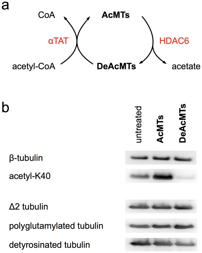
We prepared acetylated tubulin using mouse α-tubulin acetyltransferase (αTAT), which recently was discovered by two independent research groups to specifically acetylate α-tubulin K40 [12], [13] (Fig. 1a). Tubulin K40 deacetylation was performed by incubating tubulin with recombinant human histone transacetylase-like enzyme HDAC6, the role of which has been known for several years [14]. The success of acetylation and deacetylation was proven in a Western blot with antibodies specific for α-tubulin acetyl-K40 [15] (Fig. 1b). In order to rule out effects of HDAC6 or αTAT on other posttranslational tubulin modifications we additionally performed Western blots with antibodies against detyrosinated, decarboxylated (Δ2), and polyglutamylated tubulin. Neither of those modifications was affected (Fig. 1b).
Figure 1. Tubulin acetylation and deacetylation.
(a) Schematic representation of the acetylation and deacetylation of microtubules by αTAT and HDAC6, respectively. (b) Western blots of non-treated, acetylated, and deacetylated tubulin with anti-b-tubulin (SAP4G5, Abnova), anti-AcK40 (6–11B-1, Thermo Scientific), anti-detyrosinated-tubulin (AB3201, Millipore), anti-Δ2-tubulin (pab0202, covalab), and anti-polyglutamylated-tubulin (GT335, Enzo). αTAT-treated tubulin is highly acetylated, whereas HDAC6-treated tubulin is deacetylated. The levels of detyrosination, decarboxylation, and polyglutamylation are not affected by αTAT and HDAC6.
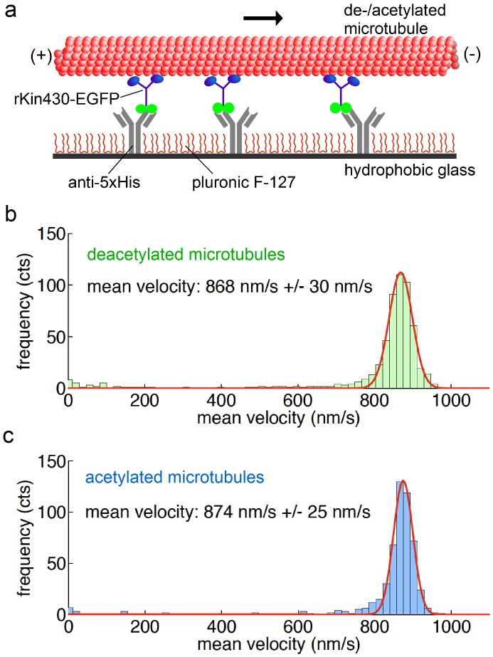
In multi-motor gliding assays [16] the velocities of rhodamine-labeled acetylated and deacetylated microtubules propelled by surface-bound, truncated rat kinesin-1 labeled by EGFP (rKin430-EGFP) [17] were determined in the presence of 1 mM ATP (Fig. 2a). We determined gliding velocities of 868+/−30 nm·s-1 (mean +/− SD, n = 568 microtubules) and 874+/−25 nm·s-1 (n = 532) for deacetylated and acetylated tubulin, respectively (Fig. 2b and c). The 0.7% difference in the mean velocities is statistically significant (p = 0.0003) due to the high number of observes microtubules. However, on the other hand this difference is equivalent to the effect of an increase in temperature by ΔT = 0.1 K (see Methods). As we are experimentally able to control the temperature only within a range of +/−0.5 K, we consider the observed velocity difference to be not significant.
Figure 2. Tubulin acetylation does not affect microtubule velocity in kinesin-1 gliding assays.
(a) Schematic representation of the experimental setup for multi-motor gliding assays. We measured the velocities of short rhodamine-labeled acetylated and deacetylated microtubules propelled by surface-bound motors in presence of 1 mM ATP. The mean gliding velocities of 568 deacetylated microtubules (b) and 532 acetylated microtubules (c) did not differ significantly. The errors represent the SD of the Gaussian distributions.
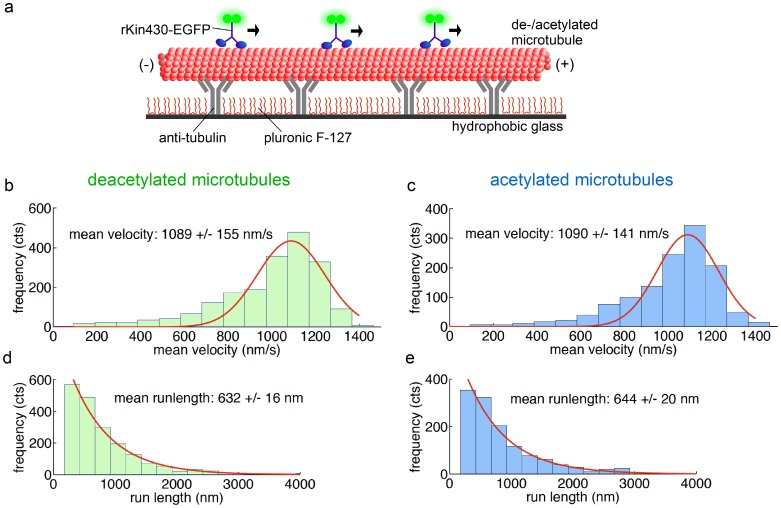
In single-motor stepping assays [18] the velocities of individual rKin430-EGFP motor molecules on acetylated and deacetylated microtubules attached to a glass surface were determined in the presence of 1 mM ATP (Fig. 3a). We determined stepping velocities of 1089+/−155 nm·s−1 (n = 1977 motors) and 1090+/−141 nm·s−1 (n = 1283) on deacetylated and acetylated microtubules, respectively (Fig. 3b and c). Again, these values did not differ significantly (p = 0.85). Moreover, independent of the acetylation status, the run lengths (632+/−16 nm, n = 1949, on deacetylated and 644+/−20 nm, n = 1270, on acetylated microtubules, measured at a modest laser illumination intensity) did not differ significantly (p = 0.44) (Fig. 3d and 3e).
Figure 3. Tubulin acetylation does not affect the kinetics of kinesin-1 stepping on microtubules.
(a) Schematic representation of the experimental setup for single-motor stepping assays. Single rKin430-EGFP motors walk on a microtubule attached to the surface via anti-tubulin antibodies. By tracking the positions of the motors we determined the mean velocities and mean run lengths for deacetylated and acetylated microtubules. The mean velocities of (b) 1977 motors on deacetylated and (c) 1283 motors on acetylated microtubules did not differ significantly. The errors represent the SD of the Gaussian distributions. Mean run lengths of (d) 1949 single motors on deacetylated and (e) 1270 motors on acetylated microtubules were determined by fitting the cumulative probability distributions. The run lengths on deacetylated and acetylated microtubules did not differ significantly. The errors were estimated with the bootstrap technique.
In both, multi-motor gliding and single-motor stepping assays with microtubules reconstituted from acetylated and deacetylated tubulin we did not observe a direct effect of K40 acetylation on kinesin-1 velocity or run length. We thus hypothesize that the differences in motor protein motility previously observed in vivo [4], [6], [9] might simply correlate with - but not be induced by – microtubule acetylation. On one hand, acetylation often coexists with other posttranslational modifications. in vivo experiments indicate that detyrosination of α-tubulin in axonal microtubules might be responsible for the selective translocation of kinesin-1 [4]. Also, chemically inhibited tubulin acetylation in vivo may have effected the acetylation of additional putative acetylation sites in the tubulin dimer [19] or the acetylation of other proteins but tubulin. On the other hand, posttranslational modifications are not the only microtubule features that potentially interfere with molecular motors. Within the cell microtubules display a range of accessory proteins, so called microtubule-associated proteins (MAPs). Although the K40 acetylation site is located in the inner lumen of the microtubule [20], rendering a direct electrostatic effect on MAPs interacting with the outer surface of the microtubule unlikely, small conformational changes induced by K40 acetylation may allosterically propagate to the respective MAP binding sites. Previous results might thus be secondary effects provoked by the interaction of kinesin-1 with MAPs that potentially recognize tubulin acetylation. In particular, the protein tau, which has been described to interfere with kinesin-1 stepping [21] and tubulin acetylation [22], might play a significant role.
In summary, by performing single- and multi-motor in vitro motility assays, we found that tubulin acetylation alone does not affect kinesin-1 velocity and run length. Rather than the acetylation state of microtubules as such, its combination with additional posttranslational modifications or MAPs, may thus be responsible for guiding kinesin-1 towards axonal transport in vivo. Further experiments – including in vitro measurements with controlled populations of posttranslationally modified microtubules and additional binding partners – will be required to fully understand the selective translocation of kinesin-1.
Materials and Methods
Tubulin Acetylation and Deacetylation
Murine αTAT was recombinantly expressed in the E. coli strain BL21 as a GST fusion protein and purified as described previously [13]. The αTAT expression plasmid was a generous gift by Prof. Jacek Gaertig (University of Georgia, Georgia, USA). 25 µM porcine tubulin was acetylated by ∼3 mM αTAT (50 mM Tris-HCl, 50 mM PIPES, 0.5 mM acetyl-CoA, pH 7.6) or deacetylated by 50 µM recombinant human HDAC6 (Enzo Life Sciences, USA) (50 mM Tris-HCl, 50 mM PIPES, pH 7.6) for 90 min at 28°C. Microtubules were polymerized for 90 min at 37°C in presence of 1.25 mM GMPCPP and 1.25 mM MgCl2. Polymerized microtubules were pelleted at 200,000 g for 10 min and resuspended in BRB80 (80 mM PIPES, 1 mM MgCl2, 1 mM EGTA, pH 6.9) with 10 µM taxol.
Gliding and Stepping Motility Assays
All experiments were performed with truncated, EGFP-labeled kinesin-1 constructs (rKin430-EGFP), which contained the first 430 aa of kinesin-1 fused to a EGFP and a His tag at the tail domain [15]. Multi-motor gliding motility assays and imaging were performed at room temperature (22–23°C) as described previously [23] except for the final assay solution (35 mM PIPES, 1 mM MgCl2, 1 mM EGTA, 20 mM KCl, 1 mM ATP, 0.2 mg/ml catalase, 0.1 mg/ml glucose oxidase, 40 mM D-glucose, 10 mM DTT, 10 µM taxol, pH 7.4). Single-motor stepping assays and imaging at modest laser illumination intensity were performed as described previously [24] under the same buffer conditions as the gliding assays.
Data Analysis
Velocities of microtubules and single motors, as well as the run lengths of single motors were obtained using FIESTA tracking software [25]. Mean velocities were determined by fitting the velocity histograms to Gaussian functions. In the analysis of the stepping assays only motor molecules moving over longer distances than 200 nm along the microtubule axis were considered. The significance of velocity differences was tested using t-test statistics. The temperature change ΔT = 0.1 K required to increase the mean velocity from v1 = 868 nm·s−1 to v2 = 868 nm·s−1 was estimated assuming a two-fold velocity increase every 10 K [26] using: ΔT = 10 K · log(v2/v1)/log(2) derived from: v2 = v1·2̂(ΔT/10 K).
To obtain bin-size independent values of the mean run lengths we analyzed the cumulative probability distributions [27]. These distributions were fitted with functions 1 - exp[(x 0−x)/t], where the only fitted parameter t is the mean run length of the distribution and x 0 the lower limit for runs included into the analysis. As an error estimate for the mean run length, we considered 200 bootstrap samples [28] and fitted them as described above. Standard deviations of the sampled sets were used as error estimates. The significance of the differences in the run lengths was tested using Kolmogorov–Smirnov statistics [29]. Repeating the run lengths measurements at 40% and 80% increased laser intensities allowed us to extrapolate to run lengths free of photobleaching effects [30]. For both, deacetylated and acetylated microtubules, we found that these photobleach-free run lengths (though approximately 8% longer than the values given in the main text) did not differ significantly. All fitting was performed in MATLAB (MathWorks, USA).
Funding Statement
This work has been supported by funding from the Volkswagen Foundation (grant I/84 087–090), the Deutsche Forschungsgemeinschaft (DFG Heisenberg Program), the Max-Planck-Society, the Technische Universität Dresden and the European Research Council (ERC starting grant 242933). The funders had no role in study design, data collection and analysis, decision to publish, or preparation of the manuscript.
References
- 1. Vale RD, Reese TS, Sheetz MP (1985) Identification of a novel force-generating protein, kinesin, involved in microtubule-based motility. Cell 42: 39–50. [DOI] [PMC free article] [PubMed] [Google Scholar]
- 2. Nakata T, Hirokawa N (2003) Microtubules provide directional cues for polarized axonal transport through interaction with kinesin motor head. J Cell Biol 162: 1045–55. [DOI] [PMC free article] [PubMed] [Google Scholar]
- 3. Jacobsen C, Schnapp B, Banker GA (2006) A change in the selective translocation of the Kinesin-1 motor domain marks the initial specification of the axon. Neuron 49: 797–804. [DOI] [PubMed] [Google Scholar]
- 4. Konishi Y, Setou M (2009) Tubulin tyrosination navigates the kinesin-1 motor domain to axons. Nat Neurosci 12: 559–67. [DOI] [PubMed] [Google Scholar]
- 5. Cambray-Deakin MA, Burgoyne RD (1987) Acetylated and detyrosinated alpha-tubulins are co-localized in stable microtubules in rat meningeal fibroblasts. Cell Motil Cytoskeleton 8: 284–91. [DOI] [PubMed] [Google Scholar]
- 6. Reed NA, Cai D, Blasius TL, Jih GT, Meyhofer E, et al. (2006) Microtubule acetylation promotes kinesin-1 binding and transport. Curr Biol 16: 2166–72. [DOI] [PubMed] [Google Scholar]
- 7. MacDonald ME, Gines S, Gusella JF, Wheeler VC (2003) Huntington’s disease. Neuromolecular Med 4: 7–20. [DOI] [PubMed] [Google Scholar]
- 8. Gauthier LR, Charrin BC, Borrell-Pagès M, Dompierre JP, Rangone H, et al. (2004) Huntingtin controls neurotrophic support and survival of neurons by enhancing BDNF vesicular transport along microtubules. Cell 118: 127–38. [DOI] [PubMed] [Google Scholar]
- 9. Dompierre JP, Godin JD, Charrin BC, Cordelières FP, King SJ, et al. (2007) Histone deacetylase 6 inhibition compensates for the transport deficit in Huntington’s disease by increasing tubulin acetylation. J Neurosci 27: 3571–83. [DOI] [PMC free article] [PubMed] [Google Scholar]
- 10. Cai D, McEwen DP, Martens JR, Meyhofer E, Verhey KJ (2009) Single molecule imaging reveals differences in microtubule track selection between Kinesin motors. PLoS Biol 7: e1000216. [DOI] [PMC free article] [PubMed] [Google Scholar]
- 11. Hammond JW, Huang CF, Kaech S, Jacobson C, Banker G, et al. (2010) Posttranslational modifications of tubulin and the polarized transport of kinesin-1 in neurons. Mol Biol Cell 21: 572–83. [DOI] [PMC free article] [PubMed] [Google Scholar]
- 12. Shida T, Cueva JG, Xu Z, Goodman MB, Nachury MV (2010) The major alpha-tubulin K40 acetyltransferase alphaTAT1 promotes rapid ciliogenesis and efficient mechanosensation. Proc Natl Acad Sci U S A 107: 21517–22. [DOI] [PMC free article] [PubMed] [Google Scholar]
- 13. Akella JS, Wloga D, Kim J, Starostina NG, Lyons-Abbott S, et al. (2010) MEC-17 is an alpha-tubulin acetyltransferase. Nature 467: 218–22. [DOI] [PMC free article] [PubMed] [Google Scholar]
- 14. Hubbert C, Guardiola A, Shao R, Kawaguchi Y, Ito A (2002) HDAC6 is a microtubule-associated deacetylase. Nature 417: 455–8. [DOI] [PubMed] [Google Scholar]
- 15. Piperno G, Fuller MT (1985) Monoclonal antibodies specific for an acetylated form of alpha-tubulin recognize the antigen in cilia and flagella from a variety of organisms. J Cell Biol 101: 2085–94. [DOI] [PMC free article] [PubMed] [Google Scholar]
- 16. Nitzsche B, Bormuth V, Bräuer C, Howard J, Ionov L, et al. (2010) Studying kinesin motors by optical 3D-nanometry in gliding motility assays. Methods Cell Biol 95: 247–71. [DOI] [PubMed] [Google Scholar]
- 17. Rogers KR, Weiss S, Crevel I, Brophy PJ, Geeves M, et al. (2001) KIF1D is a fast non-processive kinesin that demonstrates novel K-loop-dependent mechanochemistry. EMBO J 20: 5101–5113. [DOI] [PMC free article] [PubMed] [Google Scholar]
- 18. Gell C, Bormuth V, Brouhard GJ, Cohen DN, Diez S, et al. (2010) Microtubule dynamics reconstituted in vitro and imaged by single-molecule fluorescence microscopy. Methods Cell Biol 95: 221–45. [DOI] [PubMed] [Google Scholar]
- 19. Choudhary C, Kumar C, Gnad F, Nielsen ML, Rehman M, et al. (2009) Lysine acetylation targets protein complexes and co-regulates major cellular functions. Science 325: 834–840. [DOI] [PubMed] [Google Scholar]
- 20. Nogales E, Wolf SG, Downing KH (1998) Structure of the alpha beta tubulin dimer by electron crystallography. Nature 391: 199–203. [DOI] [PubMed] [Google Scholar]
- 21. McVicker DP, Chrin LR, Berger CL (2011) The nucleotide-binding state of microtubules modulates kinesin processivity and the ability of Tau to inhibit kinesin-mediated transport. J Biol Chem 286: 42873–80. [DOI] [PMC free article] [PubMed] [Google Scholar]
- 22. Perez M, Santa-Maria I, Gomez de Barreda E, Zhu X, Cuadros R, et al. (2009) Tau – an inhibitor of deacetylase HDAC6 function. Neurochem 109: 1756–66. [DOI] [PubMed] [Google Scholar]
- 23. Kerssemakers J, Howard J, Hess H, Diez S (2006) The distance that kinesin-1 holds its cargo from the microtubule surface measured by fluorescence interference contrast microscopy. Proc Natl Acad Sci U S A 103: 15812–7. [DOI] [PMC free article] [PubMed] [Google Scholar]
- 24. Varga V, Leduc C, Bormuth V, Diez S, Howard J (2009) Kinesin-8 motors act cooperatively to mediate length-dependent microtubule depolymerization. Cell 138: 1174–83. [DOI] [PubMed] [Google Scholar]
- 25. Ruhnow F, Zwicker D, Diez S (2011) Tracking single particles and elongated filaments with nanometer precision. Biophys J 100: 2820–28. [DOI] [PMC free article] [PubMed] [Google Scholar]
- 26. Kawaguchi K, Ishiwata S (2000) Temperature dependence of force, velocity, and processivity of single kinesin molecules. Biochem Biophys Res Commun. 272: 895–9. [DOI] [PubMed] [Google Scholar]
- 27. Thorn KS, Ubersax JA, Vale RD (2000) Engineering the processive run length of the kinesin motor. J Cell Biol 151: 1093–1100. [DOI] [PMC free article] [PubMed] [Google Scholar]
- 28.Press WH, Teukolsky SA, Vetterling WT, Flannery BP (1992) Numerical recipes in C. Cambridge, UK, Cambridge University Press. [Google Scholar]
- 29.Wessa P (2012) Free Statistics Software, Office for Research Development and Education, version 1.1.23-r7, URL. http://www.wessa.net/.
- 30. Mashanov GI, Tacon D, Peckham M, Molloy JE (2004) The Spatial and Temporal Dynamics of Pleckstrin Homology Domain Binding at the Plasma Membrane Measured by Imaging Single Molecules in Live Mouse Myoblasts. J Biol Chem 279: 15274–80. [DOI] [PubMed] [Google Scholar]


