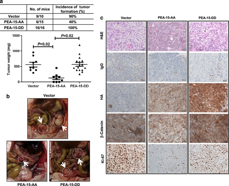
Figure 3.
PEA-15-AA has a strong antitumor effect in an ovarian cancer xenograft model. (a) Upper panel, incidence of tumor formation. Lower panel, tumor weights were significantly smaller with PEA-15-AA than with vector and PEA-15-DD. (b) Representative images showed tumor formation of SKOV3.ip1-vector, SKOV3.ip1-PEA-15-AA and SKOV3.ip1-PEA-15-DD cells in mouse peritoneal area. White arrows indicate tumors. (c) Immunohistochemical staining of HA, β-catenin and Ki-67 in tumor samples from the xenograft model. H&E, hematoxylin-eosin. Scale bars: 50 μm. Statistical significance was evaluated by paired t-test using GraphPad Prism software.

