Abstract
Killian-Jamieson diverticulum is a rare diverticular disease. This disease differs from Zenker's diverticulum in its location and mechanism. Various treatment modality have been attempted, but traditional surgical treatment has been recommended for a symptomatic Killian-Jamieson diverticulum due to the concern of possible nerve injury. We performed surgical treatment by cervical incision. We report here on a case of Killian-Jamieson diverticulum and we briefly review the relevant literature.
Keywords: Esophageal disease; Surgery, esophagus; Killian-Jamieson diverticulum
CASE REPORT
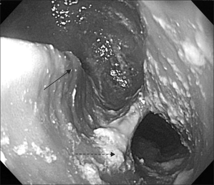
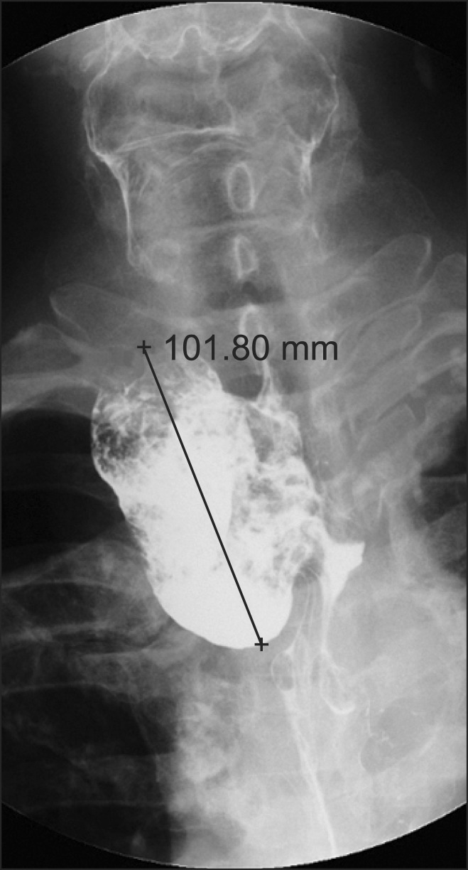
A 68-year-old male patient visited Konkuk University Hospital due to epigastric pain lasting 2 to 3 seconds 2 months before hospitalization. According to his anamnesis, he was taking 50 mg of Atenolol because he had been diagnosed with hypertension 2 years before. He had been diagnosed with esophageal diverticulum 10 years before; since then he had experienced no symptoms. He underwent a cardiac computed tomography (CT) scan 3 weeks prior to hospitalization. The CT scan showed that mild stenosis (20% to 30%) had formed in left anterior descending artery, left circumflex artery, and right coronary artery. An exercise stress test had negative results. He underwent gastroesophageal endoscopy; 5 cm of diverticulum was found 20 cm from the incisor tooth (Fig. 1). There were no other findings except for erosive gastritis in the antrum of the stomach. The esophagography 2 weeks prior to the hospitalization showed that there was 10 cm of diverticulum projecting to anterolateral side; Killian-Jamieson diverticulum (KJD) was considered (Fig. 2). He received surgical treatment for esophagedal diverticulum.
Fig. 1.
Endoscopic finding of Killian-Jamieson diverticulum. Large diverticulum was noticed at 20 cm from incisor. Continous arrow line indicates diverticular opening, and dotted arrow line indicates esophageal opening.
Fig. 2.
Esophagogram shows a large diverticulum, anterolateral side at cervical esophagus.
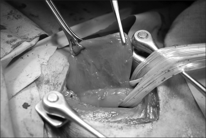
Although the location of the diverticulum on the esophagogram was right side, we decided left cervical approach for surgeon's convenience. Left cervical incision was conducted under general anesthesia and the cervical esophagus was exposed following dissection along the sternocleidomastoid muscle. Diverticulum (10×5 cm sized) was found with a wide base and which contained a small amount of necrotic tissue (Fig. 3). The diverticulum adhered to circumjacent tissues; in particular, it strongly adhered to the prevertebral fascia in the rear of the trachea. Cervical esophagus proximal to the diverticulum was dissected cautiously and looped with a silastic drain. The diverticulum was dissected from adjacent tissues. The diverticulum was excised with a TA 60 stapler (Ethicon Endo-Surgery, Cincinnati, OH, USA). Esophageal myotomy of about 3 cm was conducted along the distal part of the esophagus after the excision of the diverticulum. Reinforcement sutures were inserted with 3-0 silk along the excision area of diverticulum. The surgery was completed after a Jackson-Pratt drainage tube was inserted. The patient fasted for 3 days after the surgery with a nasogastric tube. He recovered without complications such as nerve damage or hemorrhage. The esophagography on the fourth day after the surgery showed that there were not abnormalities such as leakage or stenosis; therefore, dietary treatment was initiated. The patient was discharged from Konkuk University Hospital on the sixth day after the surgery. At the time of discharge, there was no special problem. Follow-up observation has been performed for 6 months, during which the patient has not shown any abnormalities such as diverticulum relapse, dysphagia, or stenosis.
Fig. 3.
Operative finding showing a large diverticulum which originated from the posterolateral side of the cervical esophagus.
DISCUSSION
KJD is a rare form of esophageal diverticulum which appears through the Killian's dehiscence, a mucosal protrusion below the cricopharyngeal muscle [1]. KJD is similar to Zenker's diverticulum (ZD) due to the protrusion of esophageal mucosa. KJD is rare in that the incidence of KJD is a fourth of that of ZD. The two diseases have similar symptoms (e.g., dysphagia, coughing, and chest pain). However, according to previous studies, KJD has more non-specific symptoms than ZD. This is related to the location of these two diseases. Location is a standard for distinguishing them: ZD occurs mainly in the rear of the esophagus in the upper cricopharyngeal muscle, and KJD occurs mainly in the front or side of the esophagus 2 cm away from the lower cricopharyngeal muscle. Gastroesophageal reflux occurs more frequently in ZD than in KJD [2]. Cervical cellulitis is a rare but severe complication [3]. The two diseases, ZD and KJD, have been observed simultaneously. In rare cases, bilateral Killian-Jamieson diverticula have been discovered [4]. Cervical ultrasonography is conducted as a diagnostic technique. Ultrasonography can differential diagnose between thyroid nodule and KJD [5].
Because of its symptom or greater size, endoscopic treatment and surgical treatment are used for KJD. Generally, endoscopic treatment is considered preferable in treating Zenker's diverticula smaller than 3 cm [6]. Endoscopic treatment has the possibility of occluded view when there is food or a foreign body in the diverticulum. The treatment of the KJD is more closely adjacent to the recurrent laryngeal nerves than ZD. For KJD treatments that do not come into contact with the recurrent nerves, treatments other than endoscopic treatments are preferable. According to the previous studies, the safety of endoscopic treatment has not been established for KJD, due largely to the rarity of cases, and for ZD the recurrence rate of endoscopic treatment is 10 times higher than that of surgical treatment [7,8]. Furthermore, myotomy should be adopted as a treatment for KJD, since its treatment is closely related to the prevention of the recurrence of ZD from the perspective of the features of diverticular disease.
In conclusion, Konkuk University Hospital experienced the successful treatment of a case of KJD that had been accompanied by rare symptom and discussed the result together with the review of the relevant literary works.
References
- 1.Han KN, Kim YT, Nam J, Kang CH, Kim JH. Surgical experience with Killian-Jamieson diverticulum: a case report. Korean J Thorac Cardiovasc Surg. 2010;43:324–327. [Google Scholar]
- 2.Rubesin SE, Levine MS. Killian-Jamieson diverticula: radiographic findings in 16 patients. AJR Am J Roentgenol. 2001;177:85–89. doi: 10.2214/ajr.177.1.1770085. [DOI] [PubMed] [Google Scholar]
- 3.Kitazawa M, Koide N, Saito H, Kamimura S, Uehara T, Miyagawa S. Killian-Jamieson diverticulitis with cervical cellulitis: report of a case. Surg Today. 2010;40:257–261. doi: 10.1007/s00595-009-4048-z. [DOI] [PubMed] [Google Scholar]
- 4.Kim MH, Kim EK, Kwak JY, Kim MJ, Moon HJ. Bilateral Killian-Jamieson diverticula incidentally found on thyroid ultrasonography. Thyroid. 2010;20:1041–1042. doi: 10.1089/thy.2010.0038. [DOI] [PubMed] [Google Scholar]
- 5.Kim SJ, Lee MW. Killian-Jamieson diverticula mimicking thyroid nodule on ultrasound: radiographic findings in two patients. J Korean Radiol Soc. 2006;55:443–446. [Google Scholar]
- 6.Veenker E, Cohen JI. Current trends in management of Zenker diverticulum. Curr Opin Otolaryngol Head Neck Surg. 2003;11:160–165. doi: 10.1097/00020840-200306000-00006. [DOI] [PubMed] [Google Scholar]
- 7.Lee CK, Chung IK, Park JY, et al. Endoscopic diverticulotomy with an isolated-tip needle-knife papillotome (Iso-Tome) and a fitted overtube for the treatment of a Killian-Jamieson diverticulum. World J Gastroenterol. 2008;14:6589–6592. doi: 10.3748/wjg.14.6589. [DOI] [PMC free article] [PubMed] [Google Scholar]
- 8.Chang CY, Payyapilli RJ, Scher RL. Endoscopic staple diverticulostomy for Zenker's diverticulum: review of literature and experience in 159 consecutive cases. Laryngoscope. 2003;113:957–965. doi: 10.1097/00005537-200306000-00009. [DOI] [PubMed] [Google Scholar]


