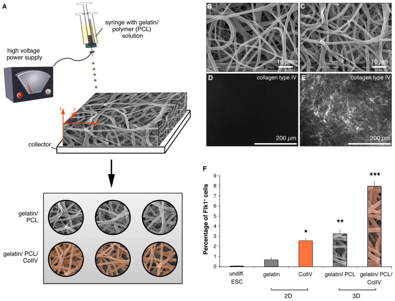
Figure 4.
(A) Schematic of the electrospinning set-up and protocol of 2D versus 3D experiments. (B-C) Scanning electron micrographs of electrospun gelatin-PCL hybrid substrates without (B) or with (C) ColIV-coating. (D-E) Compared to the non-coated controls (D), immunofluorescence staining reveals the successful ColIV-coating of the electrospun material (E). (F) FACS analyses revealed an increased percentage of Flk1+ cells from 3D gelatin/PCL/ColIV cultures. *P=0.004 versus undiff ES cells and 2D gelatin cultures. **P<0.0001 versus undiff. ES cells and 2D gelatin and 2D ColIV cultures. ***P<0.0001 versus undiff. ES cells and gelatin, 2D ColIV and 3D gelatin/PCL cultures.

