Abstract
Anterior Cruciate Ligament (ACL) rupture is a common sporting injury that frequently affects young, athletic patients. Apart from the functional problems of instability, patients with ACL deficient knees also develop osteoarthritis. Although this is frequently cited as an indication for ACL reconstruction, the relationship between ACL rupture, reconstruction and the instigation and progression of articular cartilage degenerative change is controversial.
The purpose of this paper is to review the published literature with regards ACL rupture and the multifactorial causes for osteoarthritis progression, and whether or not this is slowed or stopped by ACL reconstruction.
There is no evidence in the published literature to support the view that ACL reconstruction prevents osteoarthritis, although it may prevent further meniscal damage. It must be recognised that this conclusion is based on the current literature which has substantial methodological limitations.
Keywords: Anterior cruciate ligament, anterior cruciate ligament reconstruction, cartilage damage, knee surgery, meniscus, osteoarthritis.
INTRODUCTION
The anterior cruciate ligament (ACL) is an important stabiliser of the knee, functioning as the primary constraint preventing anterior translation of the tibia on the femur, and also stabilising the knee against rotational and valgus stresses [1]. Rupture of the ACL, unfortunately, is a common sports injury, with a reported incidence of 0.38 per 100,000 individuals [2]. It frequently affects young, active people with long working futures and sporting ambitions. Functional problems in the ACL deficient knee arise from instability, particularly in activities requiring pivoting and side stepping. This can cause levels of disability ranging from limitation of sporting activity to restricting activities of daily living. Recurrent knee injuries secondary to instability can result in intraarticular damage, in particular meniscal tears and subsequent osteoarthritis (OA).
The majority of patients with ACL deficient knees are able to walk normally and perform straight line activities. Studies in nonoperatively managed patients who remain active [3], however, have indicated that up to 44% of patients will develop significant functional disability affecting their ability to perform activities of daily living. Many studies have shown that ACL deficient knees will deteriorate radiologically and functionally over time due to advancing osteoarthritis [3, 4].
As a result the ACL is the most commonly injured ligament undergoing surgical intervention, aiming to return the patient to his or her preinjury level of activity and preventing osteoarthritis. In the United States alone around 175,000 ACL reconstructions were performed in the year 2000 [5], costing around $2 billion. Meta analyses of the published literature [6, 7] have shown that between 67 and 76 % patients of patients undergoing ACL reconstruction are able to return to preinjury levels of activity, with up to 80% of patients having normal or near normal functional International Knee Documentation Committee (IKDC) scores.
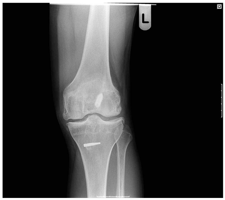
What is still a matter of controversy, however, is the ability of ACL reconstruction to prevent or delay the onset of degenerative change within the knee. Fig. (1) is an example of ACL reconstruction not preventing the development of advanced osteoarthritis from developing in a young patient. The purpose of this paper is to review the concepts surrounding the pathogenesis of osteoarthritis in the ACL deficient knee and whether early or late reconstruction can delay or prevent degenerative change.
Fig. (1).
Despite ACL reconstruction and restoration of knee stability, radiographic evidence and clinical signs of degenerative change have developed.
OSTEOARTHRITIS AFTER ACL RUPTURE
The relationship between osteoarthritis and ACL rupture is one of the main reasons cited for advocating ACL reconstruction in the young, active patient but is difficult to define. There is a great variability in the reported rate of osteoarthritis after ACL rupture in the published literature, ranging between 24% [8] to 86% [9]. It is difficult to produce a definitive figure based on published results due to the variability in study design and multiple ways in which osteoarthritis is graded radiographically and functionally, making pooling of data and meta analysis difficult [10].
This is partly because degeneration of articular cartilage is difficult to measure, particularly in the early stages. The lack of a universal measurement tool with adequate sensitivity and specificity makes it impossible to state the true prevalence or rate of advancement of degenerative change after ACL rupture or reconstruction. Plain radiographs [11] are not capable of detecting early change. Over 10 different grading systems for radiographic evidence of osteoarthritis are used in the published literature, some of which are poorly described and not validated [10]. MRI, although useful for assessing ligamentous and meniscal damage, has poor sensitivity and specificity for articular cartilage lesions [12]. Arthroscopy does have a high sensitivity and specificity in assessing articular cartilage, but is expensive, invasive and requires a general anaesthetic. Some studies [13,14] have used radioisotope bone scans to detect subchondral metabolic osseous activity in early degenerative change, but this would not be able to measure progression of degenerative change. When assessing for advancing degenerative changes it should also be noted that clinical and functional symptoms do not always correlate with imaging or arthroscopic assessment.
THE PATHOGENESIS OF OSTEOARTHRITIS IN THE ACL DEFICIENT KNEE
It is unclear what causes osteoarthritis after ACL rupture. Degenerative change may be caused by recurrent injury secondary to knee instability, but may also be due to injuries sustained by the articular cartilage, subchondral bone and menisci during the original injury. Intra-articular bleeding at the time of rupture can also affect articular cartilage, activating inflammatory pathways [10].
Subchondral Bone Damage
The substantial force required to rupture a healthy ACL can also produce other injuries to the subchondral bone. At the time of ACL rupture, MRI scans frequently show marrow signal change consistent with subchondral bone damage [15]. Similarly, a biomechanical study on cadavers [16] has demonstrated significant subchondral bone and articular cartilage damage occurring during the initial injury causing ACL rupture, particularly if the mechanism of injury involves compression loading.
These traumatic bone marrow lesions may represent the footprint of significant compressive forces applied to joint surfaces at the moment of injury [17]. The compressive mechanical forces required to produce these lesions would also affect the overlying articular cartilage. This can contribute to osteoarthritis even in the absence of joint instability, altering the cartilaginous matrix and damaging chondrocytes [18]. MRI studies investigating the natural history of these subchondral bone lesions suggest that osteochondral abnormalities do develop at the site of severe bone bruising [15, 19-21].
Meniscal Injury
There is strong evidence that meniscal damage can lead to osteoarthritic changes in knees with and without a functioning ACL. Follow up studies after partial [9, 22] or total meniscectomy with an intact ACL have shown increased radiological signs of degenerative changed when compared with the contralateral knee. In some studies the prevalence of osteoarthritis after meniscectomy was as high as 71% after 21 years [23], and up to 7 times higher than the contralateral, uninjured knee [9]. Similarly, when a control group was used, the rate of radiologically evident degenerative change was seven times higher after partial meniscectomy [24].
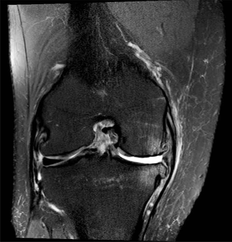
Multiple studies have shown that although meniscal tears can occur at the time of ACL rupture, the rate of subsequent meniscal injury is increased after ACL rupture. This may be due to continued attrition secondary to abnormal loading and shear forces [25]. A study arthroscopically assessing patients with ACL deficient knees found meniscal tears in 27% in the acutely injured, which increased to 90% in the chronically unstable [26]. Another arthroscopic study in ACL deficient knees suggested continued, progressive meniscal degeneration over time [25]. Thus the risk of osteoarthritis can be increased in chronic ACL deficiency at least in part due to the development of subsequent meniscal injury. Fig. (2) is an example of osteoarthritis developing in a patient with a chronic ACL tear and a medial meniscal tear.
Fig. (2).
Coronal T2 weighted MRI image of a knee with a chronic ACL tear with a deficient medial meniscus and medial compartment osteoarthritis.
There is also evidence suggesting that meniscal injury can be prevented by ACL reconstruction. A retrospective study of over 6500 active duty army personnel with ruptured ACL’s showed the incidence of subsequent meniscal damage was two times higher in ACL deficient knees compared to patients where the ACL had been reconstructed [27]. Similarly, studies following up the results of ACL reconstruction have noted a decreased in the rate of subsequent meniscal injury [28, 29].
Continued Instability and Altered Knee Biomechanics
In addition to the initial trauma to the knee at the time of ACL rupture, continued instability in the ACL deficient knee also is a contributor to the development of osteoarthritis. In the absence of a functioning ACL, the static and dynamic loading of forces across the knee is altered, increasing the forces applied to the articular cartilage [30], which can both initiate and perpetuate osteoarthritis progression. The loss of anterior-posterior translational stability and rotational stability results in a spatial shift in areas of tibiofemoral contact. This change in the load bearing area and contact mechanics, particularly in rotational malalignment, has been associated with an increased in degenerative change [31, 32]. This is likely to be perpetuated by the loss of the ACL’s proprioceptive function.
Apart from the altered contact mechanics of the articular cartilage, continued instability also increases the likelihood of further injury to the knee, injuring other structures, including the menisci [27], which in turn would accelerate degenerative change.
ACL RECONSTRUCTION AND OA PROGRESSION
In order to determine if reconstruction of the ACL can prevent or delay progression of OA, we should look at studies comparing patients with ACL deficient and ACL reconstructed knees. Unfortunately, when reviewing the literature it must be noted that long-term studies on knee injuries are very heterogeneous in methodology, in particular with regards outcome measures, type of graft, timing of surgery and rehabilitative regimes. There is also great variability and poor reporting of variables that might be expected to influence outcome, such as age, patient sex, activity levels, and other patient-associated factors. The limited size, short follow up and design flaws within many studies prevent definitive conclusions from being made and make pooling of data or meta analysis difficult.
Early studies including that by Dale Daniel and colleagues indicated a higher incidence of radiographic OA in patients with ACL reconstructed knees compared to those treated non-operatively, at mean follow-up of 64 months [33]. Although the ACL reconstruction techniques described at the time are largely no longer used, a more recent study from the same institution showed a similarly greater incidence of radiographic arthrosis in patients treated operatively with BPTB allograft compared to those managed conservatively [34]. However, in the same study, Tegner scores (grading activity level) were significantly higher in the patients treated with early reconstruction (within 3 months of the ACL injury). The main criticism of this study is the lack of randomisation, with ACL reconstruction being performed in the more severely injured knees.
Another study reviewed outcomes in Swedish soccer players who had sustained a ruptured ACL and were followed up at 7 [35], 12 [36] and 14 years post injury. Reconstruction was performed in 60% of the players studied. At each time point, there was no significant difference seen between surgically and conservatively managed players in terms of radiographic evidence of osteoarthritis, presence of symptoms, and level of sporting activity.
Similarly, another study observed 79 professional Norwegian handball players, at a mean follow-up of 7 years post ACL rupture [37]. Greater antero-posterior laxity was demonstrated in the conservatively treated subjects compared to those treated surgically. This did not, however, correlate with functional recovery as the authors found that a higher percentage of patients returned to the same level of sport activity when treated conservatively. There was no difference in the prevalence of radiologically abnormal or severe arthritic change between the two groups.
Fink and colleagues reported their findings in 2001 demonstrating no significant difference in radiographic narrowing of the joint space in patients treated with reconstruction compared to those treated conservatively at mean follow-up of 11 years (48% vs 50% respectively) [38]. In this study all the reconstructions were performed using a bone-patella tendon-bone graft. The study was somewhat weakened by the fact that the decision to proceed with operative or non-operative treatment was made by the patients, introducing significant bias. The authors also demonstrated a correlation between participation in pivoting sports and development of early osteoarthritic changes in those treated non-operatively.
A retrospective study from 2008 compared the outcomes of treatment of isolated ACL ruptures with bone-patella tendon-bone graft reconstruction with that of conservative management, demonstrating a significantly higher prevalence of radiographic joint space narrowing in the ACL reconstructed group (45% vs 24%), at average follow-up of 11.1 years [39].
One recent paper has, however, shown reduced rates of osteoarthritis after ACL reconstruction. A recent retrospective, non-randomised study with 17-20 year follow up showed a significantly lower rate of severe osteoarthritis after reconstruction when compared to a conservatively managed control group (16.5% vs 56%). Although over 50% of reconstructed patients did have mild degenerative changes, in the conservatively managed patient group there were no normal knees [40].
The continued progression of osteoarthritis after ACL reconstruction is perhaps not surprising given the multifactorial causes for degenerative change. Although the continually improving methods for ACL reconstruction restore stability and much of the normal mechanics of the knee, the reconstructed knee is not a normal knee. Altered mechanics in the reconstructed knee can be expected to produce abnormal loading patterns across the joint, significantly increasing the risk of OA [10].
Multiple studies have also investigated potential risk factors for the development of osteoarthritis after ACL reconstruction. One such study identified Female sex, Body Mass Index (BMI), time from injury to surgery, medial and patellofemoral compartment chondrosis, prior medial or lateral meniscectomy, concurrent medial meniscectomy, and length of follow-up as risk factors for developing radiographic evidence of degenerative change, with medial meniscectomy followed by pre-existing chondral damage the strongest predictors [41]. Similarly, a prospective cohort study by Keays and colleagues found five factors to be predictive of tibiofemoral osteoarthritis. These were meniscectomy, chondral damage, the use of patella tendon as a graft, weak quadriceps and poor quadriceps to hamstring strength ratios. Of these meniscectomy, followed by chondral damage, was again the strongest predictor of degenerative change [41].
When looking specifically at the effect of meniscal and chondral damage on the results of ACL reconstruction, a retrospective study found that patients with no evidence of meniscal or chondral damage at ACL reconstruction had good outcomes, with 97% having normal or near normal radiographic appearances, with a mean follow up of over 7 years [42]. Conversely, abnormal or severely abnormal radiographs were seen in 9%, 23% and 25% of patients who had undergone a lateral, medial or bilateral meniscectomy respectively.
In addition, post operative range of movement in the knee has been shown to be another risk factor for progression of osteoarthritis. In a recent paper by Shelbourne, patients were retroactively reviewed at a mean of 10 years post reconstruction. In these patients the rehabilitation regime aimed to regain a full range of motion as early as possible. Patients who regained a full range of movement had a significantly lower incidence of osteoarthritis when compared to patients who had restrictions in flexion or extension (39% vs 53%) [43].
TIMING OF ACL RECONSTRUCTION
When considering the impact of ACL reconstruction on osteoarthritis, it is difficult to compare or pool the results of multiple studies due to the great variability in surgical technique, graft choice, rehabilitation regimes and timing of surgery. The timing of surgery, and whether this may have an impact on subsequent development of arthritis, is an area of controversy.
Some studies comparing ACL reconstruction for acute ruptures with reconstruction after a period of chronic instability do show improved subjective and objective results [44-46]. This includes a reduced rate of osteoarthritis on x-rays [47]. A study by Jarvela in 1999 [48] assessed knee radiographs after early (mean 6 days) or late (mean 3.7 years) ACL reconstruction, with the late reconstruction group having a significantly higher incidence of medial compartment degenerative changes, although no difference was seen in the patellofemoral joint or lateral compartment. Other studies, however [42, 49], have shown no significant difference.
Multiple papers have noted that the need for meniscectomy at ACL reconstruction increases with delayed surgery, arguing that there is a greater incidence of meniscal injury in functionally unstable knees, which in turn may lead to subsequent tibiofemoral osteoarthritis [25, 26, 47, 50, 51]. One multicentre study quotes the relative risk of meniscal injury at 5 years being almost 6 times that of the first year post injury [51]. Furthermore, some studies have produced results suggestive that ACL reconstruction would have a protective effect, preventing further meniscal damage [27, 34, 48], and therefore make the argument that early ACL reconstruction can reduce the development of subsequent meniscal pathology and osteoarthritis.
A meta analysis of the published literature, however, did not support this hypothesis, finding no significant difference in the incidence of meniscal pathology or osteoarthritis after early or late ACL reconstruction [49]. This paper, did, however accept that this finding was underpowered due to small sample sizes. There was also considerable variability in what was felt to be an early of late reconstruction. Clearly more research is required, as the current evidence base is hindered by methodological limitations.
CONCLUSION
ACL rupture is a common sporting injury frequently affecting young patients. Although reconstruction of the ACL can improve the functional outcome, particularly in patients with a high functional demand, there is currently little data supporting the concept that ACL reconstruction prevents or delays degeneration of the knee joint.
Patients with ACL ruptures with concomitant meniscal, and to a lesser extent, chondral damage are at greater risk of developing osteoarthritis, whether or not the ACL has been reconstructed. It has been shown that ACL reconstruction prevents further meniscal damage, and the argument has been put forward that this could reduce further chondral injury. There is, however, limited evidence to support this.
Choice of treatment remains very much patient guided in many institutions and is often dependent on the level of sports participation. It is difficult to make meaningful comparisons between outcome studies due to the heterogeneity of methodology, which includes, severity of initial injury, surgical technique, graft choice, timing of surgery and rehabilitation. Pooling data and meta analyses is therefore difficult due to the weaknesses of the published studies. Randomised, controlled trials could provide a definitive answer but, to be sufficiently powered, would require recruitment of a large number of young patients who would be willing to be treated conservatively.
ACKNOWLEDGEMENTS
None declared.
CONFLICT OF INTEREST
Neither of the authors have a conflict of interest to declare in relation to the production of this manuscript.
REFERENCES
- 1. Levy IM, Torzilli PA, Warren RF. The effect of medial meniscectomy on anterior-posterior motion of the knee. J Bone Joint Surg Am. 1982;64-A:883–8. [PubMed] [Google Scholar]
- 2. Miyasaka KC, Daniel DM, Stone ML, Hirshman P. The incidence of knee ligament injuries in the general population. Am J Knee Surg. 2001;4:3–8. [Google Scholar]
- 3. Noyes FR, Mooar PA, Matthews DS, Butler DL. The symptomatic anterior cruciate-deficient knee. Part I: the long term functional disability in athletically active individuals. J Bone Joint Surg Am . 1983;65-A:154–62. doi: 10.2106/00004623-198365020-00003. [DOI] [PubMed] [Google Scholar]
- 4. Øiestad BE, Holm I, Engebretsen L, Risberg MA. The association between radiographic knee osteoarthritis and knee symptoms, function and quality of life 10-15 years after anterior cruciate ligament reconstruction. Br J Sports Med. 2011;45:583–8. doi: 10.1136/bjsm.2010.073130. [DOI] [PubMed] [Google Scholar]
- 5. Spindler KP, Wright RW. Clinical practice: anterior cruciate ligament tear. N Engl J Med. 2008;359(20):2135–42. doi: 10.1056/NEJMcp0804745. [DOI] [PMC free article] [PubMed] [Google Scholar]
- 6. Biau DJ, Tournoux C, Katsahian S, Schranz P, Nizard R. ACL reconstruction: a meta-analysis of functional scores. Clin Orthop Relat Res. 2007;458:180–7. doi: 10.1097/BLO.0b013e31803dcd6b. [DOI] [PubMed] [Google Scholar]
- 7. Biau DJ, Tournoux C, Katsahian S, Schranz PJ, Nizard RS. Bone patellar tendon-bone autografts versus hamstring autografts for reconstruction of anterior cruciate ligament: meta-analysis. Br Med J. 2008;332(7548):995–1001. doi: 10.1136/bmj.38784.384109.2F. [DOI] [PMC free article] [PubMed] [Google Scholar]
- 8. Casteleyn PP, Handelberg F. Non-operative management of anterior cruciate ligament injuries in the general population. J Bone Joint Surg. 1996;78-B:446–51. [PubMed] [Google Scholar]
- 9. Neyret P, Donell ST, Dejour H. Results of partial meniscectomy related to the state of the anterior cruciate ligament. Review at 20 to 35 years. J Bone Joint Surg. 1993;75-B:36–40. doi: 10.1302/0301-620X.75B1.8421030. [DOI] [PubMed] [Google Scholar]
- 10. Lohmander LS, Englund M, Dahl LL, Roos EM. The long term consequence of anterior cruciate ligament and meniscus injuries: Osteoarthritis. Am J Sports Med. 2007;35:1756–69. doi: 10.1177/0363546507307396. [DOI] [PubMed] [Google Scholar]
- 11. Thomas RH, Resnick D, Alazraki NP, Daniel D, Greenfield R. Compartmental evaluation of osteoarthritis of the knee: a comparative study of available diagnostic modalities. Radiology . 1975;116:585–94. doi: 10.1148/116.3.585. [DOI] [PubMed] [Google Scholar]
- 12. Polly DW, Jr, Callaghan MJ, Sikes RA, McCabe JM, McMahon K, Savory CG. The accuracy of selective MRI compared with the findings of arthroscopy of the knee. J Bone Joint Surg Am. 1988; 70-A:192–8. [PubMed] [Google Scholar]
- 13. Mooar PA, Gregg J, Jacobstein J. Radionuclide imaging in internal derangement of the knee. Am J Sports Med. 1987;15:132–7. doi: 10.1177/036354658701500207. [DOI] [PubMed] [Google Scholar]
- 14. Hart AJ, Buscombe J, Malone A, Dowd GSE. Assessment of osteoarthritis after reconstruction of the anterior cruciate ligament. J Bone Joint Surg Br. 2005;87-B:1483–7. doi: 10.1302/0301-620X.87B11.16138. [DOI] [PubMed] [Google Scholar]
- 15. Costa-Paz M, Muscolo L, Ayerza M, Makino A, Aponte-Tinao L. Magnetic resonance imaging follow-up study of bone bruises associated with anterior cruciate ligament ruptures. Arthroscopy . 2001;17:445–9. doi: 10.1053/jars.2001.23581. [DOI] [PubMed] [Google Scholar]
- 16. Meyer EG, Baumer TG, Slade JM, Smith WE, Haut RC. Tibiofemoral contact pressures and osteochondral microtrauma during anterior cruciate ligament rupture due to excessive compressive loading and internal torque of the human knee. Am J Sports Med. 2008;36:1966–77. doi: 10.1177/0363546508318046. [DOI] [PubMed] [Google Scholar]
- 17. Sanders TG, Medynski MA, Feller JF, Lawhorn KW. Bone contusion patterns of the knee at MR imaging: footprint of the mechanism of injury. Radiographics. 2000;20:S135–51. doi: 10.1148/radiographics.20.suppl_1.g00oc19s135. [DOI] [PubMed] [Google Scholar]
- 18. Buckwalter JA. Articular cartilage injuries. Clin Orthop Relat Res . 2002;402:21–37. doi: 10.1097/00003086-200209000-00004. [DOI] [PubMed] [Google Scholar]
- 19. Faber KJ, Dill JR, Amendola A, Thain L, Spouge A, Fowler PJ. Occult osteochondral lesions after anterior cruciate ligament rupture. Six-year magnetic resonance imaging follow-up study. Am J Sports Med. 1999;27:489–94. doi: 10.1177/03635465990270041301. [DOI] [PubMed] [Google Scholar]
- 20. Nakamae A, Engebretsen L, Bahr R, Krosshaug T, Ochi M. Natural history of bone bruises after acute knee injury: clinical outcome and histopathological findings. Knee Surg Sports Traumatol Arthrosc. 2006;14:1252–8. doi: 10.1007/s00167-006-0087-9. [DOI] [PubMed] [Google Scholar]
- 21. Vellet AD, Marks PH, Fowler PJ, Munro TG. Occult posttraumatic osteochondral lesions of the knee. Prevalence, classification, and short term sequelae evaluated with MR imaging. Radiology. 1991;178:271–6. doi: 10.1148/radiology.178.1.1984319. [DOI] [PubMed] [Google Scholar]
- 22. Bolano LE, Grana WA. Isolated arthroscopic partial meniscectomy. Functional radiographic evaluation at five years. Am J Sports Med . 1993;21:432–7. doi: 10.1177/036354659302100318. [DOI] [PubMed] [Google Scholar]
- 23. Roos EM, Ostenberg A, Roos H, Ekdahl C, Lohmander LS. Long-term outcome of meniscectomy: symptoms, function, and performance tests in patients with or without radiographic osteoarthritis compared to matched controls. Osteoarthr Cartil . 2001;9:316–24. doi: 10.1053/joca.2000.0391. [DOI] [PubMed] [Google Scholar]
- 24. Roos H, Lauren M, Adalberth T, Roos EM, Jonsson K, Lohmander LS. Knee osteoarthritis after meniscectomy. Prevalence of radiographic changes after twenty-one years, compared with matched controls. Arthritis Rheum. 1998;41:687–93. doi: 10.1002/1529-0131(199804)41:4<687::AID-ART16>3.0.CO;2-2. [DOI] [PubMed] [Google Scholar]
- 25. Irvine GB, Glasgow MMS. The natural history of the meniscus in anterior cruciate insufficiency: arthroscopic analysis. J Bone Joint Surg Br. 1992;74-B:403–5. doi: 10.1302/0301-620X.74B3.1587888. [DOI] [PubMed] [Google Scholar]
- 26. Hart JAL. Meniscal injury associated with acute and chronic ligamentous instability of the knee joint. J Bone Joint Surg Br . 1982;64-B:119. [Google Scholar]
- 27. Dunn WR, Lyman S, Lincoln AE, Amoroso PJ, Wickiewicz T, Marx RG. The effect of anterior cruciate ligament reconstruction on the risk of knee reinjury. Am J Sports Med. 2004;32:1906–13. doi: 10.1177/0363546504265006. [DOI] [PubMed] [Google Scholar]
- 28. Pernin J, Verdonk P, Aıt Si Selmi T, Massin P, Neyret P. Long-termfollow-up of 24.5 years after intra-articular anterior cruciate ligament reconstruction with lateral extra-articular augmentation. Am J Sports Med. 2010;38:1094–102. doi: 10.1177/0363546509361018. [DOI] [PubMed] [Google Scholar]
- 29. Lebel B, Hulet C, Galaud B, Burdin G, Locker B, Vielpeau C. Arthroscopic reconstruction of the anterior cruciate ligament using bone-patellar tendon-bone autograft. A minimum 10-year follow-up. Am J Sports Med. 2008;36:1275–82. doi: 10.1177/0363546508314721. [DOI] [PubMed] [Google Scholar]
- 30. Andriacchi TP, Mundermann A, Smith RL, Alexander EJ, Dyrby CO, Koo S. A framework for the in vivo pathomechanics of osteoarthritis at the knee. Ann Biomed Eng. 2004;32:447–57. doi: 10.1023/b:abme.0000017541.82498.37. [DOI] [PubMed] [Google Scholar]
- 31. Nagao N, Tachibana T, and Mizuno K. The rotational angle in osteoarthritic knees. Int Orthop. 1998;22:282–7. doi: 10.1007/s002640050261. [DOI] [PMC free article] [PubMed] [Google Scholar]
- 32. Yagi T. Tibial torsion in patients with medial-type osteoarthritic knees. Clin Orthop. 1994;302:52–6. [PubMed] [Google Scholar]
- 33. Daniel DM, Stone ML, Dobson BE, Fithian DC, Rossman DJ, Kaufman KR. Fate of the ACL-injured patient. A prospective outcome study. Am J Sports Med. 1994;22(5):632–44. doi: 10.1177/036354659402200511. [DOI] [PubMed] [Google Scholar]
- 34. Fithian DC, Paxton EW, Stone ML, et al. Prospective trial of a treatment algorithm for the management of the anterior cruciate ligament-injured knee. Am J Sports Med. 2005;33(3):335–46. doi: 10.1177/0363546504269590. [DOI] [PubMed] [Google Scholar]
- 35. Roos H, Ornell M, Gardsell P, Lohmander LS, Lindstrand A. Soccer after anterior cruciate ligament injury--an incompatible combination? A national survey of incidence and risk factors and a 7-year follow-up of 310 players. Acta Orthop Scand. 1995;66(2): 107–12. doi: 10.3109/17453679508995501. [DOI] [PubMed] [Google Scholar]
- 36. Lohmander LS, Ostenberg A, Englund M, Roos H. High prevalence of knee osteoarthritis, pain, and functional limitations in female soccer players twelve years after anterior cruciate ligament injury. Arthritis Rheum. 2004;50(10):3145–52. doi: 10.1002/art.20589. [DOI] [PubMed] [Google Scholar]
- 37. Myklebust G, Holm I, Maehlum S, Engebretsen L, Bahr R. Clinical, functional, and radiologic outcome in team handball players 6 to 11 years after anterior cruciate ligament injury: a follow-up study. Am J Sports Med. 2003;31(6):981–9. doi: 10.1177/03635465030310063901. [DOI] [PubMed] [Google Scholar]
- 38. Fink C, Hoser C, Hackl W, Navarro RA, Benedetto KP. Long-term outcome of operative or nonoperative treatment of anterior cruciate ligament rupture--is sports activity a determining variable? Int J Sports Med. 2001;22(4):304–9. doi: 10.1055/s-2001-13823. [DOI] [PubMed] [Google Scholar]
- 39. Kessler MA, Behrend H, Henz S, Stutz G, Rukavina A, Kuster MS. Function, Osteoarthritis and activity after ACL-rupture: 11 years follow-up, results of conservative versus reconstructive treatment. Knee Surg Sports Traumatol Arthrosc. 2008;16:442–8. doi: 10.1007/s00167-008-0498-x. [DOI] [PubMed] [Google Scholar]
- 40. Li T, Lorenz S, Xu Y, Harner CD, Fu FH, Irrgang JJ. Predictors of radiographic knee osteoarthritis after anterior cruciate ligament reconstruction. Am J Sports Med. 2010;39:2595–603. doi: 10.1177/0363546511424720. [DOI] [PubMed] [Google Scholar]
- 41. Keays SL, Newcombe PA, Bullock-Saxton JE, Bullock MI, Keays AC. Factors involved in the development of osteoarthritis after anterior cruciate ligament surgery. Am J Sports Med. 2010;38: 455–63. doi: 10.1177/0363546509350914. [DOI] [PubMed] [Google Scholar]
- 42. Shelbourne KD, Gray T. Results of anterior cruciate ligament reconstruction based on meniscus and articular cartilage status at the time of surgery: five- to fifteen-year evaluations. Am J Sports Med. 2000;28:446–52. doi: 10.1177/03635465000280040201. [DOI] [PubMed] [Google Scholar]
- 43. Shelbourne KD, Urch SE, Gray T, Freeman H. Loss of normal knee motion after anterior cruciate ligament reconstruction is associated with radiographic arthritic changes after surgery. Am J Sports Med. 2012;40:108–13. doi: 10.1177/0363546511423639. [DOI] [PubMed] [Google Scholar]
- 44. Noyes FR, Barber-Westin SD. A comparison of results in acute and chronic anterior cruciate ligament ruptures of arthroscopically assisted autogenous patellar tendon reconstruction. Am J Sports Med. 1997;25:460–71. doi: 10.1177/036354659702500408. [DOI] [PubMed] [Google Scholar]
- 45. Jomha NM, Borton DC, Clingeleffer AJ, Pinczewski LA. Long term osteoarthritic changes in anterior cruciate ligament reconstructed knees. Clin Orthop. 1999;358:188–93. [PubMed] [Google Scholar]
- 46. Shelbourne KD, Gray T. Anterior cruciate ligament reconstruction with autogenous patellar tendon graft followed by accelerated rehabilitation. A two to nine-year follow up. Am J Sports Med . 1997;25:786–95. doi: 10.1177/036354659702500610. [DOI] [PubMed] [Google Scholar]
- 47. Jomha N, Pinczewski LA, Clingeleffer A, Otto DD. Arthroscopic reconstruction of the anterior cruciate ligament with patellar-tendon autograft and interference screw fixation. J Bone Joint Surg Br . 1999;81-B:775–9. doi: 10.1302/0301-620x.81b5.8644. [DOI] [PubMed] [Google Scholar]
- 48. Jarvela T, Nyyssonen M, Kannus P, Paakkala T, Jarvinen M. Bone-patellar tendon-bone reconstruction of the anterior cruciate ligament. A long-term comparison of early and late repair. Int Orthop. 1999;23:227–31. doi: 10.1007/s002640050357. [DOI] [PMC free article] [PubMed] [Google Scholar]
- 49. Smith TO, Davies L, Hing CB. Early versus delayed surgery for anterior cruciate ligament reconstruction: a systematic review and meta-analysis. Knee Surg Sports Traumatol Arthrosc. 2010;18: 304–11. doi: 10.1007/s00167-009-0965-z. [DOI] [PubMed] [Google Scholar]
- 50. Kannus P, Jarvinen M. Conservatively treated tears of the anterior cruciate ligaments. Long term results. J Bone Joint Surg Am. 1987; 69(7):1007–12. [PubMed] [Google Scholar]
- 51. Tandogan RN, Taser O, Kayaalp A, et al. Analysis of meniscal and chondral lesions accompanying anterior cruciate ligament tears: relationship with age, time from injury, and level of sport. Knee Surg Sports Traumatol Arthrosc. 2004;12:262–70. doi: 10.1007/s00167-003-0398-z. [DOI] [PubMed] [Google Scholar]


