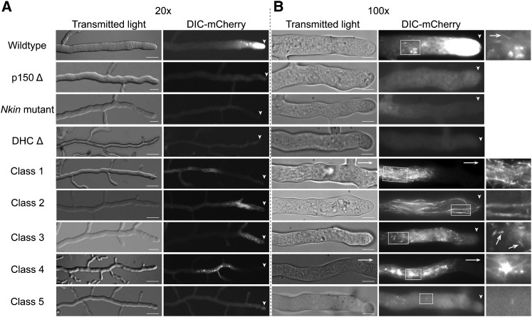
Figure 1 .
Localization of dynein in mutant strains of N. crassa. (A) Hyphae from the colony edge of different N. crassa strains were visualized at intermediate magnification to show the distance between dynein signal and the hyphal tip (arrowheads). (B) Hyphae visualized using higher magnification show details of dynein localization. The wild-type dynein localization includes a prominent hyphal-tip gradient as well as localization to short linear tracks (white arrows) and to other structures. In the mutant strains, dynein was mislocalized to particular hyphal structures and/or into a disperse signal. (A and B, left) Transmitted light. (A and B, right) mCherry fluorescence. Arrowheads indicate position of the hyphal tip. White arrows indicate that the hyphal tip was located outside the frame. Enlarged views of the boxed regions are shown on the right in B; white arrows indicate short linear tracks in wild-type and class 3 mutant strains. Bars in A, 50 µm; in B, 10 µm.

