

Abstract
Discoloration of acrylic resin denture base when it comes in contact with various food materials and beverages in the oral cavity may cause aesthetic concern to a denture wearer. The objective of this study was to evaluate the effect of tea, coffee and turmeric solutions on the colour of different brands of heat cure acrylic resin denture base materials commonly used in India. Spectrophotometer was used to evaluate the colour change. A significant difference was found when change in colour was statistically analysed.
Keywords: Acrylic resin denture base, Spectrophotometer, Colour stability
Introduction
The most important dimension of aesthetics is colour. Apart from the other properties of acrylic resin, developing its colour to match with the colour of oral mucosa and teeth makes it the material of choice for its universal application in denture prosthesis. In denture prosthesis though the denture teeth are aesthetically more important and are noticed significantly the denture base is equally important for its aesthetics in many patients if not all. Denture base has to be in contact with various food materials and beverages in the oral cavity. Its material (acrylic resin) is likely to absorb various contaminants. Also it is subjected to sorption, a process of absorption and adsorption of liquids dependent on environmental conditions should a contacting solution be pigmented leading to possible discoloration [1]. In the present study the food articles, turmeric, tea and coffee were included to see their effect on the colour of denture base material. There are related studies but unlike other food, turmeric being an essential ingredient of Indian food, its effect would be more relevant while studying the brands of acrylic resin used by most of the laboratories in India.
Aims and Objectives
The aim of the study was to evaluate the colour change of certain brands (commonly used in India) of denture base acrylic resin when they were contacted with tea, coffee and turmeric solutions. The objective of the study was to compare the effect of these solutions on the different brands of denture base acrylic resin in terms of change in their colour.
Materials and Methods
Four brands of heat cure acrylic resin (Ashvin, Lucitone-199, DPI and Travelon-HI) were used to prepare the specimens. The size of each cuboidal shape specimen was 20 × 20 × 3 mm. Metal die (Fig. 1) was used for making the wax pattern of the specimens and patterns were processed following the manufacturer instructions. Fifteen specimens of each brand were prepared and polished using no. 600 silicone–carbide sand paper.
Fig. 1.
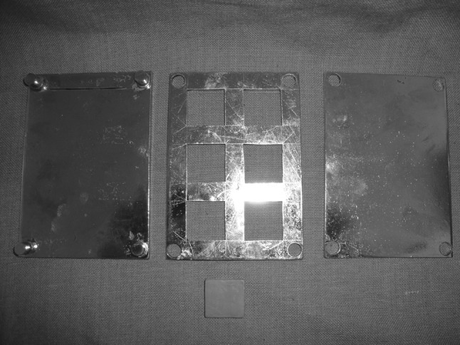
Metal die and sample
Solutions of tea, coffee, and turmeric were prepared by adding 8 g of colorant to 400 ml boiling distil water. It was allowed to cool for 10 min and then filtered through a piece of gauze. Each solution was divided into four parts so that 5 specimens of each brand of acrylic resin were immersed into the specific solutions (Fig. 2). Specimens were designated according to the acrylic resin and solution. Total 12 closed containers were taken to accommodate the twelve groups of specimens. Each container with 5 specimens was kept in incubator at 37 ± 1 °C temperature. Fresh solutions were taken after 10 and 20 days.
Fig. 2.
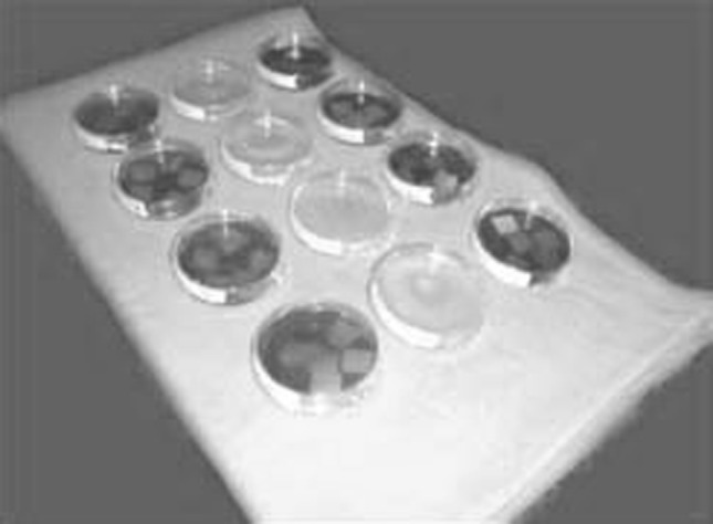
Dipped samples
The specimens were washed under distil water and dried before measuring the colour on 0, 10, 20 and 30 days of immersion. Colour measurement of the specimens from each brand of denture base acrylic resin recorded by spectrophotometer (Minolta cd-3200; Fig. 3). The mean of colour change of each material was calculated with the use of C.I.E. L* a* b* uniform colour scale. The magnitude of total colour difference is formulated by ΔE.
Fig. 3.

Spectrophotometer
ΔE* = [(ΔL*)2 + (Δa*)2 + (Δb*)2]½, where
ΔL* is L*specimen − L*standard, Δa* is a*specimen − a*standard, Δb* is b*specimen − b*standard.
The stained specimens (Fig. 4) evaluated for the standard colour value was tested with white as a standard, which is applicable for testing the colour variants. The data collected for the change in colour were evaluated and compared for the different groups using One-way Analysis of Variance and Bonferroni (post hoc) test.
Fig. 4.

Stained specimens
Result
Spectrophotometer readings reveal the difference in the colour of each material after 10, 20 and 30 days of immersion in tea, coffee and turmeric solution (Table 1). Statistically the colour change was significant between and within the groups of different heat cure denture base acrylic resins (Tables 2, 3).
Table 1.
The mean values and standard deviations (SD) of the colour change (ΔE) of the resins in tea, coffee and turmeric after 10, 20 and 30 days
| Lucitone | Travelon-HI | DPI | Ashvin | |
|---|---|---|---|---|
| Tea | ||||
| 10 Days | 3.53 ± 0.31 | 3.60 ± 0.44 | 2.78 ± 0.37 | 2.34 ± 0.30 |
| 20 Days | 4.60 ± 0.37 | 4.23 ± 0.28 | 3.74 ± 0.23 | 3.04 ± 0.11 |
| 30 Days | 5.28 ± 0.39 | 4.64 ± 1.13 | 4.67 ± 0.23 | 3.81 ± 0.09 |
| Coffee | ||||
| 10 Days | 2.76 ± 0.41 | 2.17 ± 0.37 | 1.48 ± 0.29 | 1.38 ± 0.22 |
| 20 Days | 3.48 ± 0.35 | 2.94 ± 0.41 | 2.44 ± 0.46 | 2.03 ± 0.07 |
| 30 Days | 3.97 ± 0.19 | 3.45 ± 0.49 | 3.57 ± 0.21 | 2.82 ± 0.02 |
| Turmeric | ||||
| 10 Days | 11.27 ± 0.32 | 08.18 ± 0.17 | 16.17 ± 0.21 | 15.54 ± 0.31 |
| 20 Days | 14.53 ± 0.4 | 14.56 ± 0.33 | 19.43 ± 0.25 | 21.27 ± 0.13 |
| 30 Days | 18.29 ± 0.36 | 20.09 ± 0.32 | 23.65 ± 0.28 | 24.09 ± 0.29 |
Table 2.
One way analysis of variance (tea, coffee, turmeric)
| SS | dF | MS | F | P | |
|---|---|---|---|---|---|
| Tea | |||||
| ΔE (10 days) | |||||
| B/w groups | 5.524 | 3 | 1.841 | 14.239 | 0 |
| Within groups | 2.069 | 16 | 0.129 | ||
| Total | 7.593 | 19 | |||
| ΔE (20 days) | |||||
| B/w groups | 6.864 | 3 | 2.288 | 32.967 | 0 |
| Within groups | 1.11 | 19 | 0.069 | ||
| Total | 7.974 | 22 | |||
| ΔE (30 days) | |||||
| B/w groups | 5.468 | 3 | 1.823 | 4.931 | 0.013 |
| Within groups | 5.914 | 16 | 0.37 | ||
| Total | 11.382 | 19 | |||
| Coffee | |||||
| ΔE (10 days) | |||||
| B/w groups | 6.242 | 3 | 2.081 | 18.938 | 0 |
| Within groups | 1.758 | 16 | 0.11 | ||
| Total | 8 | 19 | |||
| ΔE (20 days) | |||||
| B/w groups | 5.876 | 3 | 1.959 | 21.195 | 0 |
| Within groups | 1.478 | 16 | 0.092 | ||
| Total | 7.354 | 19 | |||
| ΔE (30 days) | |||||
| B/w groups | 3.376 | 3 | 1.125 | 36.33 | 0 |
| Within groups | 0.496 | 16 | 0.031 | ||
| Total | 3.872 | 19 | |||
| Turmeric | |||||
| ΔE (10 days) | |||||
| B/w groups | 212.675 | 3 | 1.592 | 1035.6 | 0 |
| Within groups | 1.095 | 16 | 0.068 | ||
| Total | 213.77 | 19 | |||
| ΔE (20 days) | |||||
| B/w groups | 176.918 | 3 | 55.973 | 683.4 | 0 |
| Within groups | 1.381 | 16 | 0.056 | ||
| Total | 178.299 | 19 | |||
| ΔE (30 days) | |||||
| B/w groups | 118.354 | 3 | 39.453 | 392.03 | 0 |
| Within groups | 1.61 | 16 | 0.1 | ||
| Total | 119.964 | 19 | |||
B/w between, SS sum of squares, MS mean square, df degree of freedom, F variance ratio
P < 0.05 shows significant result
Table 3.
Bonferroni (post hoc) test (tea, coffee and turmeric)
| Dependent | Groups | Groups | Tea | Coffee | Turmeric |
|---|---|---|---|---|---|
| P | P | P | |||
| ΔE (after 10 days) | Travelon-Hi | 1.000 | 0.011 | 0.000 | |
| DPI | 0.027 | 0.000 | 0.000 | ||
| Lucitone | Ashvin | 0.000 | 0.000 | 0.000 | |
| DPI | 0.014 | 0.029 | 0.000 | ||
| Travelon-Hi | Ashvin | 0.000 | 0.010 | 0.000 | |
| DPI | Ashvin | 0.434 | 1.000 | 0.010 | |
| ΔE (after 20 days) | Travelon-Hi | 0.243 | 0.475 | 1.000 | |
| DPI | 0.001 | 0.002 | 0.000 | ||
| Lucitone | Ashvin | 0.000 | 0.000 | 0.000 | |
| DPI | 0.680 | 0.092 | 0.000 | ||
| Travelon-Hi | Ashvin | 0.000 | 0.002 | 0.000 | |
| DPI | Ashvin | 0.004 | 0.495 | 0.000 | |
| ΔE (after 30 days) | Travelon-Hi | 0.679 | 0.000 | 0.000 | |
| DPI | 0.777 | 0.243 | 0.000 | ||
| Lucitone | Ashvin | 0.009 | 0.000 | 0.000 | |
| DPI | 1.000 | 0.001 | 0.000 | ||
| Travelon-Hi | Ashvin | 0.283 | 1.000 | 0.000 | |
| DPI | Ashvin | 0.244 | 0.005 | 0.265 |
P < 0.05 shows significant result
Discussion
Spectrophotometer was used to measure the colour. It is the better option when compared with Munsell colour order as instrumental measurement eliminates the subjective interpretation of visual colour comparison [2]. The use of Commission International de L’Eclairage (CIE Lab) uniform colour scale has advantage of having its arrangement in an approximately uniform three dimensional colour space whose elements are equally spaced on the basis of visual colour perception parameters.
Staining of the specimens reaches the plateau after certain period of time [3]. Like Scotti et al. [4], we also kept the time interval of 10 days for measuring the colour change. The effect of turmeric solution was maximum in terms of change in colour of all the acrylic resin specimens. ΔE values are maximum followed by tea and coffee (Table 1). It appears that colorant of turmeric is more polar. Um and Ruyter [5] mentioned in their study that ‘whenever the colorant is more polar and there by more hydrophilic it stains more as denture base resins are hydrophilic attracting more water soluble dyes on the surface’.
Staining becomes more intense with time but after 10 days of immersion it differed significantly from all succeeding time interval (Table 2). This could probably due to the sorption property of the resin which gets saturated with the pigments. Lucitone 199 was found most colour stable in turmeric followed by Travelon-HI, DPI and Ashvin but least in tea and coffee solution followed by DPI, Travelon-HI and Ashvin (Table 1). One way analysis of variance was applied to check the equality of means of colour change between the groups (Ashvin, DPI, Lucitone 199, Travelon-HI) related to tea, coffee and turmeric solution (Table 2). The variation between the groups related to three solutions after 10, 20 and 30 days were found to be statistically significant because at a confidence level of 95 %, the P value obtained was less than 0.05. To further delineate the significant variation between the means of colour change between the four brands of acrylic resins in tea, coffee and turmeric solution, the bonferroni (post hoc) test was performed (Table 3). The colour change was found to be variable. It could be due to different absorption property of materials and different polarity and hydrophilic nature of staining solutions [5]. In actual oral environment there is formation of pellicles of proteins and glucoproteins present in saliva leading to plaque formation on the acrylic resin surface. These soft materials are affected by the colorants in food more quickly and undergo colour changes [6]. In the present study, the changes in colour was examined in a saliva and diet free medium, so the result may not be the same as in oral environment.
Conclusions
All the brands of heat cure denture base acrylic resin tested in this study showed statistical significant colour change in tea, coffee and turmeric solution. Ashvin showed the highest colour variation in turmeric followed by DPI, Travelon-HI and Lucitone-199. Where as Lucitone-199 heat cure showed the highest colour variation in tea and coffee followed by DPI, Travelon-HI and Ashvin. Among the solutions tested, turmeric showed the highest staining effect on the specimens, followed by tea and later by coffee. The staining becomes more intense with time i.e. ΔE value for colour change increases with time but the rate does not remain the same as after 10, 20 and 30 days, the value does not increases in the same ratio. So while selecting the brand of denture base material its stability to colorants present in the food taken by the patient should also be an important criteria and the manufacturer should also use some scale which shows the stain resistance.
References
- 1.Hersek N, Canay S, Uzun G, Yildiz F. Colour stability of denture base acrylic resins in three food colourants. J Prosthet Dent. 1999;81:375–379. doi: 10.1016/S0022-3913(99)80001-8. [DOI] [PubMed] [Google Scholar]
- 2.Khokhar ZA, Riazzoog ME, Yaman P. Colour stability of restorative resin. Quintessence Int. 1991;22:733–737. [PubMed] [Google Scholar]
- 3.Chan KC, Fuller JL, Hormati AL. The ability of foods to stain two composite resins. J Prosthet Dent. 1980;43:542–545. doi: 10.1016/0022-3913(80)90328-5. [DOI] [PubMed] [Google Scholar]
- 4.Scotti R, Mascellani SC, Forniti F. The in vitro colour stability of acrylic resins for provisional restorations. Int J Prosthodont. 1997;10:164–168. [PubMed] [Google Scholar]
- 5.Um CM, Ruyter IE. Staining of resin-based veneering materials with coffee and tea. Quintessence Int. 1991;22:377–386. [PubMed] [Google Scholar]
- 6.Caranza FA, Newman MG. Clinical periodontology. 8. Philadelphia: WB Saunders; 1996. pp. 87–155. [Google Scholar]