Abstract
Standard of care for venous leg ulcers (VLUs) consists of the application of compression bandages or stockings and of local moist wound care. While the majority of patients heal with the above mentioned treatments some ulcers become refractory to treatment causing significant disability and costs. The authors present the observation made on two patients with VLUs who had failed to respond to a comprehensive state of the art wound care approach for 11 and 3 years respectively. Both patients were treated with a poly-N-acetyl glucosamine-derived membrane (pGlcNAc) (Talymed, Marine Polymer Technologies, Danvers, Massachusetts, USA) in addition to compression bandaging. Both patients healed within 6 weeks of the first application of pG1cNAc. The authors present two cases of VLUs that had been considered non-healable that were successfully treated in a very short period of time with the application of a novel technology.
Background
Venous leg ulcers (VLUs) are prevalent, affecting up to 1% of the older population. These ulcers are associated with substantial morbidity and decrements in quality of life.1 Healing rates of these ulcers ranges between 30% and 60% with 24 weeks of therapy and 70%–85% with up to a year of therapy. A good number of patients 15%–30% are considered un-healable. We present two cases of patients with venous stasis ankle ulcerations of 11 and 3 years, respectively. Both had failed to respond to the standard of care2 consisting in compression bandaging, debridement and local wound care and to advanced treatment modalities such as semisynthetic skin substitutes and other biological treatments. In both situations, the ulcers had been considered un-healable. The patients were treated with the usual compression bandaging system and a new product consisting of the topical use of a poly-N-acetyl glucosamine, nano-fibre derived membrane material (Talymed, Marine Polymer Technologies, Danvers, Massachusetts, USA). Both patients healed completely in the extraordinary short period of only 6 weeks with the application of this new therapy.
This case illustrates that there is still hope for healing in some of the so called un-healable ulcers and urges to explore the use of this new technology in other type of leg ulcers.
Case presentation
Patient A
The patient is an 80-year-old White Man with history of postphlebitic syndrome who presented to our Wound Center with an ulcer on the left medial malleolus that had started spontaneously about 11 years ago. His medical history was unremarkable except for bilateral idiopathic deep venous thrombosis in 1964, remote history of laryngeal carcinoma treated in 1991, and shoulder surgery. The patient was treated regularly with compression bandages or stockings, multiple different wound dressings including moist gauze, antibacterial, silver containing and collagen dressings. He was also treated unsuccessfully with negative pressure therapy (VAC, Kinetic Concepts, San Antonio Texas, USA) and multiple applications of bio-engineered skin substitutes including Apligraf, Oasis Matrix and Primatrix.
Patient B
The patient is a 68-year-old African–American Woman with history of hypertension and varicose veins who developed an ulcer on the left medial ankle 3 years before consultation. The patient was treated at another Wound Center with the application of compression bandages, silver dressings, antibiotics and bio-engineered skin substitute Apligraf.3
Investigations
Both patients underwent bilateral venous reflux ultrasound studies of the lower extremities and transcutaneous oxygen mapping to assess perfusion to the lower extremities. The venous ultrasound confirmed the presence of reflux in the deep venous system in patient A and of an incompetent perforator at the ankle close to the ulcer in patient B. Both patients had excellent results in the transcutaneous oxygen mapping of the lower extremities which excluded the presence of clinically significant peripheral vascular disease. Both patients were evaluated by a vascular surgeon and were considered to be non-surgical candidates for correction of their venous anomalies.
Treatment
Both patients were treated with Talymed, poly-N-acetyl glucosamine, nano-fibre derived membrane material weekly in addition to the standard of care which consisted on the application of a four-layer-compression bandaging system (Profore, Smith & Nephew, London, UK). A secondary dressing to lower bacteria and absorb drainage was used as needed.
Outcome and follow-up
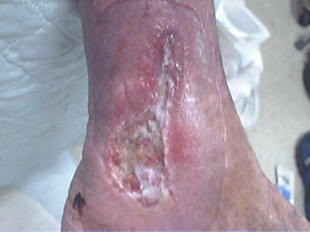
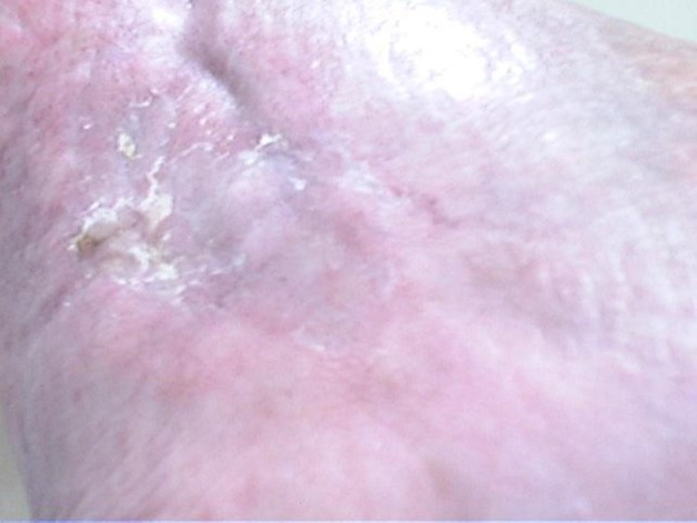
The ulcer on patient A measured 4.8×3.0 cm and was 2 mm deep at the begin of therapy with Talymed on 17 November 2011 (figure 1). The ulcer developed rapid formation of islands of new skin and rapid epithelialisation and was fully closed in 6 weeks (11 January 2012) (figure 2).
Figure 1.

Ulcer of 11 year duration before application of Talymed.
Figure 2.

Complete healing after 6 weeks of therapy.
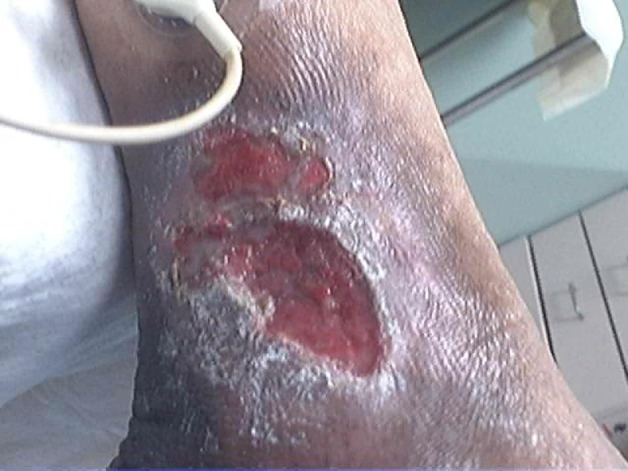
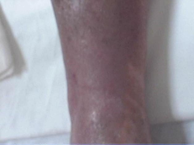
The ulcer on patient B measured 5.0×3.2 cm and was 2 mm deep on 17 November 2011 (figure 3). Full closure was obtained by 4 January 2012 (figure 4).
Figure 3.

Ulcer of 3 year duration before application of Talymed.
Figure 4.

Ulcers healed after 6 weeks of therapy.
Both patients were prescribed knee-high 30–40 mm Hg compression stockings for day-time use and have not had signs of reccurrence at a 2 month follow-up visit.
Discussion
The above described cases represent an opportunity to explore novel therapies that become available. We were compelled to try this new product based on a small randomised trial on the healing of venous stasis ulcers.4 The mechanism of action of poly-N-acetyl glucosamine-derived membran (pGlcNAc) nano-fibre-based membrane has been investigated invitro and invivo. pGlcNAc nanofibre membranes seem to increase cell metabolism and activate invitro migration of endothelial cells and fibroblasts. In mouse studies, treatment of full-thickness wounds with pGlcNAc nanofibre membranes enhanced wound healing mainly by re-epithelialisation and stimulation of angiogenesis, tissue remodelling and endothelial cell proliferation.5 6 Cases like the above mentioned are very frustrating to the patients and care givers and to the practitioners involved in their care. It is not possible to determine the total expenses these patients incurred over the course of the years without mentioning the associated pain and decrease in quality of life despite excellent wound care. Open leg ulcers represent a mayor risk factor for soft tissue and bone infection that may put a limb at risk of amputation. It is known that the longer that an ulcer remains open the higher the risk of osteomyelitis. The literature suggest that the type of dressing used on a venous stasis ulcer is of minimal importance provided that a moist wound environment and compression are maintained. For the exception of bio-engineered tissue3 there had been no mayor advances in wound care dressings for these type of ulcers that has proven to be superior. It is astonishing to see full and fast healing of ulcers that had been cared for so long with no significant improvement.
Learning points.
While compression bandaging remains the standard of care for the management of venous stasis ulcers, advanced technologies such as poly-N-acetyl glucosamine, nano-fibre derived membrane material should be further investigated and tried on recalcitrant wounds.
We learnt from this example that the physician needs to be more stubborn than the wound and that being open to new technologies may bring an answer to an old problem.
The use of compression stockings after healing of VLUs is necessary to decrease the risk of reccurrence.
Footnotes
Competing interests: None.
Patient consent: Not obtained.
References
- 1.Bergan JJ, Schmid-Schönbein GW, Smith PD, et al. Chronic venous disease. N Engl J Med 2006;355:488–98. [DOI] [PubMed] [Google Scholar]
- 2.O’Meara S, Cullum NA, Nelson EA. Compression for venous leg ulcers. Cochrane Database Syst Rev 2009;1:CD000265. [DOI] [PubMed] [Google Scholar]
- 3.Falanga V, Margolis D, Alvarez O, et al. Rapid healing of venous ulcers and lack of clinical rejection with an allogeneic cultured human skin equivalent: Human Skin Equivalent Investigators Group. Arch Dermatol 1998;134:293–300. [DOI] [PubMed] [Google Scholar]
- 4.Kelechi TJ, Mueller M, Hankin CS, et al. A randomized, investigator-blinded, controlled pilot study to evaluate the safety and efficacy of a poly-N-acetyl glucosamine-derived membrane material in patients with venous leg ulcers. J Am Acad Dermatol 2012;66:e209–15. [DOI] [PubMed] [Google Scholar]
- 5.Scherer SS, Pietramaggiori G, Matthews J, et al. Poly-N-acetyl glucosamine nanofibers: a new bioactive material to enhance diabetic wound healing by cell migration and angiogenesis. Ann Surg 2009;250:322–30. [DOI] [PubMed] [Google Scholar]
- 6.Vournakis JN, Eldridge J, Demcheva M, et al. Poly-N-acetyl glucosamine nanofibers regulate endothelial cell movement and angiogenesis: dependency on integrin activation of Ets1. J Vasc Res 2008;45:222–32. [DOI] [PMC free article] [PubMed] [Google Scholar]


