Abstract
The replication of hepatitis B virus (HBV) in hepatocytes is strongly inhibited in response to IFN-α/β and IFN-γ. Although it has been previously demonstrated that IFN-α/β eliminates HBV RNA-containing capsids from the cell in a proteasome-dependent manner, the precise cellular pathway that mediates this antiviral effect has not been identified. Because IFN-induced signal transduction involves kinase-mediated activation of gene expression, we used an immortalized hepatocyte cell line that replicates HBV in an IFN-sensitive manner to investigate the role of cellular kinase activity and the cellular transcription and translation machinery in the antiviral effect. Our results indicate that Janus kinase activity is required for the antiviral effect of IFN against HBV, but that phosphatidylinositol 3-kinase, cyclin-dependent kinase, mitogen-activated protein kinase, and NF-κB activity are not. Additionally, we found that inhibitors of cellular transcription and translation completely abolish the antiviral effect, which also appears to require cellular kinase activity downstream of signal transduction and gene expression. Collectively, these results identify IFN-regulated pathways that interrupt the HBV replication cycle by eliminating viral RNA-containing capsids from the cell, and they provide direction for discovery of the terminal effector molecules that ultimately mediate this antiviral effect.
Hepatitis B virus (HBV) replication is noncytopathically inhibited by IFN-α/β and IFN-γ (1). Studies using transgenic mouse models of HBV gene expression and replication have demonstrated that multiple mechanisms mediate this process (2, 3). First, viral DNA replicative intermediates are cleared from the liver with no change in the level of viral mRNA (3). Subsequently, HBV mRNA levels are reduced by both transcriptional and posttranscriptional mechanisms (4, 5). Viral replication is inhibited by a variety of stimuli that induce intrahepatic IFN-α/β (such as infection with adenovirus or murine cytomegalovirus, injection with polyinosinic-polycytidylic acid) and/or IFN-γ (adoptive transfer of HBsAg-specific cytotoxic T lymphocytes, injection of IL-12 or α-CD40 mAb; refs. 3 and 6-9). Whereas it has been shown that replication is inhibited by a reduction in the assembly or stability of viral pregenomic RNA-containing capsids (10), the IFN-induced molecular mechanism that mediates this inhibition is not yet defined. Notably, type I IFN-inducible genes with known antiviral activity (RNA-dependent protein kinase, RNase L, and myxovirus resistance-1) do not mediate the antiviral effect of IFN-α/β or IFN-γ in HBV-transgenic mice (11). In contrast, inducible nitric oxide synthase is required for the IFN-γ-induced antiviral effect in these animals (12).
To identify IFN-regulated genes whose induction correlates with suppressed HBV replication, gene expression profiling was performed in HBV-transgenic mouse livers and immortalized transgenic hepatocytes in response to IFN-α/β and IFN-γ (13). Multiple IFN-regulated genes, including the proteasome subunits LMP2, LMP7, MECL-1, and PA28β, were induced under conditions that correlated with the antiviral effect of both IFN-α/β and IFN-γ. By using this information, we subsequently demonstrated that proteasome activity was indeed required for the IFN-α/β- and IFN-γ-induced antiviral effects (14). In addition to the proteasome subunits, expression of a number of other genes also correlated with the antiviral effect, including IFN-regulated GTPases [T cell-specific GTPase (TGTP), IFN-γ induced GTPase] that have known antiviral activity (15, 16), as well as various genes involved in cell signaling [signal transducer and activator of transcription (STAT)-1, IP-10]. However, the role that these factors may play in the inhibition of HBV is not defined.
Although IFN-induced signal transduction and gene expression occurs primarily through the activation of Janus kinases (Jak) and STAT transcription factors, IFN-α/β and IFN-γ also activate or modulate the activity of other cellular kinases and transcriptional regulators, including phosphatidylinositol 3-kinase (PI3-kinase), mitogen-activated protein (MAP) kinase(s), cyclin-dependent kinase(s) (cdk), and NF-κB (17, 18). Furthermore, in addition to the genes reported previously, the expression of a number of other cellular kinases (or regulators of kinase activity) also correlated with IFN-induced HBV inhibition in either the transgenic mouse livers or immortalized hepatocytes, including cdk inhibitor 1A, MAP kinase-activated protein kinase 2, and hexokinase (13). Based on these results, we attempted in the current study to further define the IFN-induced cellular pathways that inhibit HBV replication, focusing primarily on the role of cellular transcription, translation, and kinase activity.
Materials and Methods
Cells and Reagents. The HBV-Met cell line (clone 1-1.4) used in this study is an immortalized hepatocyte cell line derived from HBV-transgenic mice (19). Cells were maintained in RPMI medium 1640 containing 10% heat-inactivated FCS, 2 mM l-glutamine, 100 μg of penicillin per ml, 100 units of streptomycin per ml (Invitrogen), 10 μg of insulin per ml (Sigma), 100 ng of epidermal growth factor per ml (BD Biosciences, Bedford MA), and 16 ng of insulin-like growth factor 2 per ml (Calbiochem) (Met media). All chemical inhibitors used were purchased from Calbiochem. Recombinant murine IFN-β was provided by K. Harada (Toray Industries, Chiba, Japan), and murine IFN-γ was provided by S. Kramer (Genentech).
Experimental Procedure. HBV-Met cells were grown in complete Met media to 100% confluence in collagen-coated Biocoat 60-mm dishes (Becton Dickinson Labware). The cells were induced to differentiate by culturing in Met media containing 2% DMSO for 10-12 days. On differentiation, the HBV-Met cells replicate HBV from the integrated transgene in a cytokine-sensitive manner (19). The chemical inhibitors were prepared as a 50× stock in DMSO, resulting in a final 2% DMSO concentration when added to the cell culture media. The concentration used for each inhibitor was based on previously reported effective concentrations. For the staurosporine, genistein, and cycloheximide experiments, the inhibitor was added to the cells either 1 h before or 3 h after IFN. All other inhibitors were added 1 h before IFN, except α-amanitin, which was added 2 h before IFN. Cells were then incubated with the indicated inhibitors and/or IFN for the indicated lengths of time before harvesting for total DNA and RNA preparation. Southern blots were performed to examine the level of HBV DNA-replicative intermediates, whereas Northern blots were performed to monitor the expression of representative IFN-α/β [2′5′-oligoadenylate synthetase (2′5′-OAS)]- or IFN-γ (TGTP)-induced genes relative to the level of GAPDH mRNA.
DNA Preparation and Hybridization. For DNA preparation, cells were lysed in DNA lysis buffer (50 mM Tris·HCl/20 mM EDTA/1% SDS). The cellular lysates were incubated ≈16 h at 37°C with 1 mg of proteinase K per ml. The lysates were then extracted with Tris-buffered phenol (pH 8.0), phenol/chloroform (1:1) and chloroform, followed by precipitation of total DNA with an equal volume of isopropanol. For the Southern hybridizations, 20 μg of total DNA was digested with HindIII, electrophoresed in a 1.4% agarose gel, and transferred to nylon a membrane by using standard protocols (20). Hybridizations were performed with a genome-length HBV probe as described (21).
RNA Preparation and Hybridization. For RNA preparation, cells were lysed in GTC solution (4.2 M guanidine isothiocyanate, 0.5% saponin, 25 mM sodium citrate) containing 100 mM 2-mercaptoethanol. The lysates were extracted twice with a 2.5:1 mixture of phenol (pH 4.0):chloroform, and RNA was precipitated with an equal volume of isopropanol. The RNA was then resuspended in diethyl pyrocarbonate-treated H2O, and was further extracted with buffered phenol (pH 8.0) and chloroform. The RNA was once again precipitated with an equal volume of isopropanol, washed in 80% ethanol, and resuspended in diethyl pyrocarbonate-H2O. For the Northern hybridizations, equivalent amounts (10-20 μg) of total RNA were electrophoresed through a 1% agarose/formaldehyde gel, followed by transfer to a nylon membrane by using standard protocols (22). Hybridizations were performed as described (21).
Results
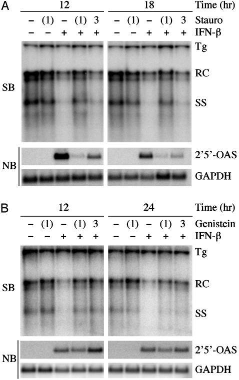
Nonspecific Kinase Inhibitors Block the Antiviral Effect of IFN. Staurosporine inhibits the activity of a broad spectrum of serine/threonine and tyrosine kinases (23). Furthermore, staurosporine inhibits IFN-induced signal transduction by both inhibition of Jak activation and by down-regulation of Jak expression (24). When added 1 h before treatment with 500 units/ml IFN-β, staurosporine (5 μM) efficiently inhibited the expression of the representative type I IFN-inducible gene 2′5′-OAS, and completely blocked the antiviral effect (Fig. 1A). However, staurosporine did not block the antiviral effect when added 3 h after IFN, even though it was able to partially inhibit IFN-induced gene expression when added to the cells at this time (Fig. 1 A). Thus, this result indicates that kinase activity is necessary for the antiviral effect of IFN, and that the requisite kinases are induced within 3 h after IFN treatment. Parenthetically, it is important to note that in this and all other experiments reported herein, the levels of the HBV 3.5- and 2.1-kb mRNAs were unchanged by either the inhibitor or IFN (data not shown), which is consistent with our previous report that IFN-α/β and IFN-γ do not alter HBV gene expression in the HBV Met cells (19).
Fig. 1.
Staurosporine and genistein inhibit IFN-induced inhibition of HBV. (A) Differentiated HBV-Met cells either were pretreated for 1 h with 5 μM staurosporine, followed by the addition of 500 units/ml IFN-β [staurosporine (stauro) (1)], or were pretreated with 500 units/ml IFN-β for 3 h before the addition of 5 μM stauro (3). Southern blot (SB) and Northern blot (NB) analyses of HBV DNA-replicative intermediates and 2′5′-OAS/GAPDH expression were performed at 12 and 18 h post-IFN-α treatment. Staurosporine blocks the antiviral effect when added 1 h before IFN-β but not 3 h after IFN-β. (B) Differentiated HBV-Met cells either were pretreated for 1 h with 100 μM genistein, followed by the addition of 500 units/ml IFN-β [genistein (1)], or were pretreated with 500 units/ml IFN-β for 3 h before the addition of 100 μM genistein [genistein (3)]. SB and NB analyses of HBV DNA-replicative intermediates and 2′5′-OAS/GAPDH expression were performed at 12 and 24 h post-IFN-β treatment. Genistein blocks the antiviral effect when added before or after IFN without altering 2′5′-OAS expression. Tg, transgene; RC and SS, relaxed circle and single-stranded DNA-replicative intermediate forms, respectively.
The broad-spectrum tyrosine kinase inhibitor genistein was also tested for its ability to block the antiviral effect of IFN in the HBV-Met cells (25). At a concentration of 300 μM, genistein inhibited IFN-induced gene expression and the antiviral effect (data not shown). As shown in Fig. 1B, however, genistein also blocked the antiviral effect of IFN at a concentration (100 μM) that does not inhibit IFN-induced expression of 2′5′-OAS (Fig. 1B) or IFN-stimulated gene 15, ubiquitin-specific protease 18, STAT1, and the proteasome subunit LMP2 (data not shown). Furthermore, unlike staurosporine, genistein blocked the antiviral effect when added either 1 h before or 3 h after IFN treatment (Fig. 1B). Thus, these results suggest the possibility that a genistein-sensitive kinase downstream of the IFN-induced signal transduction cascade is important for the inhibition of HBV.
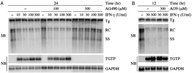
The Antiviral Effect of IFN Requires Jak Activity. IFN-mediated signal transduction occurs through the sequential activation of the Jak kinases and STAT transcription factors (26). More specifically, signaling through the IFN-α/β receptor primarily involves the activation of Tyk2, Jak1, and STAT1/STAT2 heterodimers, whereas activation of IFN-γ-induced genes requires Jak2, Jak1, and STAT1 homodimers. Therefore, we examined the role of Jak activity in the inhibition of HBV replication by using the specific Jak inhibitor AG490 (27). As this compound has primarily been reported to efficiently inhibit the activity of Jak2, we determined whether AG490 would block the antiviral effect of IFN-γ. A 1-h pretreatment of HBV-Met cells with 300 μM AG490 abrogated the inhibition of HBV replication by IFN-γ (Fig. 2A). At this concentration of AG490, expression of the representative IFN-γ-inducible gene TGTP was completely blocked over a wide range of IFN concentrations (Fig. 2 A). In contrast, treatment of the cells with a lower concentration of AG490 (100 μM) or an equivalent concentration of a similar compound that does not inhibit Jak-2 activity (AG9) failed to block IFN-induced gene expression or the antiviral effect (Fig. 2 A and B). Thus, the inhibition of HBV replication by IFN-γ requires Jak activity.
Fig. 2.
Jak kinase activity is required for IFN-γ induced inhibition of HBV. Differentiated HBV-Met cells were pretreated for 1 h with the indicated concentrations of AG490 (A) or AG9 (B) before the addition of IFN-γ. Southern blot (SB) and Northern blot (NB) analyses of HBV DNA-replicative intermediates and TGTP/GAPDH expression were performed at 12 or 24 h post-IFN-γ treatment. AG490 (300 μM) inhibits IFN-γ-induced gene expression and the antiviral effect over a wide range of IFN concentrations. AG490 (100 μM) and AG9 (300 μM) inhibit neither IFN-γ-induced gene expression nor the antiviral effect. Tg, transgene; RC and SS, relaxed circle and single-stranded DNA replicative intermediate forms, respectively.
Other Specific Kinase Inhibitors Do Not Block the Antiviral Effect of IFN. It is well established that in addition to induction of the Jak/STAT pathway, both IFN-α/β and IFN-γ induce activation of NF-κB (28, 29). Activation of NF-κB by IFN-α occurs through the PI3-kinase-mediated activation of the serine/threonine kinase Akt (30). Wortmannin and Ly-294002 are both potent and specific inhibitors of PI3-kinase, and Ly-294002 was previously shown to inhibit the IFN-α-induced activation of NF-κB (30, 31). However, these reagents did not block the antiviral effect of IFN when used at equal to or greater than previously determined effective concentrations (Table 1). Furthermore, an inhibitor of IκBα phosphorylation (Bay 11-7082) did not modulate the antiviral effect (32) (Table 1). It is noteworthy that these agents (and others that did not block the antiviral effect) inhibited the proliferation of nondifferentiated HBV-Met cells, indicating that effective concentrations of the drugs were achieved in these experiments (data not shown). We were also unable to detect an IFN-induced increase in levels of phospho-IκBα or decrease in total IκBα in the HBV-Met cells on treatment with IFN-β (data not shown). Similarly, neither TNF-α nor IL-1β inhibits HBV replication in these cells, even though they are known to activate NF-κB (ref. 19 and data not shown). Thus, it is unlikely that PI3-kinase or NF-κB mediates the signal transduction or down-stream events that inhibit HBV replication in the HBV-Met cells.
Table 1. Effect of inhibitors on IFN-mediated inhibition of HBV.
| Inhibitor | Target | Max concentration tested | Block of antiviral activity |
|---|---|---|---|
| Kinase inhibitor | |||
| Staurosporine | Ser/Thr, Tyr kinases | 5 μM | + |
| Genistein | Tyr kinases | 300 μM | + |
| AG490 | Jak kinases | 300 μM | + |
| Wortmannin | PI3-kinase | 1 μM | — |
| Ly-294002 | PI3-kinase | 25 μM | — |
| Bay 11–7082 | NF-κB | 10 μM | — |
| Olomoucine | cdk1,2,5 | 50 μM | — |
| Roscovitine | cdk1,2,5 | 50 μM | — |
| U0126 | MAP kinase kinase1/2 | 50 μM | — |
| PD 98059 | MAP kinase kinase | 25 μM | — |
| SB 203580 | p38 MAP kinase | 25 μM | — |
| Gene Expression | |||
| α-Amanitin | RNA pol II | 10 μg/ml | + |
| Cycloheximide | Translation | 100 μg/ml | + |
HBV-Met cells were pretreated with the indicated concentration of inhibitor for 1 h before the addition of IFN-β or IFN-γ. HBV replication was examined 12–24 h after IFN treatment.
In addition to PI3-kinase, IFN also influences the activity of a number of other regulatory kinases, including cdk and MAP kinases (33-35). However, as with PI3-kinase, multiple inhibitors of cdk and MAP kinases also failed to block the ability of IFN to inhibit HBV replication (Table 1), supporting the notion that these kinases are also not required for the inhibition of HBV by IFN in this model system (36-38).
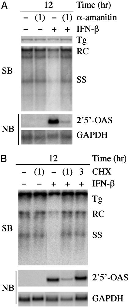
The Antiviral Effect of IFN Requires Cellular Transcription and Translation. Because IFN-induced Jak/STAT activation ultimately leads to induced expression of IFN-regulated genes, we determined whether transcription or translation inhibitors would block the antiviral effect of IFN in the HBV-Met cells. α-Amanitin is a potent inhibitor of RNA polymerase II-mediated transcription (39). When pretreated with α-amanitin, IFN-induced gene expression was blocked, and the antiviral effect was also abrogated (Fig. 3A). Similarly, treating the cells with an inhibitor of translation (cycloheximide) also blocked the antiviral effect of IFN (Fig. 3B). This finding was independent of an effect on IFN-induced gene expression because cycloheximide blocked the antiviral effect without blocking the induction of 2′5′-OAS when it was added 3 h after IFN (Fig. 3B). Therefore, IFN-induced transcription and translation are both required for the inhibition of HBV replication.
Fig. 3.
Antiviral effect requires new transcription and translation. (A) Differentiated HBV-Met cells were pretreated for 1 h with 10 μg/ml α-amanitin, followed by the addition of 500 units/ml IFN-β. Southern blot (SB) and Northern blot (NB) analyses of HBV DNA-replicative intermediates and 2′5′-OAS/GAPDH expression were performed at 12 h post-IFN-β treatment. α-Amanitin blocks both IFN-induced gene expression and the antiviral effect. (B) Differentiated HBV-Met cells either were pretreated for 1 h with 100 μg/ml cycloheximide, followed by the addition of 500 units/ml IFN-β [cycloheximide (CHX) (1)], or were pretreated with 500 units/ml IFN-β for 3 h before the addition of 100 μg/ml CHX (3). SB and NB analyses of HBV DNA-replicative intermediates and 2′5′-OAS/GAPDH expression were performed at 12 h post-IFN-β treatment. When added 1 h before IFN, cycloheximide inhibits the antiviral effect and reduces IFN-induced gene expression. When added 3 h after IFN, CHX blocks the antiviral effect without altering IFN-induced gene expression. Tg, transgene; RC and SS, relaxed circle and single-stranded DNA-replicative intermediate forms, respectively.
Discussion
To better define the mechanism whereby IFN inhibits HBV replication, we studied the impact of kinase and transcription/translation inhibition on the antiviral effect. When added before treatment with IFN, staurosporine blocked IFN-induced gene expression and the antiviral effect (Fig. 1 A). However, when added 3 h after IFN treatment, the antiviral effect was not blocked, despite substantially reduced 2′5′-OAS expression. This result indicates that the IFN-mediated signal transduction that occurs during the first 3 h of treatment is sufficient to mediate the inhibition of HBV replication.
Interestingly, unlike staurosporine, genistein blocked the antiviral effect at a concentration (100 μM) that did not inhibit the expression of five representative IFN-induced genes in the HBV-Met cells. At a higher concentration of genistein (300 μM), IFN-induced gene expression was blocked as expected, which was consistent with the recent report by Guo et al. (40) who demonstrated that genistein inhibits IFN-induced gene expression in Huh7 cells. Furthermore, unlike staurosporine, genistein blocked the antiviral effect when it was added 3 h after IFN treatment. These results are consistent with the hypothesis that a genistein-sensitive/staurosporine-insensitive kinase functions either directly or indirectly in the antiviral effect downstream of signal transduction. Because phosphorylation of the viral core protein has been shown to influence hepadnaviral capsid stability (41, 42), the possibility that IFN-induced changes in HBV core phosphorylation mediate the antiviral effect warrants further study.
Many studies have clearly established that gene expression induced through the IFN-α and IFN-γ receptors occurs primarily through activation of the Jak/STAT pathway (26). Consistent with these findings, we observed that a Jak-2 inhibitor blocks the IFN-γ-mediated antiviral effect, and that this inhibitor is accompanied by a complete abrogation of IFN-γ-induced gene expression (Fig. 2 A). Furthermore, transcription and translation inhibitors also blocked the antiviral effect (Fig. 3), indicating that IFN-induced cellular gene expression is required to inhibit HBV replication. Interestingly, IFN-induced gene expression is reduced when the cells are pretreated with cycloheximide. This result raises the possibility that translation of an IFN-induced gene (such as STAT1) might be necessary for maximal induction of gene expression (13). Importantly, we have already shown that IFN has no effect on HBV gene expression (5) or translation (10) under these conditions. Thus, these results indicate that the antiviral effect of IFN reflects the impact of IFN-induced genes on events downstream of HBV transcription and translation, which is consistent with our previous results demonstrating that IFN reduces the formation or stability of HBV RNA-containing capsids (10).
In addition to activating the Jak/STAT pathway, it is becoming increasingly clear that both IFN-α/β and IFN-γ also induce the activation of other transcription factors, including NF-κB (28, 29). Furthermore, NF-κB activation has been reported to inhibit HBV replication when induced to high levels by TNF-α (43). As NF-κB activation depends on the proteasome-mediated degradation of the IκB inhibitors, this phenomenon was of interest to us, because it is possible that the ability of proteasome inhibitors to block the antiviral effect of IFN that we reported previously may reflect their ability to block NF-κB activation (14). The induction of NF-κB by IFN-α/β is known to occur through a step that requires Akt activation, and is thus sensitive to PI3-kinase inhibition. However, we were unable to block the antiviral effect with inhibitors of PI3-kinase activity or IκBα phosphorylation (Table 1). Furthermore, we were unable to detect an IFN-induced increase in the phosphorylation or degradation of IκBα in the HBV-Met cell line. Finally, treatment of the HBV-Met cells with TNF-α or IL-1β (both inducers of NF-κB) failed to inhibit HBV replication. Whereas these results differ from those reported by Biermer et al. (43) who have reported that NF-κB activation by TNF-α inhibits HBV replication in HepG2 cells, these differences are likely explained by different sensitivities of the cell lines to the two cytokines or different mechanisms that are capable of inhibiting HBV replication. Thus, it appears unlikely that NF-κB is playing a role in the IFN-induced antiviral effect in our model system.
The replication of HBV may also depend on the activity of other cellular kinases. The HBV core protein contains phosphorylation sites with similarity to consensus cdk phosphorylation sites. Furthermore, a cdc2 kinase recognition motif regulates capsid stability in the related duck hepatitis B virus (44). In addition, the c-Raf/MAP kinase kinase pathway has been reported to be needed for efficient HBV gene expression, and activated extracellular signal-regulated kinase 1/2 has been shown to inhibit HBV replication (45, 46). Because IFN signaling is known to regulate the activity of cdk- and MAP kinases, we wanted to determine whether these pathways regulate HBV replication under baseline conditions or whether they mediate the ability of IFN to inhibit HBV replication. However, we did not detect an influence of these pathways on HBV replication or the antiviral effect of IFN in the HBV-Met cells.
Interestingly, it should also be emphasized that, in the short duration of these experiments (12-24 h), the baseline level of HBV DNA replicative intermediates in the absence of IFN was not affected by any of the kinase or gene expression inhibitors, indicating the relatively stable nature of the HBV DNA-containing nucleocapsids. This result is consistent with a previous report by Pasquetto et al. (47) who demonstrated that HBV DNA-containing capsids are stable for at least 24 h in hepatocytes undergoing cytotoxic T lymphocyte-induced apoptosis. However, Bouchard et al. (48) have reported that prolonged (4 days) treatment with tyrosine kinase inhibitors impairs HBV replication in HBV genome-transfected HepG2 cells, suggesting that cellular kinases may influence one or more currently undefined aspects of the HBV replication cycle.
Determination of the mechanism whereby IFN inhibits HBV replication could lead to the therapeutic activation of the effectors of the response in chronically infected patients. Of course, this approach would require knowledge of the IFN-induced proteins that actually inhibit viral replication. By identifying the cellular events that are involved in this process (and those that are not), the current study represents an important step in the road to discovery of these effectors.
Acknowledgments
We thank John Tavis for review of the manuscript and helpful advice, Toray Industries for providing murine IFN-β, and Genentech for providing murine IFN-γ. This work was supported by National Institutes of Health Grant CA40489 (to F.V.C.) and National Research Service Award AI49665 (to M.D.R.). This is publication number 16202-MEM from the Scripps Research Institute.
Abbreviations: HBV, hepatitis B virus; PI3-kinase, phosphatidylinositol 3-kinase; MAP, mitogen-activated protein; Jak, Janus kinase; STAT, signal transducer and activator of transcription; cdk, cyclin-dependent kinase; 2′5′-OAS, 2′5′-oligoadenylate synthetase; TGTP, T cell-specific GTPase.
References
- 1.Guidotti, L. G. & Chisari, F. V. (1999) Curr. Opin. Microbiol. 2, 388-391. [DOI] [PubMed] [Google Scholar]
- 2.Guidotti, L. G., Ando, K., Hobbs, M. V., Ishikawa, T., Runkel, L., Schreiber, R. D. & Chisari, F. V. (1994) Proc. Natl. Acad. Sci. USA 91, 3764-3768. [DOI] [PMC free article] [PubMed] [Google Scholar]
- 3.Guidotti, L. G., Ishikawa, T., Hobbs, M. V., Matzke, B., Schreiber, R. & Chisari, F. V. (1996) Immunity 4, 25-36. [DOI] [PubMed] [Google Scholar]
- 4.Tsui, L. V., Guidotti, L. G., Ishikawa, T. & Chisari, F. V. (1995) Proc. Natl. Acad. Sci. USA 92, 12398-12402. [DOI] [PMC free article] [PubMed] [Google Scholar]
- 5.Uprichard, S. L., Wieland, S. F., Althage, A. & Chisari, F. V. (2003) Proc. Natl. Acad. Sci. USA 100, 1310-1315. [DOI] [PMC free article] [PubMed] [Google Scholar]
- 6.Cavanaugh, V. J., Guidotti, L. G. & Chisari, F. V. (1997) J. Virol. 71, 3236-3243. [DOI] [PMC free article] [PubMed] [Google Scholar]
- 7.Cavanaugh, V. J., Guidotti, L. G. & Chisari, F. V. (1998) J. Virol. 72, 2630-2637. [DOI] [PMC free article] [PubMed] [Google Scholar]
- 8.Kimura, K., Kakimi, K., Wieland, S., Guidotti, L. G. & Chisari, F. V. (2002) J. Immunol. 169, 5188-5195. [DOI] [PubMed] [Google Scholar]
- 9.McClary, H., Koch, R., Chisari, F. V. & Guidotti, L. G. (2000) J. Virol. 74, 2255-2264. [DOI] [PMC free article] [PubMed] [Google Scholar]
- 10.Wieland, S. F., Guidotti, L. G. & Chisari, F. V. (2000) J. Virol. 74, 4165-4173. [DOI] [PMC free article] [PubMed] [Google Scholar]
- 11.Guidotti, L. G., Morris, A., Mendez, H., Koch, R., Silverman, R. H., Williams, B. R. & Chisari, F. V. (2002) J. Virol. 76, 2617-2621. [DOI] [PMC free article] [PubMed] [Google Scholar]
- 12.Guidotti, L. G., McClary, H., Loudis, J. M. & Chisari, F. V. (2000) J. Exp. Med. 191, 1247-1252. [DOI] [PMC free article] [PubMed] [Google Scholar]
- 13.Wieland, S. F., Vega, R. G., Muller, R., Evans, C. F., Hilbush, B., Guidotti, L. G., Sutcliff, J. G., Schultz, P. G. & Chisari, F. V. (2003) J. Virol. 77, 1227-1236. [DOI] [PMC free article] [PubMed] [Google Scholar]
- 14.Robek, M. D., Wieland, S. F. & Chisari, F. V. (2002) J. Virol. 76, 3570-3574. [DOI] [PMC free article] [PubMed] [Google Scholar]
- 15.Zhang, H. M., Yuan, J., Cheung, P., Luo, H., Yanagawa, B., Chau, D., Stephan-Tozy, N., Wong, B. W., Zhang, J., Wilson, J. E., et al. (2003) J. Biol. Chem. 278, 33011-33019. [DOI] [PubMed] [Google Scholar]
- 16.Carlow, D. A., Teh, S. J. & Teh, H. S. (1998) J. Immunol. 161, 2348-2355. [PubMed] [Google Scholar]
- 17.Ramana, C. V., Gil, M. P., Schreiber, R. D. & Stark, G. R. (2002) Trends Immunol. 23, 96-101. [DOI] [PubMed] [Google Scholar]
- 18.David, M. (2002) BioTechniques 33, S58-S65. [Google Scholar]
- 19.Pasquetto, V., Wieland, S. F., Uprichard, S. L., Tripodi, M. & Chisari, F. V. (2002) J. Virol. 76, 5646-5653. [DOI] [PMC free article] [PubMed] [Google Scholar]
- 20.Brown, T. (1993) in Current Protocols in Molecular Biology, eds. Ausubel, F. M., Brent, R., Kingston, R. E., Moore, D. D., Seidmen, J. G., Smith, J. A. & Struhl, K. (Wiley, New York), Vol. 1, pp. 2.9.1-2.9.15. [Google Scholar]
- 21.Guidotti, L. G., Matzke, B., Schaller, H. & Chisari, F. V. (1995) J. Virol. 69, 6158-6169. [DOI] [PMC free article] [PubMed] [Google Scholar]
- 22.Brown, T. & Mackey, K. (1997) in Current Protocols in Molecular Biology, eds. Ausubel, F. M., Brent, R., Kingston, R. E., Moore, D. D., Seidmen, J. G., Smith, J. A. & Struhl, K. (Wiley, New York), Vol. 1, pp. 4.9.1-4.9.15. [Google Scholar]
- 23.Meggio, F., Deana, A. D., Ruzzene, M., Brunati, A. M., Cesaro, L., Guerra, B., Meyer, T., Mett, H., Fabbro, D. & Furet, P. (1995) Eur. J. Biochem. 234, 317-322. [DOI] [PubMed] [Google Scholar]
- 24.Fiorucci, G., Percario, Z. A., Marcolin, C., Coccia, E. M., Affabris, E. & Romeo, G. (1995) J. Virol. 69, 5833-5837. [DOI] [PMC free article] [PubMed] [Google Scholar]
- 25.Dixon, R. A. & Ferreira, D. (2002) Phytochemistry 60, 205-211. [DOI] [PubMed] [Google Scholar]
- 26.Stark, G. R., Kerr, I. M., Williams, B. R., Silverman, R. H. & Schreiber, R. D. (1998) Annu. Rev. Biochem. 67, 227-264. [DOI] [PubMed] [Google Scholar]
- 27.Meydan, N., Grunberger, T., Dadi, H., Shahar, M., Arpaia, E., Lapidot, Z., Leeder, J. S., Freedman, M., Cohen, A., Gazit, A., et al. (1996) Nature 379, 645-648. [DOI] [PubMed] [Google Scholar]
- 28.Deb, A., Haque, S. J., Mogensen, T., Silverman, R. H. & Williams, B. R. (2001) J. Immunol. 166, 6170-6180. [DOI] [PubMed] [Google Scholar]
- 29.Yang, C. H., Murti, A., Pfeffer, S. R., Basu, L., Kim, J. G. & Pfeffer, L. M. (2000) Proc. Natl. Acad. Sci. USA 97, 13631-13636. [DOI] [PMC free article] [PubMed] [Google Scholar]
- 30.Yang, C. H., Murti, A., Pfeffer, S. R., Kim, J. G., Donner, D. B. & Pfeffer, L. M. (2001) J. Biol. Chem. 276, 13756-13761. [DOI] [PubMed] [Google Scholar]
- 31.Ui, M., Okada, T., Hazeki, K. & Hazeki, O. (1995) Trends Biochem. Sci. 20, 303-307. [DOI] [PubMed] [Google Scholar]
- 32.Pierce, J. W., Schoenleber, R., Jesmok, G., Best, J., Moore, S. A., Collins, T. & Gerritsen, M. E. (1997) J. Biol. Chem. 272, 21096-21103. [DOI] [PubMed] [Google Scholar]
- 33.Plantanias, L. C. (2003) Pharmacol. Ther. 98, 129-142. [DOI] [PubMed] [Google Scholar]
- 34.Zhou, Y., Wang, S., Yue, B. G., Gobl, A. & Oberg, K. (2002) Cancer Invest. 20, 348-356. [DOI] [PubMed] [Google Scholar]
- 35.Dormond, O., Lejeune, F. J. & Ruegg, C. (2002) Anticancer Res. 22, 3159-3163. [PubMed] [Google Scholar]
- 36.Chang, F., Steelman, L. S., Lee, J. T., Shelton, J. G., Navolanic, P. M., Blalock, W. L., Franklin, R. A. & McCubrey, J. A. (2003) Leukemia 17, 1263-1293. [DOI] [PubMed] [Google Scholar]
- 37.Kumar, S., Boehm, J. & Lee, J. C. (2003) Nat. Rev. Drug Discov. 2, 717-726. [DOI] [PubMed] [Google Scholar]
- 38.Senderowicz, A. M. (2003) Oncogene 22, 6609-6620. [DOI] [PubMed] [Google Scholar]
- 39.Lindell, T. J., Weinberg, F., Morris, P. W., Roeder, R. G. & Rutter, W. J. (1970) Science 170, 447-449. [DOI] [PubMed] [Google Scholar]
- 40.Guo, J. T., Zhu, Q. & Seeger, C. (2003) J. Virol. 77, 10769-10779. [DOI] [PMC free article] [PubMed] [Google Scholar]
- 41.Lan, Y. T., Li, J., Liao, W. & Ou, J. (1999) Virology 259, 342-348. [DOI] [PubMed] [Google Scholar]
- 42.Gazina, E. V., Fielding, J. E., Lin, B. & Anderson, D. A. (2000) J. Virol. 74, 4721-4728. [DOI] [PMC free article] [PubMed] [Google Scholar]
- 43.Biermer, M., Puro, R. & Schneider, R. J. (2003) J. Virol. 77, 4033-4042. [DOI] [PMC free article] [PubMed] [Google Scholar]
- 44.Barrasa, M. I., Guo, J.-T., Saputelli, J., Mason, W. S. & Seeger, C. (2001) J. Virol. 75, 2024-2028. [DOI] [PMC free article] [PubMed] [Google Scholar]
- 45.Stockl, L., Berting, A., Malkowski, B., Foerste, R., Hofschneider, P. & Hildt, E. (2003) Oncogene 22, 2604-2610. [DOI] [PubMed] [Google Scholar]
- 46.Zheng, Y., Li, J., Johnson, D. L. & Ou, J. H. (2003) J. Virol. 77, 7707-7712. [DOI] [PMC free article] [PubMed] [Google Scholar]
- 47.Pasquetto, V., Wieland, S. & Chisari, F. V. (2000) J. Virol. 74, 9792-9796. [DOI] [PMC free article] [PubMed] [Google Scholar]
- 48.Bouchard, M. J., Puro, R. J., Wang, L. & Schneider, R. J. (2003) J. Virol. 77, 7713-7719. [DOI] [PMC free article] [PubMed] [Google Scholar]


