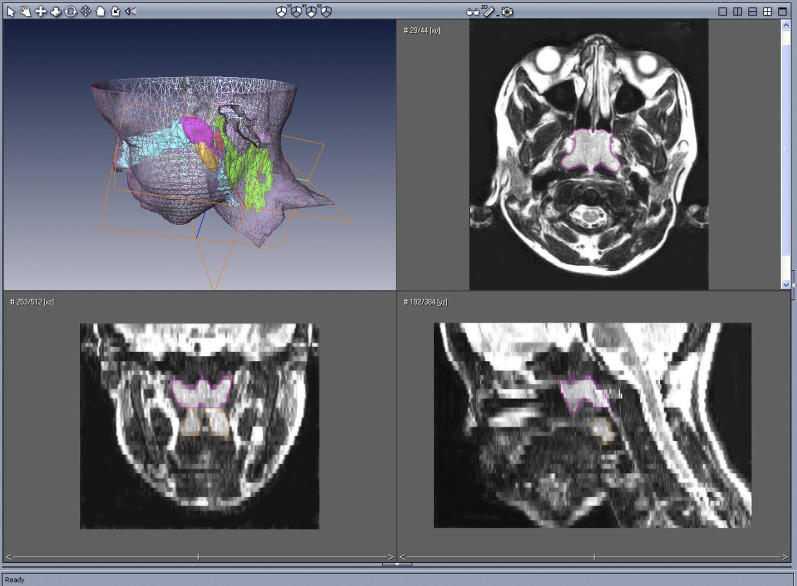
Figure 1.
Upper left, Surface rendering of the head and neck with three-dimensional reconstruction of the adenoid (magenta), tonsils (orange), retropharyngeal nodes (red), and deep cervical lymph nodes (green), of an 11.8-year-old male subject with sickle cell disease with obstructive sleep apnea syndrome using AMIRA software. Upper right, Axial T2-weighted image at the nasopharyngeal level outlining the adenoid (magenta). Lower left, Coronal reconstructed image outlining the adenoid (magenta) and tonsils (orange). Lower right, Midsagittal reconstructed image outlining the adenoid (magenta) and tonsils (orange).

