

Abstract
Translational research using animal models has traditionally involved genetically modified rodents; however there is increasing use of other novel genetically engineered species. As histology laboratories interface with researchers studying these novel species there will be many situations in which protocols will need to be adapted to the species, model and research goals. This paper gives examples of protocol adaptations to meet research needs and addresses common considerations that should be addressed for all research tissues submitted to the histotechnology laboratory. Positioning the histotechnologist, as well as the investigator, to meet the challenges associated with novel research models will help maximize research efficacy and quality.
Keywords: Genetically modified animals, Histology, Immunohistochemistry, Tissue embedding, Translational research
1) Introduction
The field of translation research has seen increasing diversity in novel animal model species. While the growth of genetically modified mouse models in recent years has been noted, biomedical investigators have progressively expanded their studies to include non-rodent transgenic species, which often recapitulate the human condition.1–4 A brief survey of novel non-rodent transgenic species used for new models of human diseases include pigs2,3,5–10, sheep11, rabbits12, zebrafish13, ferrets3, frogs14, flies15,16, and primates4. As a result of the diversity in animal species used in modeling and applications in modeling over the past decade, histotechnologists that work in the research field should expect to encounter tissues from an increasingly broad range of species asking a wealth of biologic questions. Therefore, it is important to understand specific research goals prior to tissue processing to be sure the pathologic data is best represented in quality and interpretation.
As biotechnology is ever-changing, it is impossible to fully address all situations that may be encountered when handling research tissues. This report will examine key considerations to address before beginning studies and provide examples on how to adapt histologic techniques to meet the diverse needs of novel transgenic model species which can also be applied to common genetically engineered rodent models. This paper can be a useful resource for histotechnologists and research investigators to provide a template for optimal interaction and production of quality histologic data (Table 1).
Table 1.
General considerations for handling research tissues
Project overview and specifics
|
Fixation
|
Decalcification
|
Processing
|
Embedding
|
Sectioning
|
Histochemical Staining
|
Immunohistochemical (IHC) Staining
|
2) Tissue Fixation
Many research histology laboratories and investigators have standard laboratory techniques for handling and fixation of tissues from mice, however in many cases researchers often need to be reminded of proper fixation protocol. For example, a common practice when harvesting small tissues is to place them into 50ml (or smaller) conical tubes. This practice prevents exposure of the tissue (e.g. liver) to fixative and reduces effective fixative penetration due to the conical shape of the tubes. When working with larger transgenic animals fixation problems can be compounded by the thought that “the more tissue, the better”. Often tissues from larger model species are collected in similar sized containers used for rodent species. Tissues from larger species require a large container to meet the minimum 15–20 times volumes of fixative to tissue. Additionally, some investigators experienced with small rodent tissues may not appreciate that the maximum thickness of tissues in fixative should be no thicker than about 5mm in at least one dimension. There is no substitute for proper and adequate fixation therefore educating investigators on fixation techniques can prevent suboptimal final results such as poor morphology and irregular immunohistochemical staining. While proper fixation technique is best determined before the project begins, it is often neglected until a problem arises which is when the value of optimal fixation is appreciated. One additional technique used by our lab for large and small animal tissues has been the use of rotary tables to avoid regional under-fixation commonly seen with tissues fixed in stationary jars however this technique is not a surrogate for proper fixation techniques as discussed (Figure 1).
Figure 1.
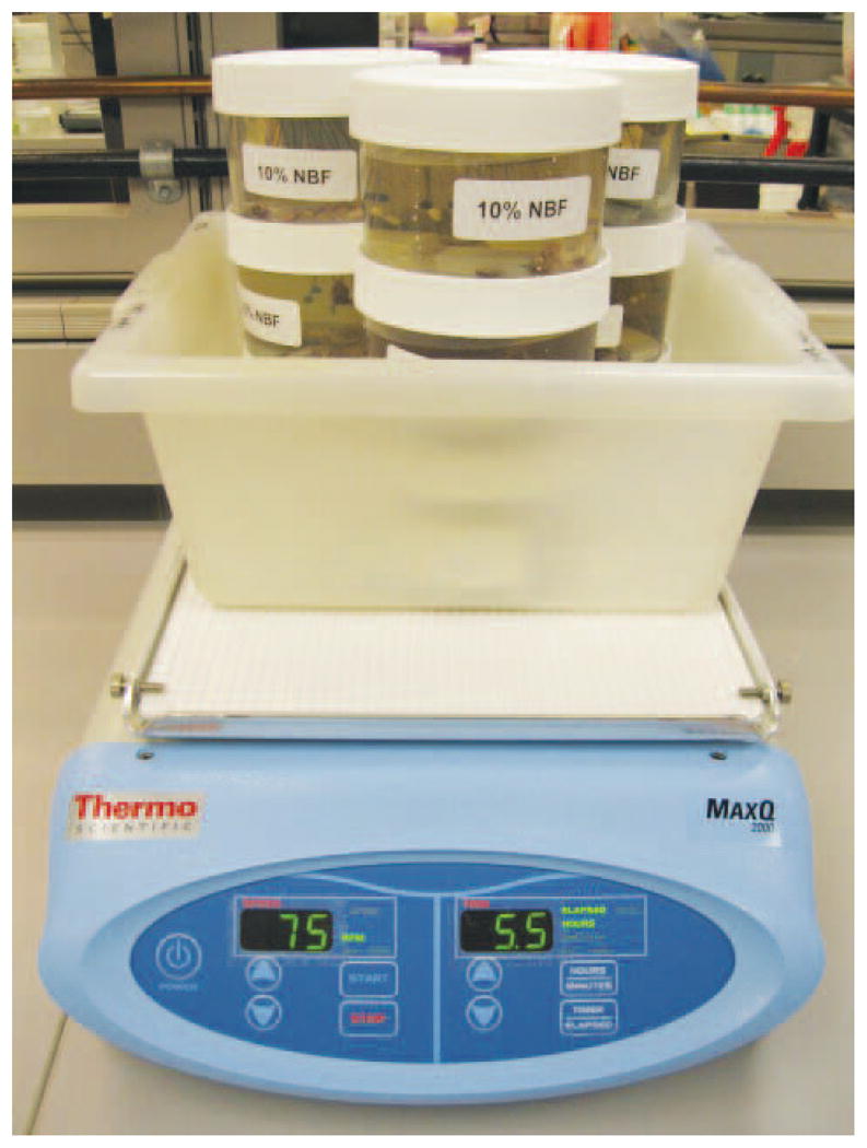
Placement of fixative containers on rotary tables helps to provide consistent fixation of all tissue surfaces.
Consideration of the type of fixative used should also be determined based on the desired analysis. The most commonly used fixative in our laboratory is 10% neutral buffered formalin (NBF). However the research goals should be discussed prior to recommendation of a fixative solution as some morphologic assessments, special stains and immunohistochemical stains require alternative fixative solutions. It is not the intent of this paper to review the various fixatives as several good reviews can be consulted but to realize that fixative choice should be considered based on predetermined endpoints necessary to answer the proposed experimental questions.17
3) Processing
Proper tissue processing is vital to sectioning and tissue evaluation. Sometimes transgenic species show a phenotype that is similar to humans, which by its very nature can be an unexpected challenge for processing. A good example is lipid-laden tissues such as the pancreas in the cystic fibrosis (CF) pig model.5,8 At birth the pancreas from CF pigs is damaged, but within a few months the pancreas is almost completely replaced by adipose tissue. Lipid-laden tissues such as these and others (ex: sheep brain) require increased processing time as compared to normal pancreas for adequate sectioning. Another example of processing challenges is that of small tissues, such as prosected intrapulmonary airways of CF pigs. Taking detailed notes and wrapping these tissues in mesh or filter paper prevents the tissue from getting lost during processing and will facilitate orientation efforts at the embedding station. When working with new phenotypes or alternative species the tissue size and composition should be taken into consideration to assure optimal processing. In some cases testing the processing run conditions prior to running all the experimental tissues is not only informative but necessary.
4) Embedding
Embedding of tissues is often a straightforward operation with the principal concern being cut side orientation. In research, scientific questions may require adaption of typical procedures to answer some important questions. For example, examination of the vas deferens (a small tube connecting the epididymis to the urethra) in the neonatal CF ferret model is required for comparative pathophysiology to the human condition.3 This is an embedding challenge as neonatal ferrets are very small (~6–8g) and their vas deferens are accordingly exceptionally small. The lack of mature collagen present in neonatal tissues also makes the vas deferens very friable and difficult to harvest. Because of this, the samples are fixed in 10% NBF to make them less friable and easier to handle. However, the standard processing protocol dehydrates and shrinks tissues, which may be counterproductive to handling these tissues. Thus embedding the CF vas deferens in OCT and snap freezing is a useful adaptation to the problem and allows for optimal handling, tissue morphology and orientation of the tissues for proper sectioning. Another example relates to embedding prosected intrapulmonary airways of pigs. Investigators may request cross sections through the length of these airways and by using deep embedding molds the whole airway can be stood on end in one block without having to section it into multiple blocks.
When encountering new procedures or tissue types, it is often best to work up the tissues to optimize the techniques. For instance, examination of Drosophila brain can give quite variable results depending on the embedding methods. Drobysheva and colleagues were able to assess variable embedding methods and found that paraffin alone was inadequate compared to a commercial product containing paraffin with dimethyl sulfoxide (DMSO) or agarose media embedding.18 Such experimental workups can be useful not only to the investigator but to the scientific community at large.
5) Sectioning
Sectioning is considered by some to be the cornerstone of the histology laboratory. Discussion with investigators about sectioning parameters (thickness, tissue levels, etc) is warranted to maximize the research potential of the tissue. Sometimes the investigator’s goal for the tissue samples is not feasible without modifications. An example of this is seen in therapeutic assessment of bone remodeling and fusion. Many times the area of tissue to be examined in such studies is so large (e.g. the interspace between transverse processes of the vertebrae) that whole fixed specimens are sectioned in a nondecalcified manner approximately 30 μm thick.19 These thick hematoxylin and eosin (HE) sections can provide good examination of morphologic features such as bone fusion; however, it does not allow for accurate assessment of cellular inflammation which is a potential adverse effect of such therapies. Thus, serial sections from the nondecalcified tissue or tissue samples from another identically treated sample will need to be decalcified, routinely processed and HE stained for appropriate examination of cellular inflammation.
6) Histochemical staining
A majority of tissues that come through the lab receive a HE stain for routine examination and special stains are typically applied on an as needed basis to identify specific morphologic changes and/or disease processes. Researching the literature may be required to arrive at new approaches to analyze the tissues from novel models species. For example, detection of mucus in airway epithelium using a nontraditional method (e.g. using alcian blue/pyronine Y stains) can be validated in novel animal models in part through comparison to known expression in mouse or human tissues.20 Comparing expression and distribution patterns in normal tissues of the species in question allows for more specific interpretation when analyzing the phenotype of the transgenic model. Such ground work on the normative expression can be published separately or included within the context of defining the phenotype of the transgenic model.
Another example is ex vivo histochemical staining. In CF, fetal disease of the trachea can cause abnormal cartilage development in neonatal humans, mice and pigs.7,21 In vivo imaging of the abnormal cartilage is limited by traditional technology, thus histochemical staining of excised trachea can be useful in analysis. As the mouse and pig tracheas are of significantly different physical size, the tissue uptake and staining qualities of the tracheal cartilage are different; thus one cannot simply follow the same protocols for slides and get the equivalent results. Often the staining time and extent of clearing with alcohols have to be titrated to the size of the trachea for optimal viewing. For the CF pig trachea, the time in alcian blue stain has to be increased and the final tissue has to stay aqueous in glycerin and not placed in serial alcohol bathes, which causes loss of the cartilage specific color distinction.7 Histotechnologists in research will be called upon to titrate and modify such techniques to make them work for novel model systems.
7) Immunohistochemistry/Immunofluorescence
Immunohistochemical (IHC) and immunofluorescent (IF) applications can be one of the most challenging aspects in working with non-rodent transgenic animal models. There are numerous reviews of immunohistochemical techniques and procedures that can be consulted.22–25 The limited availability of commercially available antibodies for non-rodent species makes antibody selection difficult. Optimizing new immunohistochemical markers in non-rodent species can be time consuming and expensive; however there are steps that can be taken to enhance the success rate.
The first step in optimizing new markers is to make sure that the tissue of interest is collected in multiple ways. If enough tissue is available, embedding and freezing in OCT along with routine fixation in paraformaldehyde or neutral buffered formalin are useful. This allows for identification of the best fixation conditions for the antibody of interest. In order to start workups, normal tissue samples from the species of interest are required which can typically be requested from the investigator’s lab. For example, if the antigen of interest is expressed by lymphocytes, then collection of the spleen, lymph nodes and thymus in addition to the tissue of interest would be useful.
The next step is a thorough literature search to determine if an antibody for the antigen of interest has been identified in the literature. If this is not successful, antibody company websites can be searched to determine if an antibody exists with a known cross-reactivity to the species of interest. In many cases this will not be successful either. At this point it is helpful to know the sequence of the antigen of interest in the species of interest. As most commercial antibodies are made against human or rodent antigens, the sequence from the species of interest can then be compared against the human and mouse sequences to determine the sequence homology. Knowing the sequence homology can help in making an educated guess to select an antibody to purchase. While some companies do not disclose the location and sequence utilized to make the antibody, we have found that contacting the customer support department of many of the major antibody companies is a great early resource. A representative in customer service can quickly search the sequence of interest against their database of known antigen sequences to identify antibodies that may be suitable. At this point if an antibody has not be identified that will cross-react or is likely to cross-react against the sequence of interest then the decision whether to empirically try commercial antibodies or to have an antibody custom made should be discussed. Often times, investigators choose to try commercial antibodies due to the expense and time involved in custom antibody generation. Taking the time to contact the antibody company to determine if they will give an aliquot free or offer a guarantee based on sequence homology can be a cost effective way to test various antibodies.
The antibody workup then depends on the tissue and antigen of interest along with the scientific goal of the project. In many ways antibody workup decisions in non-rodent tissues are similar to workups on rodent tissue including needs related to tissue morphology, antigen abundance and detection methods. Detection methods may be critical to the project goals and data analysis. The decision to pursue IHC or IF detection depends on multiple factors such as tissue fixation, antigen abundance and the desire to stain multiple antigens at the same time. Double staining techniques using IF are useful in determining correct staining patterns or identifying new staining patterns. One of the most important components in the antibody workup is the inclusion of a known positive and negative control. Although this is important regardless of the species, identification of a proper control can be more difficult in non-rodent species, in part due to the lack or unavailability of knockout strains for many antigens.
Utilizing these steps along with a firm background in immunohistochemical methods will enable histotechnologists, researchers and pathologists to increase the success rate of immunohistochemical staining of non-rodent tissue.
8) Conclusion
As histology laboratories increasingly interface with researchers, exposure to the growing repertoire of genetically modified species is inevitable. Histotechnologists will be called upon to utilize their professional skills to adapt and modify techniques to help enable effective translational research and to educate researchers of the necessary questions that should be asked prior to tissue processing.
Acknowledgments
This work was supported by the National Heart Lung and Blood Institute (grant HL51670 and HL091842), the National Institute of Diabetes and Digestive and Kidney Diseases (grant DK54759), and the Cystic Fibrosis Foundation.
References
- 1.Cardiff RD, Ward JM, Barthold SW. ‘One medicine---one pathology’: are veterinary and human pathology prepared? Lab Invest. 2008;88:18–26. doi: 10.1038/labinvest.3700695. [DOI] [PMC free article] [PubMed] [Google Scholar]
- 2.Rogers CS, Stoltz DA, Meyerholz DK, et al. Disruption of the CFTR gene produces a model of cystic fibrosis in newborn pigs. Science. 2008;321:1837–41. doi: 10.1126/science.1163600. [DOI] [PMC free article] [PubMed] [Google Scholar]
- 3.Sun X, Sui H, Fisher JT, et al. Disease phenotype of a ferret CFTR-knockout model of cystic fibrosis. J Clin Invest. 2010;120:3149–60. doi: 10.1172/JCI43052. [DOI] [PMC free article] [PubMed] [Google Scholar]
- 4.Sasaki E, Suemizu H, Shimada A, et al. Generation of transgenic non-human primates with germline transmission. Nature. 2009;459:523–7. doi: 10.1038/nature08090. [DOI] [PubMed] [Google Scholar]
- 5.Stoltz DA, Meyerholz DK, Pezzulo AA, et al. Cystic fibrosis pigs develop lung disease and exhibit defective bacterial eradication at birth. Sci Transl Med. 2010;2:29ra31. doi: 10.1126/scitranslmed.3000928. [DOI] [PMC free article] [PubMed] [Google Scholar]
- 6.Ostedgaard LS, Meyerholz DK, Chen JH, et al. The DeltaF508 mutation causes CFTR misprocessing and cystic fibrosis-like disease in pigs. Sci Transl Med. 2011;3:74ra24. doi: 10.1126/scitranslmed.3001868. [DOI] [PMC free article] [PubMed] [Google Scholar]
- 7.Meyerholz DK, Stoltz DA, Namati E, et al. Loss of cystic fibrosis transmembrane conductance regulator function produces abnormalities in tracheal development in neonatal pigs and young children. Am J Respir Crit Care Med. 2010;182:1251–61. doi: 10.1164/rccm.201004-0643OC. [DOI] [PMC free article] [PubMed] [Google Scholar]
- 8.Meyerholz DK, Stoltz DA, Pezzulo AA, Welsh MJ. Pathology of gastrointestinal organs in a porcine model of cystic fibrosis. Am J Pathol. 2010;176:1377–89. doi: 10.2353/ajpath.2010.090849. [DOI] [PMC free article] [PubMed] [Google Scholar]
- 9.Renner S, Fehlings C, Herbach N, et al. Glucose intolerance and reduced proliferation of pancreatic beta-cells in transgenic pigs with impaired glucose-dependent insulinotropic polypeptide function. Diabetes. 2010;59:1228–38. doi: 10.2337/db09-0519. [DOI] [PMC free article] [PubMed] [Google Scholar]
- 10.Whyte JJ, Samuel M, Mahan E, et al. Vascular endothelium-specific overexpression of human catalase in cloned pigs. Transgenic Res. 2011;20:989–1001. doi: 10.1007/s11248-010-9473-7. [DOI] [PMC free article] [PubMed] [Google Scholar]
- 11.Schnieke AE, Kind AJ, Ritchie WA, et al. Human factor IX transgenic sheep produced by transfer of nuclei from transfected fetal fibroblasts. Science. 1997;278:2130–3. doi: 10.1126/science.278.5346.2130. [DOI] [PubMed] [Google Scholar]
- 12.Kondo M, Sakai T, Komeima K, et al. Generation of a transgenic rabbit model of retinal degeneration. Invest Ophthalmol Vis Sci. 2009;50:1371–7. doi: 10.1167/iovs.08-2863. [DOI] [PubMed] [Google Scholar]
- 13.Paquet D, Bhat R, Sydow A, et al. A zebrafish model of tauopathy allows in vivo imaging of neuronal cell death and drug evaluation. J Clin Invest. 2009;119:1382–95. doi: 10.1172/JCI37537. [DOI] [PMC free article] [PubMed] [Google Scholar]
- 14.Tam BM, Moritz OL. Dark rearing rescues P23H rhodopsin-induced retinal degeneration in a transgenic Xenopus laevis model of retinitis pigmentosa: a chromophore-dependent mechanism characterized by production of N-terminally truncated mutant rhodopsin. J Neurosci. 2007;27:9043–53. doi: 10.1523/JNEUROSCI.2245-07.2007. [DOI] [PMC free article] [PubMed] [Google Scholar]
- 15.Feany MB, Bender WW. A Drosophila model of Parkinson’s disease. Nature. 2000;404:394–8. doi: 10.1038/35006074. [DOI] [PubMed] [Google Scholar]
- 16.Crowther DC, Kinghorn KJ, Miranda E, et al. Intraneuronal Abeta, non-amyloid aggregates and neurodegeneration in a Drosophila model of Alzheimer’s disease. Neuroscience. 2005;132:123–35. doi: 10.1016/j.neuroscience.2004.12.025. [DOI] [PubMed] [Google Scholar]
- 17.Kiernan JA. Histological and histochemical methods: theory and practice. 4. Bloxham UK: Scion; 2008. [Google Scholar]
- 18.Drobysheva D, Ameel K, Welch B, et al. An optimized method for histological detection of dopaminergic neurons in Drosophila melanogaster. J Histochem Cytochem. 2008;56:1049–63. doi: 10.1369/jhc.2008.951137. [DOI] [PMC free article] [PubMed] [Google Scholar]
- 19.Smucker JD, Bobst JA, Petersen EB, Nepola JV, Fredericks DC. B2A peptide on ceramic granules enhance posterolateral spinal fusion in rabbits compared with autograft. Spine (Phila Pa 1976) 2008;33:1324–9. doi: 10.1097/BRS.0b013e3181732a74. [DOI] [PubMed] [Google Scholar]
- 20.Meyerholz DK, Rodgers J, Castilow EM, Varga SM. Alcian Blue and Pyronine Y histochemical stains permit assessment of multiple parameters in pulmonary disease models. Vet Pathol. 2009;46:325–8. doi: 10.1354/vp.46-2-325. [DOI] [PMC free article] [PubMed] [Google Scholar]
- 21.Bonvin E, Le Rouzic P, Bernaudin JF, et al. Congenital tracheal malformation in cystic fibrosis transmembrane conductance regulator-deficient mice. J Physiol. 2008;586:3231–43. doi: 10.1113/jphysiol.2008.150763. [DOI] [PMC free article] [PubMed] [Google Scholar]
- 22.Dabbs DJ. Diagnostic immunohistochemistry theranostic and genomic applications. 3. xviii. Philadelphia, PA: Saunders/Elsevier; 2010. ScienceDirect (Online service) p. 941. [Google Scholar]
- 23.Ramos-Vara JA, Beissenherz ME. Optimization of immunohistochemical methods using two different antigen retrieval methods on formalin-fixed paraffin-embedded tissues: experience with 63 markers. J Vet Diagn Invest. 2000;12:307–11. doi: 10.1177/104063870001200402. [DOI] [PubMed] [Google Scholar]
- 24.Beckstead JH. A simple technique for preservation of fixation-sensitive antigens in paraffin-embedded tissues. J Histochem Cytochem. 1994;42:1127–34. doi: 10.1177/42.8.8027531. [DOI] [PubMed] [Google Scholar]
- 25.Daneshtalab N, Dore JJ, Smeda JS. Troubleshooting tissue specificity and antibody selection: Procedures in immunohistochemical studies. J Pharmacol Toxicol Methods. 2010;61:127–35. doi: 10.1016/j.vascn.2009.12.002. [DOI] [PubMed] [Google Scholar]