Abstract
Development of biofilm is a key mechanism involved in Staphylococcus epidermidis virulence during device-associated infections. We aimed to investigate antibiofilm formation and mature biofilm eradication ability of ethanol and water extracts of Thai traditional herbal recipes including THR-SK004, THR-SK010, and THR-SK011 against S. epidermidis. A biofilm forming reference strain, S. epidermidis ATCC 35984 was employed as a model for searching anti-biofilm agents by MTT reduction assay. The results revealed that the ethanol extract of THR-SK004 (THR-SK004E) could inhibit the formation of S. epidermidis biofilm on polystyrene surfaces. Furthermore, treatments with the extract efficiently inhibit the biofilm formation of the pathogen on glass surfaces determined by scanning electron microscopy and crystal violet staining. In addition, THR-SK010 ethanol extract (THR-SK010E; 0.63–5 μg/mL) could decrease 30 to 40% of the biofilm development. Almost 90% of a 7-day-old staphylococcal biofilm was destroyed after treatment with THR-SK004E (250 and 500 μg/mL) and THR-SK010E (10 and 20 μg/mL) for 24 h. Therefore, our results clearly demonstrated THR-SK004E could prevent the staphylococcal biofilm development, whereas both THR-SK004E and THR-SK010E possessed remarkable eradication ability on the mature staphylococcal biofilm.
1. Introduction
Staphylococcus epidermidis, a normal inhabitant of the healthy human skin and mucosal microbial communities, has emerged as a common cause of numerous nosocomial infections, mostly occurring in immunocompromised hosts or patients with implanted medical devices [1]. Even though, it has a low pathogenic potential, data from the European surveillance indicated that coagulase-negative staphylococci were isolated up to 40% in bloodstream infections while Staphylococcus aureus was isolated less than 20% [2]. In S. epidermidis, biofilm formation is regarded as a major pathomechanism as it renders S. epidermidis highly resistant to conventional antibiotics and host defenses. This can be caused by slow diffusion of these compounds through the extracellular polymeric matrix and slow growth of the bacteria [3, 4]. Staphylococcal biofilm is therefore difficult to eradicate and is a source of many recalcitrant infections. As such, novel strategies or more effective agents exhibiting an antibiofilm ability with clinical efficacy and safety are of great interest.
Medicinal plant-derived compounds have increased widespread interest in the search of alternative antibacterial agents because of the perception that they are safe and have a long history of use in folk medicine for the treatment of infectious diseases [5]. The active constituents isolated from medicinal plants have intensively been studied for their antibacterial effects against planktonic bacteria. More importantly, some plants have been reported to be able to prevent the formation of biofilm in some pathogens such as Listeria monocytogenes [6], Pseudomonas aeruginosa [7], Streptococcus pyogenes [8], Streptococcus mutans [9], and S. aureus [10]. So far, most studies have focused on the observation of anti-biofilm activity of herbs taken as a single unit but not in combination, such as in herbal recipes. No attention has been paid to the antibacterial or anti-biofilm activity of traditionally used herbal recipes.
This study was therefore undertaken to investigate the in vitro anti-biofilm potential of selected Thai traditional herbal recipes (THRs) that have been traditionally employed for the treatment of wounds and skin infections against an important biofilm producing pathogen, S. epidermidis. The biofilm formation requires bacterial attachment to solid surfaces, the development of bacterial multilayers, and their enclosure in a large exopolymeric matrix [3]. Because of that, we investigated both anti-biofilm development and mature biofilm eradication of the recipes.
2. Material and Methods
2.1. Bacterial Strains and Their Biofilm-Forming Ability
Bacteria used in this study were clinically isolated methicillin resistant Staphylococcus aureus (MRSA) NPRC R001-R005, methicillin susceptible S. aureus (MSSA) NPRC S001-S005, S. aureus ATCC 25923, a biofilm-positive strain (Staphylococcus epidermidis ATCC 35984), and a biofilm-negative strain (S. epidermidis ATCC 12228).
In order to assess the biofilm formation ability of Staphylococcus spp., well-isolated colonies grown overnight at 37°C on tryptic soy agar (TSA, Becton, Dickinson, and Company, France) were inoculated in tryptic soy broth (TSB, Becton, Dickinson, and Company) supplemented with 2% (w/w) D-glucose (TSBGlc). Following incubation at 37°C for 24 h; culture supernatants from each isolate were diluted 1 : 200 in TSBGlc. Aliquots of bacterial suspension (200 μL; 5 × 10⁵ CFU/mL, final concentration) were transferred into a flat-bottomed 96-well polystyrene microtiter plate (Nunc, Roskilde, Denmark). The medium without the bacterial suspension was used as the negative control. The plates were incubated at 37°C for 24 h, culture supernatants from each well were then decanted and planktonic cells were removed by washing three times with phosphate-buffered saline (PBS; pH 7.4) [10].
3-(4, 5-dimethyl-2-thiazolyl)-2, 5-diphenyl-2H-tetrazolium-bromid (MTT) (Sigma-Aldrich, USA) reduction assay according to the method previously described [11] was applied to quantify the biofilm forming ability of the isolates. An aliquot of MTT solution (0.2 mg/mL; 200 μL) was added to each of the prewashed wells and the plate was then incubated for 3 h in the dark at 37°C. Following the incubation, MTT was then replaced by 200 μL of dimethylsulfoxide (DMSO; Merck, Darmstadt, Germany). Bacteria with an active electron transport system will reduce the tetrazolium salt to a water-insoluble purple formazan product. The colour intensity of DMSO dissolved formazan was determined by a microplate reader (Tecan Sunrise, Tecan Austria) at A482 nm. The absorbance values for the negative control were then subtracted from the tested wells to eliminate false results due to background interference.
2.2. Preparation of Herbal Recipes
Selected southern Thai herbal recipes that have been used for the treatment of wounds and skin infections were kindly provided by Mr. Earn Thongsongsi (THR-SK004) [12] and Mr. Somporn Chanwanisakul (THR-SK010 and THR-SK011), a traditional Thai Medical Doctor at Traditional Thai Medicine Hospital, Prince of Songkla University, Hat Yai, Thailand. Plant parts as described in Table 1 were locally collected and reference voucher specimens were deposited at Faculty of Traditional Thai Medicine, Prince of Songkla University, Hat Yai, Songkhla, Thailand. The powdered formulas (100 g) were submitted to solvent extractions by maceration with distilled water at room temperature for three days or with ethanol for seven days (500 mL each solvent). After filtrations through a Whatman no. 1 paper, aqueous filtrates were freeze-dried and ethanol filtrates were concentrated using a rotatory evaporator, and kept at 55°C until they were completely dry. Yields (%; w/w) of each extracts were calculated as the ratio of the weight of the extract to the weight of the crude herb powder and presented in Table 1. Lyophilized water extracts (THR-SK004W, THR-SK010W, and THR-SK011W) and evaporated ethanol extracts (THR-SK004E, THR-SK010E, and THR-SK011E) were dissolved in 10% dimethylsulfoxide (DMSO; Merck, Germany) before use.
Table 1.
Wound healing related-biological activities, herbal components, and extraction yields of selected southern Thai herbal recipes.
| Herbal components (Plant parts) | Yield (%) | Wound healing related-biological activities | References |
|---|---|---|---|
| THR-SK004 | 2.40/2.22a | ||
| Maranta arundinacea L. (Rhizome) | NA | ||
| Oroxylum indicum Vent. (Bark) | Ulcer protective | [17] | |
| Commelina benghalensis L. (Whole plant) | Anti-oxidant | [18] | |
|
| |||
| THR-SK010 | 6.45/3.43 | ||
| Curcuma longa L. (Rhizome) | Anti-oxidant/anti-inflammatory/wound healing | [19] | |
| Areca catechu L. (Seed) | Anti-inflammatory | [20] | |
| Oryza sativa L. (Seed) | NA | ||
| Garcinia mangostana L. (Pericarp) | Anti-inflammatory; Anti-ulcerogenic | [21, 22] | |
|
| |||
| THR-SK011 | 2.01/3.33 | ||
| Ceiba pentandra L. Gaertn. (Leaf) | Anti-oxidant; Anti-inflammatory | [23] [24] |
|
| Aloe barbadensis Mill. (Leaf) | Anti-oxidant/anti-inflammatory | [25] | |
| Coccinia grandis (L.) Voigt. (Climber) | Anti-oxidant | [26] | |
| Senna siamea (Lam.) Irwin & Barneby. (Leaf) | Anti-inflammatory | [27] | |
| Chromolaena odorata (L.) R. M. King & H. Rob. (Climber) | Anti-oxidant; Wound healing | [28, 29] | |
| Tinospora crispa (L.) Miers ex Hook. f. & Thomson. (Climber) | Anti-oxidant/anti-tumor | [30] | |
aExtraction yields of ethanol/water extracts.
2.3. Inhibition of Staphylococcal Biofilm Formation by the Herbal Recipes.
The herbal recipe extracts were tested for their potential to prevent biofilm formation of a biofilm producing strain, S. epidermidis ATCC 35984. They were added to the growth medium at the time of inoculation and the cells were allowed to form biofilm. An aliquot of twofold serial dilutions (100 μL) was prepared in the 96-well microtiter plate containing TSBGlc, with final concentrations of THR-SK004W, THR-SK010W, THR-SK011W, THR-SK004E, and THR-SK011E ranging from 15.63 to 2501 μg/mL and 0.63 to 250 μg/mL for THR-SK010E. Bacterial suspensions (100 μL; 5 × 10⁵ CFU/mL, final concentration) were then transferred into the plate. TSBGlc containing 0.2% DMSO was employed as a negative control. TSBGlc without the extract was used as the nontreated well and the medium with each concentration of the extracts was used as the blank control [13].
Following incubation at 37°C for 24 h, the effect of the extracts on the growth of S. epidermidis was evaluated using the microplate reader at optical density of 620 nm (OD620 nm). The biofilm formation of S. epidermidis ATCC 35984 in the presence of the herbal recipe extracts was subsequently determined using the MTT reduction assay as already stated.
2.4. Biofilm Time-Dependent Inhibition Assay
According to anti-biofilm inhibition, THR-SK004 ethanol extract (THR-SK004E) was selected for further experiment. The biofilm development of S. epidermidis after being treated with this effective extract at concentrations 250 and 500 μg/mL was observed every 12 h for 2 days using the MTT reduction assay as described above.
2.5. Observation on Biofilm Formation by Scanning Electron Microscopy
In addition, scanning electron microscopy (SEM) images were taken to confirm the prevention of biofilm formation by THR-SK004E [14]. Briefly, this strain was allowed to grow on squared glass slides (1 × 1 cm) placed in 24-well polystyrene plates (Greiner Bio-One, France) supplemented with TSBG1c containing the extract at 250 and 500 μg/mL followed by incubation at 37°C for 48 h. After removal of the media and washing, the samples were initially fixed in 2.5% glutaraldehyde in cacodylate buffer for 90 min, then washed twice with cacodylate buffer and dehydrated for 10 min using a graded ethanol series. A critical point drying procedure was followed, and the specimens were then sputter-coated with gold. Samples were examined with a scanning electron microscope (5800LV, JEOL, Japan).
Beside, the biofilm glass pieces were washed three times with PBS and stained with 1% (w/v) crystal violet solution [15]. The biofilm formation on the stained glass pieces was dissolved with DMSO and quantified by measuring the absorbance at 620 nm using microplate reader.
2.6. Eradication of Established Staphylococcal Biofilm by the Herbal Recipes
Static biofilms were grown for seven days as previously described [16]. As above, the bacterial culture was prepared and incubated at 37°C for 24 h. Planktonic cells in the culture medium were then removed and fresh TSBGlc was added. This procedure was repeated daily for seven consecutive days. At the end of the 7 days of biofilm growth, the medium and the planktonic cells were gently aspirated. Thereafter, 200 μL of PBS containing different concentrations of the THR-SK004E (250 and 500 μg/mL as final concentrations) or THR-SK010E (10 and 20 μg/mL as final concentrations) were added into the wells. Eradication of staphylococcal-preformed biofilm by the extract was measured at selected time intervals of 1, 3, 6, 12, and 24 h by the MTT reduction assay as explained above. The buffer with no antimicrobial agents was added to the positive biofilm control wells. The percentage of biofilm eradication in comparison with untreated wells was calculated using the equation [(OD(at 0 h) − OD(after treatment))/OD(at 0 h)] × 100.
3. Results
S. epidermidis ATCC 35984 was employed as a model isolate for primary screening of the anti-biofilm ability of water and ethanol extracts prepared from the traditional herbal recipes. Extraction yields of three selected herbal recipes including THR-SK004 and THR-SK010 used for wound healing and THR-SK011 used for abscess treatment and reported biological activities of their herbal components are summarized in Table 1.
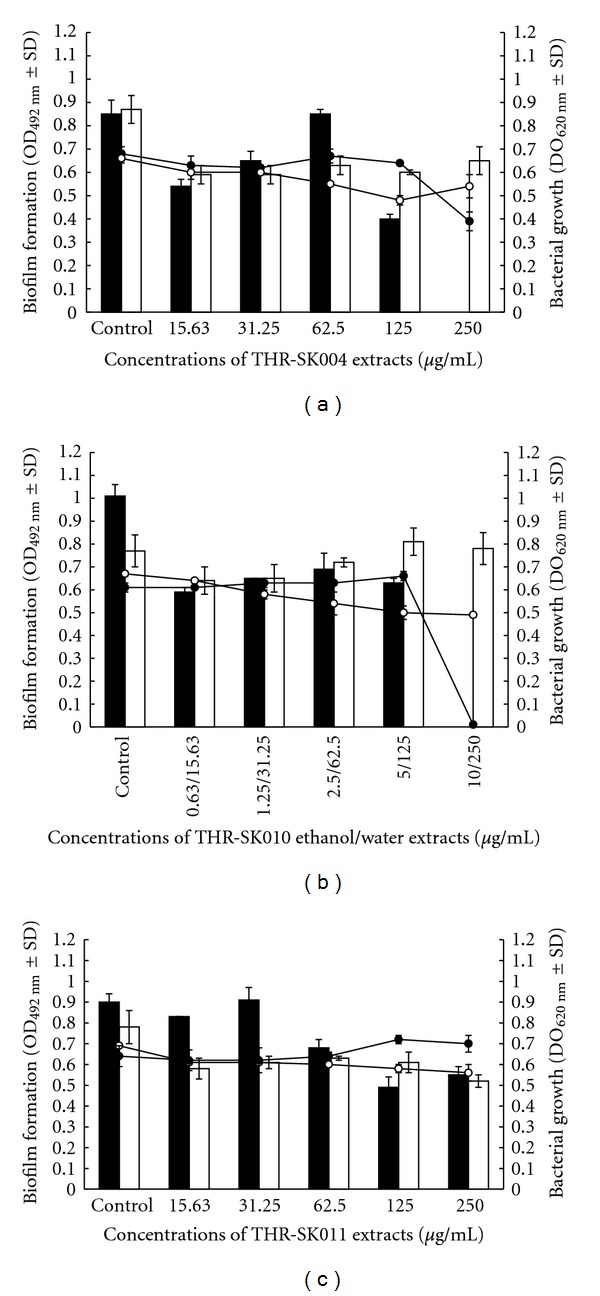
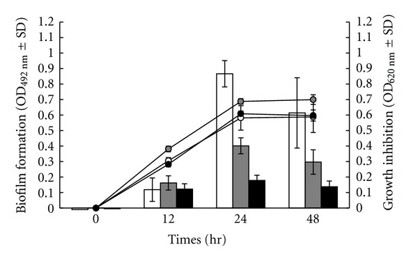
In order to investigate the effect of the recipes on S. epidermidis biofilm formation, the relationship between drug doses and the metabolic activity of cells in biofilm was monitored (Figure 1). The MTT reduction assay results showed that THR-SK004E at 250 μg/mL could inhibit the formation of S. epidermidis biofilm. THR-SK010E (5–0.63 μg/mL) could decrease 30 to 40% of staphylococcal biofilm. It is noteworthy that the effective concentrations of both THR-SK004E and THR-SK010E did not affect the growth of planktonic cells. Moreover, THR-SK004E at concentrations 125 and 250 μg/mL was able to inhibit biofilm formation of S. epidermidis on polystyrene surfaces over a 48 h period as depicted in Figure 2.
Figure 1.

Effect of different concentrations of THR-SK004 (a), THR-SK010 (b), and THR-SK011 (c) ethanol (-⚫-,■), and water (-o-, □) extracts on the bacterial growth (linear charts) and the biofilm formation (column charts) of Staphylococcus epidermidis ATCC 35984.
Figure 2.

Development of Staphylococcus epidermidis ATCC 35984 biofilm (column charts) and the bacterial growth (linear charts) after treatment with THR-SK004 ethanol extract at 125 (grey symbols) and 250 μg/mL (black symbols). Dimethylsulfoxide at 0.2% (white symbols) was used as positive control. Each symbol indicates the means ± standard error for three independent experiments performed in duplicate.
Inhibition of biofilm formation on glass surfaces by THR-SK004E was additionally visualized by both SEM and crystal violet assay which is illustrated in Figure 3. As expected, the additions of THR-SK004E at 250 or 500 μg/mL, which reduced the staphylococcal biofilm formation on polystyrene surfaces remarkably inhibited the biofilm formation of the pathogen on glass surfaces.
Figure 3.

SEM micrographs of S. epidermidis ATCC 35984 biofilm formation on glass surfaces. Biofilms were grown in TSBGlc (a) or in TSBGlc supplemented with THR-SK004 ethanol extract at 250, (b) and 500 μg/mL, (c), and all images shown were taken at magnification 2500x. The selected images were chosen as the best representatives of the amount of biofilm on the glass surfaces. Inhibition of staphylococcal biofilm development by THR-SK004 ethanol extract was additionally confirmed by crystal violet assay (d).
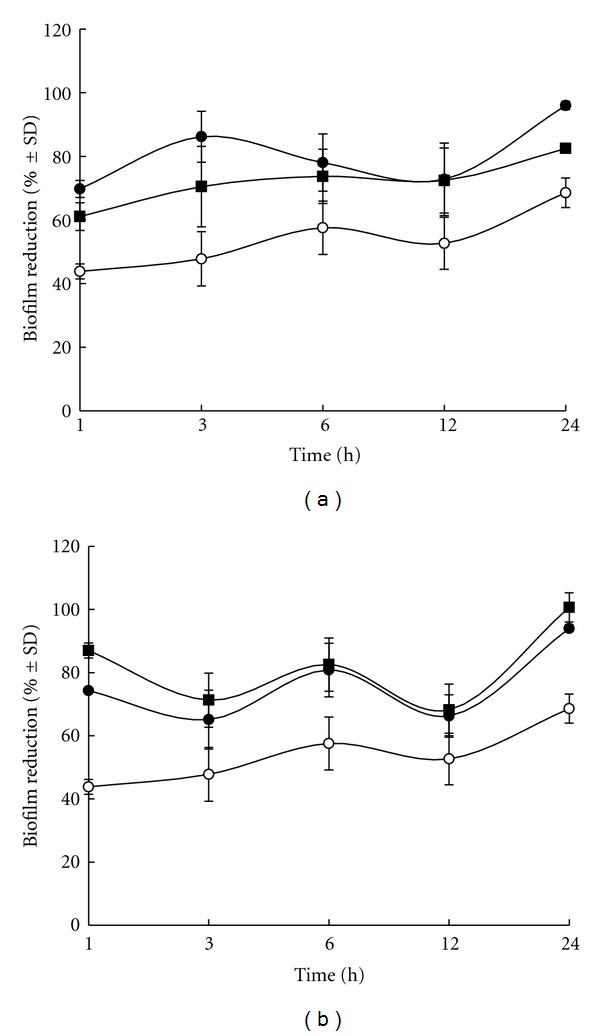
Static S. epidermidis biofilm were grown for seven days and then treated with THR-SK004E (250 and 500 μg/mL) and THR-SK010E (10 and 20 μg/mL). As presented in Figure 4, more than 60% of the static biofilm reduction was noted in biofilms treated with the extracts for 1 h. The effect of both extracts was much more pronounced with a more lengthy treatment regimen. Noticeably, more than 90% of the 7-day-old biofilms was destroyed following a 24 h treatment with THR-SK004E at 500 μg/mL and THR-SK010E at 10 and 20 μg/mL.
Figure 4.

Time-dependent eradication of the mature biofilm formed by S. epidermidis ATCC 35984 after treatment with THR-SK004 ethanol extract (a) at 250 (⚫) and 500 μg/mL (■) or THR-SK010 ethanol extract (b) at 10 μg/mL (⚫) and 20 μg/mL (■). Dimethylsulfoxide at 0.2% (o) was used as positive control. Each symbol indicates the means ± standard error for three independent experiments performed in duplicate.
4. Discussion
Impairment of bacterial adhesion and biofilm formation by a pathway that does not influence bacterial growth is a characteristic for antivirulence therapies, one of the recent promising alternatives to combat pathogenic microorganisms, particularly S. epidermidis [31]. In addition to the antibacterial activity and antibiofilm potency of individual medicinal plants, the effects of herbal recipes on S. epidermidis biofilm were studied for the first time.
THR-SK004 and its herbal constituents (Maranta arundinacea, Oroxylum indicum, and Commelina benghalensis) have never been judged for their antibiofilm ability. However, ethanol extracts of THR-SK004, Commelina benghalensis, and Oroxylum indicum were proposed to have mild-to-moderate antistaphylococcal activities [12, 32, 33]. Treatment with THR-SK004E resulted in a great inhibition of S. epidermidis biofilm formation, but the presence of the extract did not influence the bacterial growth. This study demonstrates that the recipe extract inhibits the formation of S. epidermidis on both hydrophobic surface (polystyrene) and hydrophilic surface (glass). The information suggests that intensive study on THR-SK004E active constituents may potentially be used as a tool to prevent biofilm formation on both hydrophobic and hydrophilic medicinal devices. Likewise, previous investigations have implied that coating clinical materials with antimicrobial substances successfully prevents microbial colonization and biofilm formation [10, 13, 34, 35].
Ethanol extract of THR-SK010 is composed of Oryza sativa and other well-documented medicinal plants including Curcuma longa, Areca catechu, and Garcinia mangostana. The active constituents of Curcuma longa have been proved or anti-biofilm and antiadherence potencies on Candida albicans [36], Streptococcus mutans [37], Vibrio vulnificus [38], and Pseudomonas aeruginosa [39]. However, at the tested concentrations (5–0.63 μg/mL) which are lower than the values used in the previous reports, THR-SK010 did not inhibit the bacterial growth but showed a slight antibiofilm activity. However, anti-biofilm staphylococcal formation activity of water extracts of the tested recipes and THR-SK011E was not demonstrated. Among the herbal components of the recipes, 12 (92.3%) of them have been reported to possess wound healing or related biological activities such as antioxidant and anti-inflammatory activities [40]. In addition, our results concur with literature evidence that ethanol is a more reliable extraction solvent of antimicrobial substances from medicinal plants compared to water, representative of the therapeutically effective preparations currently favoured by traditional healers [40, 41].
As there is an urgent need to identify therapeutic strategies that are directed toward the inhibition of bacterial preformed biofilm, the eradication potency of effective extracts was evaluated. This present study showed that both THR-SK004E and THR-SK010E successfully eliminated the established biofilm of S. epidermidis, even at low concentrations of THR-SK010E (10 to 20 μg/mL). Antipreformed biofilm activity of THR-SK010E is comparable with antibiotics (daptomycin, linezolid, and tigecycline) [42] or plant-derive compounds (eucalyptus oil, 1,8-cineole [43], tea tree oil [44], farnesol [16], oregano, carvacrol, and thymol [10]).
There is a critical need for the development of alternative treatment to combat the growing number of multidrug resistant pathogen-associated infections, especially in situation where biofilms are involved. THR-SK004E strongly exhibited anti-biofilm formation of S. epidermidis on both polystyrene and glass surfaces, whereas both THR-SK004E and THR-SK010E remarkably destroyed the established biofilm.
5. Conclusion
Based on our results, THR-SK004E and THR-SK010E have promising applications as alternative antibiofilm agents. Close investigations into the identification of active constituents from the effective recipes and study on mechanisms involved in the inhibition of biofilm by the recipes are therefore warranted and currently being pursued in our laboratory.
Acknowledgment
This work was supported by Grants for Development of New Faculty Staff, The Annual Income Budget of Prince of Songkla University (TTM540049S, fiscal year 2010–2012).
References
- 1.McCann MT, Gilmore BF, Gorman SP. Staphylococcus epidermidis device-related infections: pathogenesis and clinical management. Journal of Pharmacy and Pharmacology. 2008;60(12):1551–1571. doi: 10.1211/jpp/60.12.0001. [DOI] [PubMed] [Google Scholar]
- 2.Suetens C, Morales I, Savey A, et al. European surveillance of ICU-acquired infections (HELICS-ICU): methods and main results. Journal of Hospital Infection. 2007;65(2):171–173. doi: 10.1016/S0195-6701(07)60038-3. [DOI] [PubMed] [Google Scholar]
- 3.Schoenfelder SMK, Lange C, Eckart M, Hennig S, Kozytska S, Ziebuhr W. Success through diversity—how Staphylococcus epidermidis establishes as a nosocomial pathogen. International Journal of Medical Microbiology. 2010;300(6):380–386. doi: 10.1016/j.ijmm.2010.04.011. [DOI] [PubMed] [Google Scholar]
- 4.Mah TFC, O’Toole GA. Mechanisms of biofilm resistance to antimicrobial agents. Trends in Microbiology. 2001;9(1):34–39. doi: 10.1016/s0966-842x(00)01913-2. [DOI] [PubMed] [Google Scholar]
- 5.Guarrera PM. Traditional phytotherapy in Central Italy (Marche, Abruzzo, and Latium) Fitoterapia. 2005;76(1):1–25. doi: 10.1016/j.fitote.2004.09.006. [DOI] [PubMed] [Google Scholar]
- 6.Sandasi M, Leonard CM, Viljoen AM. The effect of five common essential oil components on Listeria monocytogenes biofilms. Food Control. 2008;19(11):1070–1075. [Google Scholar]
- 7.Adonizio A, Kong KF, Mathee K. Inhibition of quorum sensing-controlled virulence factor production in Pseudomonas aeruginosa by south Florida plant extracts. Antimicrobial Agents and Chemotherapy. 2008;52(1):198–203. doi: 10.1128/AAC.00612-07. [DOI] [PMC free article] [PubMed] [Google Scholar]
- 8.Limsuwan S, Voravuthikunchai SP. Boesenbergia pandurata (Roxb.) Schltr., Eleutherine americana Merr. and Rhodomyrtus tomentosa (Aiton) Hassk. as antibiofilm producing and antiquorum sensing in Streptococcus pyogenes . FEMS Immunology and Medical Microbiology. 2008;53(3):429–436. doi: 10.1111/j.1574-695X.2008.00445.x. [DOI] [PubMed] [Google Scholar]
- 9.Song JH, Yang TC, Chang KW, Han SK, Yi HK, Jeon JG. In vitro effects of a fraction separated from Polygonum cuspidatum root on the viability, in suspension and biofilms, and biofilm formation of mutans streptococci. Journal of Ethnopharmacology. 2007;112(3):419–425. doi: 10.1016/j.jep.2007.03.036. [DOI] [PubMed] [Google Scholar]
- 10.Nostro A, Roccaro AS, Bisignano G, et al. Effects of oregano, carvacrol and thymol on Staphylococcus aureus and Staphylococcus epidermidis biofilms. Journal of Medical Microbiology. 2007;56(4):519–523. doi: 10.1099/jmm.0.46804-0. [DOI] [PubMed] [Google Scholar]
- 11.Tang HJ, Chen CC, Ko WC, Yu WL, Chiang SR, Chuang YC. In vitro efficacy of antimicrobial agents against high-inoculum or biofilm-embedded meticillin-resistant Staphylococcus aureus with vancomycin minimal inhibitory concentrations equal to 2 μg/mL (VA2-MRSA) International Journal of Antimicrobial Agents. 2011;38(1):46–51. doi: 10.1016/j.ijantimicag.2011.02.013. [DOI] [PubMed] [Google Scholar]
- 12.Chusri S, Chaicoch N, Thongza-ard W, Limsuwan S. In vitro antibacterial activity of ethanol extracts of nine herbal formulas and its plant components used for skin infections in Southern Thailand. Journal of Medicinal Plant Research. 2012;6 [Google Scholar]
- 13.Lin MH, Chang FR, Hua MY, Wu YC, Liu ST. Inhibitory effects of 1,2,3,4,6-penta-O-galloyl-β-D-glucopyranose on biofilm formation by Staphylococcus aureus . Antimicrobial Agents and Chemotherapy. 2011;55(3):1021–1027. doi: 10.1128/AAC.00843-10. [DOI] [PMC free article] [PubMed] [Google Scholar]
- 14.Chaieb K, Kouidhi B, Jrah H, Mahdouani K, Bakhrouf A. Antibacterial activity of Thymoquinone, an active principle of Nigella sativa and its potency to prevent bacterial biofilm formation. BMC Complementary and Alternative Medicine. 2011;11, article 29 doi: 10.1186/1472-6882-11-29. [DOI] [PMC free article] [PubMed] [Google Scholar]
- 15.Nithya C, Pandian SK. The in vitro antibiofilm activity of selected marine bacterial culture supernatants against Vibrio spp. Archives of Microbiology. 2010;192(10):843–854. doi: 10.1007/s00203-010-0612-6. [DOI] [PubMed] [Google Scholar]
- 16.Jabra-Rizk MA, Meiller TF, James CE, Shirtliff ME. Effect of farnesol on Staphylococcus aureus biofilm formation and antimicrobial susceptibility. Antimicrobial Agents and Chemotherapy. 2006;50(4):1463–1469. doi: 10.1128/AAC.50.4.1463-1469.2006. [DOI] [PMC free article] [PubMed] [Google Scholar]
- 17.Hari Babu T, Manjulatha K, Suresh Kumar G, et al. Gastroprotective flavonoid constituents from Oroxylum indicum Vent. Bioorganic and Medicinal Chemistry Letters. 2010;20(1):117–120. doi: 10.1016/j.bmcl.2009.11.024. [DOI] [PubMed] [Google Scholar]
- 18.Hasan SMR, Hossain MM, Akter R, Jamila M, Mazumder MEH, Rahman S. DPPH free radical scavenging activity of some Bangladeshi medicinal plants. Journal of Medicinal Plant Research. 2009;3(11):875–879. [Google Scholar]
- 19.Sidhu GS, Mani H, Gaddipati JP, et al. Curcumin enhances wound healing in streptozotocin induced diabetic rats and genetically diabetic mice. Wound Repair and Regeneration. 1999;7(5):362–374. doi: 10.1046/j.1524-475x.1999.00362.x. [DOI] [PubMed] [Google Scholar]
- 20.Lee KK, Choi JD. The effects of Areca catechu L extract on anti-inflammation and anti-melanogenesis. International Journal of Cosmetic Science. 1999;21(4):275–284. doi: 10.1046/j.1467-2494.1999.196590.x. [DOI] [PubMed] [Google Scholar]
- 21.Mahendran P, Vanisree AJ, Shyamala Devi CS. The antiulcer activity of Garcinia cambogia extract against indomethacin induced gastric ulcer in rats. Phytotherapy Research. 2002;16(1):80–83. doi: 10.1002/ptr.946. [DOI] [PubMed] [Google Scholar]
- 22.Chen LG, Yang LL, Wang CC. Anti-inflammatory activity of mangostins from Garcinia mangostana . Food and Chemical Toxicology. 2008;46(2):688–693. doi: 10.1016/j.fct.2007.09.096. [DOI] [PubMed] [Google Scholar]
- 23.Alagawadi KR, Shah AS. Anti-inflammatory activity of Ceiba pentandra L. seed extracts. Journal of Cell and Tissue Research. 2011;11:2781–2784. [Google Scholar]
- 24.Loganayaki N, Siddhuraju P, Manian S. Antioxidant activity and free radical scavenging capacity of phenolic extracts from Helicteres isora L. and Ceiba pentandra L. doi: 10.1007/s13197-011-0389-x. Journal of Food Science and Technology. In press. [DOI] [PMC free article] [PubMed] [Google Scholar]
- 25.Eshun K, He Q. Aloe vera: a valuable ingredient for the food, pharmaceutical and cosmetic industries—a review. Critical Reviews in Food Science and Nutrition. 2004;44(2):91–96. doi: 10.1080/10408690490424694. [DOI] [PubMed] [Google Scholar]
- 26.Umamaheswari M, Chatterjee TK. In vitro antioxidant activities of the fractions of Coccinia grandis L. leaf extract. African Journal of Traditional and Complementary Medicine. 2008;5:61–73. [PMC free article] [PubMed] [Google Scholar]
- 27.Nsonde Ntandou GF, Banzouzi JT, Mbatchi B, et al. Analgesic and anti-inflammatory effects of Cassia siamea Lam. stem bark extracts. Journal of Ethnopharmacology. 2010;127(1):108–111. doi: 10.1016/j.jep.2009.09.040. [DOI] [PubMed] [Google Scholar]
- 28.Phan TT, Wang L, See P, Grayer RJ, Chan SY, Lee ST. Phenolic compounds of Chromolaena adorata protect cultured skin cells from oxidative damage: implication for cutaneous wound healing. Biological and Pharmaceutical Bulletin. 2001;24(12):1373–1379. doi: 10.1248/bpb.24.1373. [DOI] [PubMed] [Google Scholar]
- 29.Thang PT, Patrick S, Teik LS, Yung CS. Anti-oxidant effects of the extracts from the leaves of Chromolaena odorata on human dermal fibroblasts and epidermal keratinocytes against hydrogen peroxide and hypoxanthine-xanthine oxidase induced damage. Burns. 2001;27(4):319–327. doi: 10.1016/s0305-4179(00)00137-6. [DOI] [PubMed] [Google Scholar]
- 30.Zulkhairi HA, Abdah MA, Kamal NHM, et al. Biological properties of Tinospora crispa (Akar Patawali) and its antiproliferative activities on selected human cancer cell lines. Malaysian Journal of Nutrition. 2008;14(2):173–187. [PubMed] [Google Scholar]
- 31.Escaich S. Antivirulence as a new antibacterial approach for chemotherapy. Current Opinion in Chemical Biology. 2008;12(4):400–408. doi: 10.1016/j.cbpa.2008.06.022. [DOI] [PubMed] [Google Scholar]
- 32.Kaisarul Islam M, Zahan Eti I, Ahmed Chowdhury J. Phytochemical and antimicrobial analysis on the extracte of Oroxylum indicum Linn. Stem-Bark. Iranian Journal of Pharmacology and Therapeutics. 2010;9(1):25–28. [Google Scholar]
- 33.Khan MAA, Islam MT, Rahman MA, Ahsan Q. Antibacterial activity of different fractions of Commelina benghalensis L. Der Pharmacia Sinica. 2011;2:320–326. [Google Scholar]
- 34.Wang Y, Wang T, Hu J, et al. Anti-biofilm activity of TanReQing, a traditional Chinese Medicine used for the treatment of acute pneumonia. Journal of Ethnopharmacology. 2011;134(1):165–170. doi: 10.1016/j.jep.2010.11.066. [DOI] [PubMed] [Google Scholar]
- 35.Wu EC, Kowalski RP, Romanowski EG, Mah FS, Gordon YJ, Shanks RMQ. AzaSite inhibits Staphylococcus aureus and coagulase-negative Staphylococcus biofilm formation in vitro . Journal of Ocular Pharmacology and Therapeutics. 2010;26(6):557–562. doi: 10.1089/jop.2010.0097. [DOI] [PMC free article] [PubMed] [Google Scholar]
- 36.Kassab N, Mustafa E, Al-Saffar M. The ability of different curcumine solutions on reducing Candida albicans bio-film activity on acrylic resin denture base material. Al-Rafidain Dental Journal. 2007;7:32–37. [Google Scholar]
- 37.Lee KH, Kim BS, Keum KS, et al. Essential oil of Curcuma longa inhibits Streptococcus mutans biofilm formation. Journal of Food Science. 2011;76(9):226–230. doi: 10.1111/j.1750-3841.2011.02427.x. [DOI] [PubMed] [Google Scholar]
- 38.Na HS, Cha MH, Oh DR, Cho CW, Rhee JH, Kim YR. Protective mechanism of curcumin against Vibrio vulnificus infection. FEMS Immunology & Medical Microbiology. 2011;63:355–362. doi: 10.1111/j.1574-695X.2011.00855.x. [DOI] [PubMed] [Google Scholar]
- 39.Rudrappa T, Bais HP. Curcumin, a known phenolic from Curcuma longa, attenuates the virulence of Pseudomonas aeruginosa PAO1 in whole plant and animal pathogenicity models. Journal of Agricultural and Food Chemistry. 2008;56(6):1955–1962. doi: 10.1021/jf072591j. [DOI] [PubMed] [Google Scholar]
- 40.Adetutu A, Morgan WA, Corcoran O. Antibacterial, antioxidant and fibroblast growth stimulation activity of crude extracts of Bridelia ferruginea leaf, a wound-healing plant of Nigeria. Journal of Ethnopharmacology. 2011;133(1):116–119. doi: 10.1016/j.jep.2010.09.011. [DOI] [PubMed] [Google Scholar]
- 41.Steenkamp V, Mathivha E, Gouws MC, Van Rensburg CEJ. Studies on antibacterial, antioxidant and fibroblast growth stimulation of wound healing remedies from South Africa. Journal of Ethnopharmacology. 2004;95(2-3):353–357. doi: 10.1016/j.jep.2004.08.020. [DOI] [PubMed] [Google Scholar]
- 42.Raad I, Hanna H, Jiang Y, et al. Comparative activities of daptomycin, linezolid, and tigecycline against catheter-related methicillin-resistant Staphylococcus bacteremic isolates embedded in biofilm. Antimicrobial Agents and Chemotherapy. 2007;51(5):1656–1660. doi: 10.1128/AAC.00350-06. [DOI] [PMC free article] [PubMed] [Google Scholar]
- 43.Hendry ER, Worthington T, Conway BR, Lambert PA. Antimicrobial efficacy of eucalyptus oil and 1,8-cineole alone and in combination with chlorhexidine digluconate against microorganisms grown in planktonic and biofilm cultures. Journal of Antimicrobial Chemotherapy. 2009;64(6):1219–1225. doi: 10.1093/jac/dkp362.dkp362 [DOI] [PubMed] [Google Scholar]
- 44.Kwieciński J, Eick S, Wójcik K. Effects of tea tree (Melaleuca alternifolia) oil on Staphylococcus aureus in biofilms and stationary growth phase. International Journal of Antimicrobial Agents. 2009;33(4):343–347. doi: 10.1016/j.ijantimicag.2008.08.028. [DOI] [PubMed] [Google Scholar]


