Abstract
Classical swine fever virus (CSFV) is the causative agent of classical swine fever (CSF), a highly contagious disease of pigs. There are numerous CSFV strains that differ in virulence, resulting in clinical disease with different degrees of severity. Low-virulent and moderately virulent isolates cause a mild and often chronic disease, while highly virulent isolates cause an acute and mostly lethal hemorrhagic fever. The live attenuated vaccine strain GPE− was produced by multiple passages of the virulent ALD strain in cells of swine, bovine, and guinea pig origin. With the aim of identifying the determinants responsible for the attenuation, the GPE− vaccine virus was readapted to pigs by serial passages of infected tonsil homogenates until prolonged viremia and typical signs of CSF were observed. The GPE−/P-11 virus isolated from the tonsils after the 11th passage in vivo had acquired 3 amino acid substitutions in E2 (T830A) and NS4B (V2475A and A2563V) compared with the virus before passages. Experimental infection of pigs with the mutants reconstructed by reverse genetics confirmed that these amino acid substitutions were responsible for the acquisition of pathogenicity. Studies in vitro indicated that the substitution in E2 influenced virus spreading and that the changes in NS4B enhanced the viral RNA replication. In conclusion, the present study identified residues in E2 and NS4B of CSFV that can act synergistically to influence virus replication efficiency in vitro and pathogenicity in pigs.
INTRODUCTION
Classical swine fever (CSF) is an economically important, highly contagious disease of pigs caused by classical swine fever virus (CSFV). The virus belongs to the genus Pestivirus of the family Flaviviridae, together with bovine viral diarrhea virus (BVDV) and border disease virus (30). CSFV possesses a single-stranded positive-sense RNA genome of approximately 12.3 kb with one large open reading frame flanked by 5′ and 3′ untranslated regions (UTRs) and coding for a polyprotein of approximately 4,000 amino acids (30). Co- and posttranslational processing of the polyprotein by cellular and viral proteases yields the 12 cleavage products Npro, C, Erns, E1, E2, p7, NS2, NS3, NS4A, NS4B, NS5A, and NS5B (2, 3, 4, 6, 16, 17, 26, 45, 53, 59, 60). The structural components of the CSFV virion include the capsid (C) protein and the glycoproteins Erns, E1, and E2. The nonstructural proteins NS3 through NS5B are essential for pestivirus RNA replication, while Npro, p7, NS2, and all structural proteins are dispensable (1, 30).
Most countries have eradicated CSFV successfully using stamping out and vaccination (57). The areas of the world in which the virus is still endemic represent a constant threat for disease-free countries. In Japan, which is currently free of CSF, a live attenuated vaccine based on the GPE− strain was employed. The GPE− vaccine virus was derived from the virulent CSFV strain ALD through multiple passages and biological cloning in swine testicle cells, bovine testicle cells, and primary guinea pig kidney cells (48, 51). Pigs inoculated with the GPE− vaccine were completely protected from challenge virus infection and replication (34, 49). Comparison of the genomes of the GPE− vaccine virus and the parent ALD virus revealed 225 nucleotide substitutions and 46 amino acid differences (22), which did not provide any conclusive information on the molecular basis of the attenuation. Numerous reports suggest that the viral proteins Npro, C, Erns, E1, E2, p7, and NS4B may be involved in determining the virulence of CSFV in pigs (7, 8, 9, 10, 11, 12, 13, 32, 36, 37, 38, 39, 40, 43, 46, 54). So far, however, all conclusions on CSFV virulence determinants rely on mutations that result in downregulation of pathogenicity.
In the present study, the live attenuated GPE− vaccine virus was readapted to its natural host by multiple passages in pigs in order to rescue virus with increased replication efficiency, with the aim of identifying determinants of CSFV virulence. After 11 serial inoculations of tonsil homogenate from infected pigs, prolonged viremia with clinical signs typical of CSF was observed. The genome of the GPE−/P-11 virus isolated after the 11th passage had acquired a substitution in E2 (T830A) and 2 substitutions in NS4B (V2475A and A2563V). The relevance of these loci for virulence was confirmed by reverse genetics in vivo and by viral spreading and RNA replication assays in vitro.
MATERIALS AND METHODS
Viruses and cells.
The GPE− strain was generated from the virulent ALD strain by multiple passages and cloning in swine testicle cells, bovine testicle cells, and primary guinea pig kidney cells (48, 51). The ALD/A76 strain was derived from the ALD strain by serial passages in swine testicle cells (25). The ALD/A76 and GPE− strains were kindly provided by the National Institute of Animal Health, Japan, and propagated in the swine kidney cell lines SK-L and CPK (25). These cell lines were maintained in Eagle's minimum essential medium (MEM) (Nissui, Tokyo, Japan) supplemented with 0.295% tryptose phosphate broth (TPB) (Becton Dickinson, San Jose, CA) and 10% horse serum (Invitrogen, Carlsbad, CA) at 37°C in the presence of 5% CO2. The primary guinea pig kidney (GPK) cells were isolated from kidney tissue from specific-pathogen-free (SPF) guinea pigs (Japan SLC, Shizuoka, Japan) by treatment with 350 protease units/ml of dispase II (EIDIA, Tokyo, Japan) in MEM and slow stirring for 8 h at 4°C. After treatment, the cells were seeded in culture flasks in MEM supplemented with 0.295% TPB and 10% BVDV-free fetal calf serum (Mitsubishi Chemical, Tokyo, Japan) and incubated at 37°C.
Sequencing.
Viral RNA was extracted from infected SK-L cells with TRIzol Reagent (Invitrogen) and subsequently reverse transcribed (RT) using random primer (N)9 (TaKaRa Bio, Shiga, Japan) and SuperScript III Reverse Transcriptase (Invitrogen). The viral cDNA, without the 5′ and 3′ ends, was amplified by PCR with CSFV-specific primers. The 5′ ends of the viral cDNA were amplified using the 5′ rapid amplification of cDNA ends (RACE) kit, version 2.0 (Invitrogen), and TaKaRa Ex Taq polymerase (TaKaRa Bio) according to the manufacturer's protocol. For the amplification of the 3′ ends, RNA was extracted from the supernatant of infected SK-L cells with TRIzol LS reagent (Invitrogen) and polyadenylated by using 50 mM Tris-HCl (pH 7.9), 10 mM MgCl2, 250 mM NaCl, 1 mM dithiothreitol (DTT) (Invitrogen), 2.5 mM MnCl2, 0.1% bovine serum albumin (BSA), 1 mM ATP (TaKaRa Bio), and poly(A) polymerase (TaKaRa Bio). The polyadenylated RNA was purified on S-300 HR columns (GE Healthcare, Buckinghamshire, United Kingdom) and amplified using the 3′-RACE kit (Invitrogen) and TaKaRa Ex Taq polymerase. Nucleotide sequencing was carried out with the BigDye Terminator v3.1 cycle sequencing kit (Applied Biosystems, Foster City, CA) and a 3500 Genetic Analyzer (Applied Biosystems). The sequencing data were analyzed using the GENETYX-MAC version 13 software (Genetyx, Tokyo, Japan).
For preparation of a bead-bound clonal amplicon of the original inoculum of serial passages (GPE− strain), emulsion PCRs (emPCRs) were carried out with the GS Junior Titanium emPCR kit (Roche, Basel, Switzerland) according to the manufacturer's instructions, with 2 copies per bead. After bead recovery and enrichment, the beads were sequenced using the GS Junior Titanium sequencing kit (Roche) and a GS Junior bench top system (Roche) according to the appropriate instrument run protocol. The resulting reads were sorted and assembled using the CLC Genomics Workbench software (CLC bio, Aarhus, Denmark).
Plasmid constructs.
The cDNA fragments of the GPE− strain obtained by RT-PCR were cloned into the pBR322 vector (TaKaRa Bio) between the restriction sites SalI and BamHI. The cDNA sequence was flanked by a modified T7 promoter sequence at the 5′ end and an SrfI restriction site at the 3′ end in analogy to a construct described previously (44). The cDNA was analyzed by nucleotide sequencing as described above. Subclones were assembled to the full-length clone, termed pGPE−, in the low-copy-number plasmid pACNR1180 (44) by using appropriate restriction enzymes. Details of the constructions may be obtained on request. The 7 full-length cDNA clones with all possible combinations of the 3 amino acid substitutions T830A, V2475A, and A2563V were constructed in the pGPE− backbone using the QuikChange XL site-directed mutagenesis kit (Stratagene, Heidelberg, Germany). The plasmids pGPE−/T830A, pGPE−/V2475A, and pGPE−/A2563V have single amino acid substitutions at positions 830, 2475, and 2563 of pGPE−, respectively. The plasmids pGPE−/T830A; V2475A, pGPE−/T830A; A2563V, and pGPE−/V2475A; A2563V carry 2 amino acid substitutions. The plasmid pGPE−/T830A; V2475A; A2563V harbors all 3 amino acid substitutions.
The replicon cDNA clone pGPE−-Npro-Luc-IRES-NS3 was constructed by replacing the C, Erns, E1, E2, p7, and NS2 genes in the pGPE− backbone with a DNA cassette containing the firefly luciferase gene and the internal ribosome entry site (IRES) of encephalomyocarditis virus (EMCV) obtained from the replicon cDNA clone pA187-Npro-Luc-IRES-C-delErns (52). The cDNA cloning was performed using the In-Fusion HD cloning kit according to the manufacturer's protocol (Clontech, Mountain View, CA). The 3 replicon cDNA clones carrying all possible combinations of the amino acid substitutions in NS4B were constructed by site-directed mutagenesis of the pGPE−-Npro-Luc-IRES-NS3 backbone as described above. The plasmids pGPE−-Npro-Luc-IRES-NS3/V2475A and pGPE−-Npro-Luc-IRES-NS3/A2563V have a single amino acid substitution in NS4B, at positions 2475 and 2563 of pGPE−-Npro-Luc-IRES-NS3, respectively. The plasmid pGPE−-Npro-Luc-IRES-NS3/2475A; A2563V carries the 2 amino acid substitutions in NS4B.
Rescue of vGPE− and mutant viruses.
The cDNA-derived viruses were rescued as described previously (33). Briefly, full-length genomic pGPE− and mutant cDNA clones were linearized with SrfI and used as a template for runoff RNA transcription using the MEGAscript T7 kit (Ambion, Huntingdon, United Kingdom) according to the manufacturer's protocol. The in vitro-transcribed RNA was purified on S-400 HR columns (GE Healthcare). For electroporation, SK-L cells were washed twice with ice-cold phosphate-buffered saline (PBS). Approximately 107.3 cells were mixed with 1 μg of RNA, transferred to a 0.2-cm electroporation cuvette (Bio-Rad, Hercules, CA), and electroporated immediately by using a Gene Pulser Xcell electroporation system (Bio-Rad) set at 200 V and 500 μF. After electroporation, the cells were plated in 60-mm dishes and incubated at 37°C and 5% CO2. After 3 days, the supernatants were harvested and used to infect fresh SK-L cells. The viruses were termed according to the plasmid they were rescued from, replacing “p” with “v” in the nomenclature. The viral antigens were detected by immunoperoxidase staining using the anti-NS3 monoclonal antibody (MAb) 46/1 as described previously (23, 47). The entire genomes of rescued viruses were verified by nucleotide sequencing to exclude any accidental mutation. The rescued viruses were stored at −80°C.
Virus titration.
The virus titers were determined in SK-L cells. For the titration of virus from SK-L or GPK cell supernatants, 10-fold dilutions of the supernatants were mixed with trypsinized SK-L cells, and the mixtures were seeded in 96-well plates and incubated for 4 days at 37°C and 5% CO2. For the titration of virus from porcine blood and tissue samples, 10-fold dilutions of the samples were used to inoculate confluent monolayers of SK-L cells. After 1 h of incubation at 37°C, the inocula were replaced with fresh medium, and the cells were incubated at 37°C and 5% CO2. After 4 days of incubation, viral NS3 was detected by immunoperoxidase staining using MAb 46/1. The virus titers were calculated using the formula of Reed and Muench (35) and expressed as 50% tissue culture infective doses (TCID50) per ml or gram.
Serial passages of viruses in pigs.
The serial passage of the GPE− strain in pigs was initiated by intramuscular injection of 1 ml of cell culture supernatant containing 107.6 TCID50 of GPE− virus into two 4-week-old crossbred Landrace × Duroc × Yorkshire SPF pigs (Yamanaka Chikusan, Hokkaido, Japan). The pigs were euthanized on day 4 postinoculation (p.i.), and the tonsils were collected aseptically. The tonsils were homogenized using a pestle and mortar and mixed to make a pool of 10% tissue suspension to be used for subsequent passaging. The tonsil suspensions were injected intramuscularly into 2 new naïve pigs. This process was repeated for a total of 15 times. The viruses isolated after each passage were identified by the name of the parental strain and the passage number. For instance, GPE−/P-15 indicates that the GPE− virus was passaged 15 times in pigs. The GPE−/P-3 virus could be isolated from the tonsils of 1 pig only, since the other pig died accidentally before the experiment.
All animal experiments were carried out in self-contained isolator units (Tokiwa Kagaku, Tokyo, Japan) in a biosafety level 3 (BSL3) facility of the Graduate School of Veterinary Medicine, Hokkaido University, Sapporo, Japan. The institutional animal care and use committee of the Graduate School of Veterinary Medicine authorized this animal experiment (approval number 10-0106), and all experiments were performed according to the guidelines of this committee.
Experimental infection of pigs.
In order to assess the pathogenicity of the GPE−, GPE−/P-11, and ALD/A76 isolates and of the recombinant vGPE− and mutants thereof, 5 pigs per group were inoculated by intramuscular injection of 1 ml of virus from cell culture supernatant. The pigs were monitored for clinical signs over a period of 14 days and their blood collected in tubes containing EDTA (Terumo, Tokyo, Japan) on days 0, 3, 5, 7, 9, 11, and 14 p.i. Total white blood cell and platelet numbers were counted using a pocH-100iV Diff apparatus (Sysmex, Hyogo, Japan). The pigs that survived the infections were euthanized on day 14 p.i., and tissues from tonsils, kidneys, and mesenteric lymph nodes were collected aseptically. The tissues were also collected for virus recovery from pigs that died of disease during the course of the experiments. The tissue samples were homogenized in MEM to obtain a 10% suspension for virus titration. The virus titers were expressed as TCID50 per ml (blood) or gram (tissue).
Virus replication kinetics.
The replication kinetics of the parent and mutant viruses in SK-L or GPK cells were determined by inoculation of confluent cell monolayers at a multiplicity of infection (MOI) of 0.001 and collection of cell culture supernatants at different times p.i. After inoculation, the SK-L cells were incubated at 40°C in the presence of 5% CO2 and the GPK cells at 30°C. The supernatants were collected on days 0, 1, 2, 3, 4, 5, 6, and 7 p.i., and the virus titers were determined in SK-L cells.
Focus formation assay.
SK-L cells were seeded in Lab-Tek II chamber slides (Thermo Fisher Scientific, Waltham, MA) and infected with parent and mutant viruses at an MOI of 0.0001. After incubation at 37°C for 1 h, the cells were washed once with PBS, overlaid with MEM containing 0.295% TPB, 1.0% Bacto agar (Becton, Dickinson), and 10% horse serum, and incubated at 37°C in the presence of 5% CO2. After 72 h, the cells were fixed with 100% acetone for 10 min and incubated for 1 h with MAb 46/1. The cells were then washed with PBS and incubated for 1 h with a fluorescein isothiocyanate (FITC)-conjugated goat anti-mouse IgG (MP Biomedicals, Irvine, CA). After incubation with the conjugate, the cells were washed again and the NS3-positive foci of infected cells were counted using a fluorescence microscope (Axiovert 200; Carl Zeiss, Oberkochen, Germany). The number of infected cells per focus was determined for 30 independent foci (n = 30), and the statistical significance was calculated using the t test.
Viral RNA replication assay.
For RNA replication kinetics of the parental virus and mutant viruses carrying amino acid changes in NS4B, SK-L cell monolayers were inoculated at an MOI of 1.5. After 1 h, the cells were washed twice with PBS and incubated at 37°C in the presence of 5% CO2. Total RNA was extracted from the cells at 2, 7, and 12 h p.i. using TRIzol reagent and reverse transcribed as described above. The cDNA of CSFV was analyzed with quantitative PCR (qPCR) using the Kapa Probe Fast qPCR kit (Kapa Biosystem, Woburn, MA), a Light Cycler 480 II (Roche), and the primers CSF-100F (sense), CSF-190R (antisense), and CSF-Probe 1 (probe) reported by Hoffmann et al. (18). Cycling consisted of denaturation at 95°C for 10 s, followed by 42 amplification cycles (94°C for 15 s, 57°C for 30 s, and 68°C for 30 s). The results representing the relative copy number of viral RNA compared with the copy number at 2 h postinoculation were recorded from 3 independent experiments, and each experiment was performed in duplicate. The statistical significance was determined with the t test.
Luciferase assay.
Replicon RNA was transcribed in vitro from linearized plasmid DNA with SrfI as described above. In order to assess the efficiency of viral genome replication by luciferase expression, 106.8 CPK cells were electroporated with 1 μg of replicon RNA and 100 ng of pRL-TK (Int−) reporter plasmid (Promega, Madison, WI). Electroporation was performed at 200 V and 500 μF as described above. The cells were seeded in 6-well plates and incubated at 37°C in the presence of 5% CO2. After 6 h, 12 h, and 24 h of transfection, cell extracts were prepared with 250 μl of passive lysis buffer, and firefly and Renilla luciferase activities were measured using the dual-luciferase reporter assay system (Promega) and a Lumat LB9507 luminometer (Berthold, Freiburg, Germany). The firefly luciferase activity was normalized with the corresponding Renilla luciferase activity. The results were recorded from 3 independent experiments, and each experiment was performed in duplicate. The statistical significance was calculated using the t test.
RESULTS
Serial passages of the GPE− strain in pigs.
The live attenuated GPE− vaccine virus was obtained after multiple passages of the virulent ALD virus in nonnatural host cells (48, 51). Under field conditions, the GPE− vaccine virus is not transmitted from pig to pig (48). In order to assess whether the attenuated GPE− strain can readapt to pigs and recover pathogenicity when transmission from pig to pig is forced, the virus was serially passaged in pigs by injection of virus recovered from infected animals. The serial propagation of the virus was initiated in 2 pigs by intramuscular injection of 1 ml cell culture supernatant containing 107.6 TCID50 of the GPE− strain. On day 4 p.i., the tonsils were collected, homogenized, and reinoculated intramuscularly into 2 naïve pigs. Passaging of tonsil homogenates from infected pigs was performed a total of 15 times. After the first passage of tonsil homogenate, the GPE−/P-1 titers in the tonsils of the infected pigs were 103.8 and 104.0 TCID50/g (Table 1). The virus titers increased after the third and sixth passages to 106.3 TCID50/g and 106.8/107.5 TCID50/g, respectively, and remained stable for the following passages.
Table 1.
Amino acid substitutions during serial passages of the GPE− strain and virus recovery from the tonsils of infected pigs
| Virus | Amino acid at the indicated position in proteina: |
Virus recovery (log10 TCID50/g) | ||||||||||
|---|---|---|---|---|---|---|---|---|---|---|---|---|
| Ems |
E2 |
p7 |
NS3 |
NS4B |
NS5A |
|||||||
| 480 | 748 | 779 | 794 | 830 | 1085 | 1597 | 2447 | 2475 | 2563 | 3136 | ||
| GPE− | K | N | S | S | T | V | V | T | V | A | A | NTb |
| GPE−/P-1 | . | . | . | . | . | . | . | . | A | . | . | 3.8/4.0 |
| GPE−/P-2 | . | . | . | . | . | . | . | . | A | . | . | 4.6/4.3 |
| GPE−/P-3 | . | . | . | . | . | . | . | . | A | . | . | 6.3 |
| GPE−/P-4 | . | D | . | . | . | . | . | A | A | . | . | 5.8/7.5 |
| GPE−/P-5 | . | . | . | . | A | . | . | . | A | . | A/V | 6.6/6.3 |
| GPE−/P-6 | . | . | . | . | A | . | . | . | A | A/V | . | 6.8/7.5 |
| GPE−/P-7 | . | . | . | . | A | . | . | . | A | V | . | 6.8/7.3 |
| GPE−/P-8 | . | . | . | . | A | . | . | . | A | V | . | 6.8/7.8 |
| GPE−/P-9 | . | . | . | . | A | . | . | . | A | V | . | 7.3/7.8 |
| GPE−/P-10 | . | . | . | . | A | . | . | . | A | V | . | 7.8/6.6 |
| GPE−/P-11 | . | . | . | . | A | . | . | . | A | V | . | 7.8/8.0 |
| GPE−/P-12 | K/Nc | . | S/P | . | A | . | . | . | A | V | . | 7.0/7.0 |
| GPE−/P-13 | . | . | S/P | . | A | . | . | . | A | V | . | 7.1/7.3 |
| GPE−/P-14 | . | . | S/P | . | A | . | . | . | A | V | . | 7.6/7.3 |
| GPE−/P-15 | . | . | . | S/F | A | V/A | V/I | . | A | V | . | 7.8/6.8 |
| ALD/A76 | . | . | . | F | A | . | . | . | A | V | . | NT |
The methionine encoded by the AUG start codon is defined as position 1. The periods indicate identity with the GPE− sequence.
NT, not tested.
Two amino acids were found (quasispecies).
Amino acid changes acquired during serial passages in pigs.
In order to determine whether the serial passages of the GPE− virus in pigs resulted in selection of mutant viruses, the complete genome sequences of the viruses recovered after each passage were analyzed by direct sequencing of DNA fragments amplified by RT-PCR. A total of 11 amino acid changes occurred during the 15 passages. Three amino acid substitutions were retained until the end of the serial passages (Table 1). A valine-to-alanine substitution at position 2475 in NS4B (V2475A) occurred already after the first passage and was maintained during all subsequent passages. After the fifth passage, a threonine-to-alanine change at position 830 in E2 (T830A) emerged and was also maintained for the 15 passages. In NS4B, a second substitution, alanine to valine at position at position 2563 (A2563V), emerged after the sixth passage and also remained stable.
Pathogenicities of the viruses passaged in pigs.
In order to determine the effect on pathogenicity of the 3 mutations T830A, V2475A, and A2563V acquired by the GPE− virus during adaptation in pigs, 5 pigs were inoculated intramuscularly with 1 ml of GPE−/P-11 virus, a 10% tonsil suspension recovered from the pigs infected with the GPE−/P-10 virus. The inoculum contained 106.9 TCID50 of infectious virus. As control, 2 groups of 5 pigs were inoculated with 107.0 TCID50 of GPE− and ALD/A76, respectively. In the pigs inoculated with the GPE− strain, transient leukocytopenia and thrombocytopenia were observed without clinical signs (Fig. 1A and B and Table 2). Small amounts of viruses were recovered from blood samples of 3 of the 5 pigs on days 3 and 5 p.i. and from tonsils of 1 pig on day 14 p.i. In contrast, 2 of the 5 pigs inoculated with the GPE−/P-11 virus had diarrhea and dysstasia, and 1 pig died on day 11 p.i. All 5 pigs had severe leukocytopenia and thrombocytopenia. Accordingly, virus was detectable in blood of all pigs on day 3 p.i. and could be detected in 1 pig for a period of 14 days. Infectious virus could also be isolated from tonsils, kidney, and/or mesenteric lymph nodes in all 5 pigs and from all 3 organs in 2 pigs (Table 2). With the ALD/A76 strain, diarrhea, cyanosis, and/or weight loss was observed in 4 of the 5 pigs, and all pigs had severe leukocytopenia and thrombocytopenia. High virus titers were detected in the blood and tissue of all pigs. These data demonstrate that the GPE− strain acquired pathogenicity by readaptation in pigs. However, the pathogenicity of the readapted virus did not reach that of the ALD/A76 strain.
Fig 1.
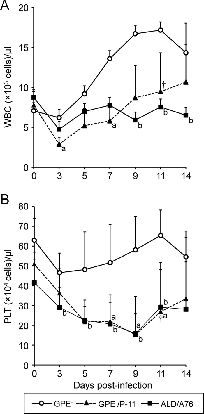
White blood cell and platelet counts in pigs inoculated with the GPE−, GPE−/P-11, or ALD/A76 strain. Groups of 5 pigs were inoculated with the indicated viruses, and blood was collected on days 0, 3, 5, 7, 9, 11, and 14 p.i. The white blood cell (WBC) (A) and platelet (PLT) (B) counts were determined for each time point and are shown as mean values, with error bars representing the standard deviations. The statistical significance of the differences between the cell counts from the pigs inoculated with GPE− virus and the pigs inoculated with the other viruses was calculated with the t test. a, P < 0.05 between GPE− and GPE−/P-11; b, P < 0.05 between GPE− and ALD/A76. One pig died on day 11 p.i. (†). All remaining pigs were euthanized on day 14 p.i.
Table 2.
Clinical signs in and virus recovery from pigs inoculated with the GPE−, GPE−/P-11, and ALD/A76 strains
| Inoculated virus | Clinical signs | Virus recovery from: |
||||||||
|---|---|---|---|---|---|---|---|---|---|---|
| Blood (log10 TCID50/ml) on day p.i.: |
Tissue (log10 TCID50/g) |
|||||||||
| 3 | 5 | 7 | 9 | 11 | 14 | Tonsil | Kidney | Mesenteric lymph node | ||
| GPE− | —a | <0.8 | —b | — | — | — | — | —b | — | — |
| — | — | <0.8 | — | — | — | — | <1.8 | — | — | |
| — | — | — | — | — | — | — | — | — | — | |
| — | — | <0.8 | — | — | — | — | — | — | — | |
| — | — | — | — | — | — | — | — | — | — | |
| GPE−/P-11 | — | ≤1.6 | NTc | ≤1.5 | NT | — | — | 4.0 | — | — |
| Diarrhea, dysstasia, death | 2.5 | NT | 4.0 | NT | NT | NT | 4.3 | 3.3 | 5.0 | |
| — | 2.0 | 2.3 | <0.8 | — | — | — | ≤2.8 | — | <1.8 | |
| — | 2.8 | 3.0 | 3.8 | 3.3 | 4.0 | 3.6 | 4.3 | <1.8 | 3.8 | |
| Diarrhea, dysstasia | 2.3 | 2.8 | 3.3 | 3.0 | ≤1.0 | — | — | — | ≤2.0 | |
| ALD/A76 | Diarrhea, weight loss | 3.0 | 4.8 | 7.0 | 6.6 | 7.0 | 6.0 | 9.5 | 9.3 | 8.8 |
| — | 3.6 | 4.5 | 6.0 | 5.8 | 6.8 | 6.0 | 8.3 | 7.3 | 7.5 | |
| Weight loss | 3.8 | 4.3 | 6.3 | 5.3 | 6.8 | 6.0 | 8.7 | 8.8 | 8.8 | |
| Diarrhea, cyanosis | 3.6 | 5.0 | 6.6 | 7.1 | 6.8 | 6.8 | 8.8 | 9.1 | 9.3 | |
| Diarrhea, weight loss | 3.1 | 4.0 | 6.0 | 6.0 | 6.3 | 6.6 | 8.0 | 7.1 | 7.5 | |
—, not observed.
—, not isolated.
NT, not tested.
Pathogenicity of the vGPE− and mutant viruses in pigs.
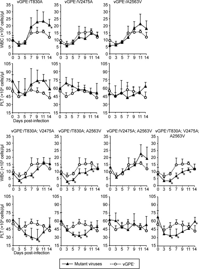
The pig-adapted viruses isolated from the tonsils are likely to possess some degree of heterogeneity, as suggested by the genome quasispecies distribution observed in the RNA preparations from passages 12 to 15 (Table 1). Therefore, in order to determine which amino acid changes are responsible for the increased pathogenicity acquired during passaging, recombinant viruses were generated by site-directed mutagenesis in the vGPE− backbone using reverse genetics (Fig. 2A and B). The pathogenicities of the reconstructed mutant viruses were determined in pigs and compared with that of the parent vGPE− virus. For each recombinant virus, 5 pigs were infected by intramuscular injection of 107.0 TCID50 of virus. Clinical signs and virus content in blood and organs were monitored. Infection with the parent vGPE− did not result in any clinical sign, with no or only very little virus detection in blood and tonsils (Table 3). The virus with the single alanine-to-valine change in NS4B at position 2563 (vGPE−/A2563V) behaved similarly to the parent vGPE− virus. Also, the 3 viruses with a single amino acid change did not cause any major leukocytopenia and thrombocytopenia (Fig. 3). Slightly better replication in pigs than the parent virus, also in the absence of any clinical signs, was observed with the virus carrying the single mutation at position 2475 in NS4B (vGPE−/V2475A). None of the other mutant viruses induced any obvious clinical signs either, despite higher titers in the tonsils, except for the virus with the 3 amino acid substitutions (vGPE−/T830A; V2475A; A2563V), which resulted in cyanosis, diarrhea, and/or weight loss in 4 of the 5 pigs, in severe leukocytopenia and thrombocytopenia in all 5 animals, and in prolonged virus shedding for up to 11 days after infection (Table 3 and Fig. 3). Prolonged virus shedding was also observed up to day 9 p.i. in pigs infected with viruses carrying the T830A substitution alone and in combination with the V2475A or the A2563V mutation in NS4B. In pigs inoculated with the latter 2 viruses (vGPE−/T830A; V2475A and vGPE−/T830A; A2563V), the highest titers in blood and tonsils were observed, with severe leukocytopenia and thrombocytopenia, similar to the case for the infection with the triple mutant virus. In addition, after infection with the triple mutant virus, infectious virus was also detected in the mesenteric lymph nodes of 2 out of 5 pigs, in contrast to infection with any of the other mutants. No additional amino acid change was detected in viruses recovered from tonsils of pigs inoculated with the triple mutant virus on day 14 p.i. Altogether, these results demonstrate that the 3 amino acid substitutions act in concert in the acquisition of pathogenicity after adaptation of the GPE− virus in pigs.
Fig 2.
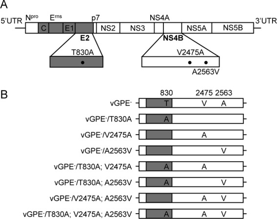
Schematic representation of the cDNA clones of the GPE−/P-11 and mutant vGPE− viruses. (A) Three amino acid substitutions, T830A, V2475A, and A2563V, were found in the GPE−/P-11 virus. (B) Seven recombinant viruses with all possible combinations of amino acid substitutions were generated by site-directed mutagenesis of the GPE− backbone. The white and gray boxes indicate the nonstructural and structural proteins, respectively.
Table 3.
Clinical signs in and virus recovery from pigs inoculated with the vGPE− and mutant viruses
| Inoculated virus | Clinical signs | Virus recovery from: |
||||||||
|---|---|---|---|---|---|---|---|---|---|---|
| Blood (log10 TCID50/ml) on day p.i.: |
Tissue (log10 TCID50/g) |
|||||||||
| 3 | 5 | 7 | 9 | 11 | 14 | Tonsil | Kidney | Mesenteric lymph node | ||
| vGPE− | —a | —b | — | — | — | — | — | —b | — | — |
| — | <0.8 | — | — | — | — | — | <1.8 | — | — | |
| — | — | — | — | — | — | — | <1.8 | — | — | |
| — | — | <0.8 | — | — | — | — | <1.8 | — | — | |
| — | — | <0.8 | — | — | — | — | <1.8 | — | — | |
| vGPE−/T830A | — | <0.8 | ≤1.0 | <0.8 | — | — | — | <1.8 | — | — |
| — | <0.8 | 2.6 | ≤2.1 | <0.8 | — | — | 3.8 | <1.8 | — | |
| — | — | ≤1.0 | <0.8 | — | — | — | <1.8 | — | — | |
| — | <0.8 | ≤1.5 | — | — | — | — | <1.8 | — | — | |
| — | <0.8 | <0.8 | — | — | — | — | <1.8 | — | — | |
| vGPE−/V2475A | — | — | <0.8 | — | — | — | — | <1.8 | — | — |
| — | <0.8 | <0.8 | — | — | — | — | <1.8 | — | — | |
| — | — | <0.8 | <0.8 | — | — | — | 4.0 | — | — | |
| — | — | <0.8 | — | — | — | — | 2.5 | — | — | |
| — | — | <0.8 | — | — | — | — | <1.8 | — | — | |
| vGPE−/A2563V | — | — | — | — | — | — | — | <1.8 | — | — |
| — | — | — | — | — | — | — | <1.8 | — | — | |
| — | — | — | — | — | — | — | ≤2.0 | — | — | |
| — | <0.8 | <0.8 | — | — | — | — | — | — | — | |
| — | <0.8 | — | — | — | — | — | — | — | — | |
| vGPE−/T830A; V2475A | — | 2.6 | 3.8 | 3.6 | — | — | — | 4.8 | — | — |
| — | <0.8 | ≤1.0 | — | — | — | — | 3.3 | — | — | |
| — | 2.8 | 3.8 | 2.3 | — | — | — | <1.8 | — | — | |
| — | 2.6 | 3.3 | ≤1.5 | <0.8 | — | — | 2.1 | — | — | |
| — | ≤1.0 | 3.0 | <0.8 | <0.8 | — | — | 4.0 | — | — | |
| vGPE−/T830A; A2563V | — | ≤1.0 | ≤1.6 | ≤1.0 | — | — | — | 4.3 | — | — |
| — | 2.8 | 3.3 | ≤1.0 | — | — | — | 3.8 | — | — | |
| — | <0.8 | 2.8 | 3.0 | — | — | — | 2.8 | — | — | |
| — | 2.5 | 3.8 | 3.8 | ≤2.3 | <0.8 | — | 3.6 | — | — | |
| — | ≤2.1 | 2.3 | ≤1.8 | — | — | — | 3.9 | — | — | |
| vGPE−/V2475A; A2563V | — | <0.8 | ≤1.0 | — | — | — | — | ≤2.3 | — | — |
| — | ≤1.0 | ≤1.1 | — | — | — | — | <1.8 | — | — | |
| — | ≤1.0 | ≤1.1 | — | — | — | — | 3.3 | — | — | |
| — | — | ≤1.0 | <0.8 | — | — | — | ≤2.3 | — | — | |
| — | <0.8 | <0.8 | — | — | — | — | 3.5 | — | — | |
| vGPE−/T830A; V2475A; A2563V | Cyanosis | ≤1.0 | ≤2.1 | ≤1.0 | ≤1.0 | — | — | 4.0 | — | — |
| — | ≤1.0 | 2.6 | 3.6 | ≤2.6 | <0.8 | — | 3.3 | — | <1.8 | |
| Weight loss, cyanosis | ≤1.1 | 2.8 | ≤2.3 | ≤1.3 | — | — | 3.7 | — | — | |
| Cyanosis | ≤1.0 | 2.6 | 2.0 | ≤1.0 | — | — | 3.0 | — | — | |
| Diarrhea, cyanosis | ≤2.1 | 2.3 | 2.6 | 3.1 | <0.8 | — | 4.1 | — | ≤2.3 | |
—, not observed.
—, not isolated.
Fig 3.
White blood cell and platelet counts in pigs inoculated with the vGPE− parent and mutant viruses. Groups of 5 pigs were inoculated with the indicated viruses, and blood was collected on days 0, 3, 5, 7, 9, 11, and 14 p.i. The WBC and PLT counts were determined for each time point and are shown as mean values, with error bars representing the standard deviations. The statistical significance of the differences between the cell counts from the pigs inoculated with vGPE− virus and the pigs infected with the other viruses was calculated with the t test. *, P < 0.05. In each graph, the mean WBC and PLT counts of the pigs inoculated with the vGPE− parent are shown for comparison purposes. The pigs were euthanized on day 14 p.i.
Comparison of in vitro growth kinetics of the mutant viruses.
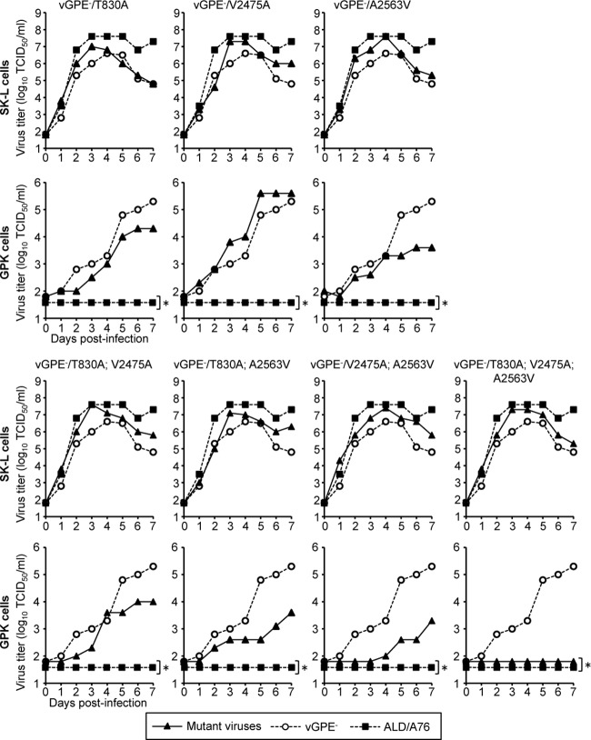
At 30°C, the GPE− strain replicates efficiently in guinea pig cells in which it has been adapted, as opposed to the ALD and other wild-type strains, which grow very poorly in these cells (48). Therefore, multistep growth curves of the recombinant viruses carrying the different amino acid substitutions in the vGPE− backbone were evaluated in SK-L cells and in the primary guinea pig (GPK) cell cultures in comparison with the vGPE− and ALD/A76 viruses (Fig. 4). Viruses that carry a single amino acid change in NS4B (A2563V) replicated better than the GPE− virus in the porcine cell lines at 40°C at all time points. Two mutant viruses carrying double amino acid substitutions (vGPE−/T830A; V2475A and vGPE−/V2475A A2563V) and the virus carrying the triple mutations (vGPE−/T830A; V2475A; A2563V) also replicated more efficiently than the vGPE− virus in swine cells. Except for the vGPE−/V2475A virus, the mutant viruses with single and double mutations in the vGPE− backbone lost replication efficiency in the GPK cells compared with the vGPE− virus. Both the ALD/A76 virus and the virus with 3 adaptive mutations in the GPE− backbone (vGPE−/T830A; V2475A; A2563V) did not replicate at all in the GPK cells, in contrast to the case for the parent vGPE− virus. These replication kinetics were confirmed with kinetics of parallel samples analyzed by qPCR (data not shown). Thus, these results show that the 3 amino acid substitutions found during adaptation in pigs act together to enhance virus replication in swine cells and to abolish replication in GPK cells, which is in agreement with the effect of the 3 adaptive mutations on pathogenicity in pigs.
Fig 4.
Growth kinetics of the vGPE− parent and mutant viruses and of the ALD/A76 strain in swine cells and guinea pig cells. Swine kidney cell lines (SK-L cells) and the primary guinea pig kidney (GPK) cells were inoculated at a multiplicity of infection (MOI) of 0.001 with the parent and mutant viruses as indicated. The SK-L and GPK cells were incubated at 40°C in the presence of 5% CO2 and at 30°C, respectively. The supernatants were collected at the indicated times. The titers were determined in SK-L cells and expressed as 50% tissue culture infective doses (TCID50) per ml. In each graph, the titers of the vGPE− and ALD/A76 strains are included for comparison purposes. *, virus titer was below detection the limit of 101.8 TCID50/ml.
Effect of the adaptive mutations on virus focus formation.
The adaptive mutation in E2 is likely to influence virus spreading, while the mutations acquired in NS4B may determine the RNA replication efficiency. In order to determine the effects of these amino acid substitutions on viral spreading in swine cells, monolayers of SK-L cells were infected with the mutant and parent viruses and focus formation was monitored under a semisolid overlay using viral antigen detection by immunofluorescence. The number of infected cells per focus was counted for 30 randomly selected foci, and the data were analyzed statistically. The recombinant GPE− virus carrying all 3 adaptive mutations produced the largest foci, comparable in size to the foci obtained with the ALD/A76 virus (Fig. 5). The smallest foci were observed with the vGPE− virus and with the mutants carrying 1 or 2 mutations in NS4B. All mutants that carried the T830A substitution alone or together with one of the substitutions in NS4B resulted in foci of intermediate size. These results indicate that the T830A substitution acquired in E2 during adaptation in pigs is a major determinant of virus spreading in swine cells.
Fig 5.
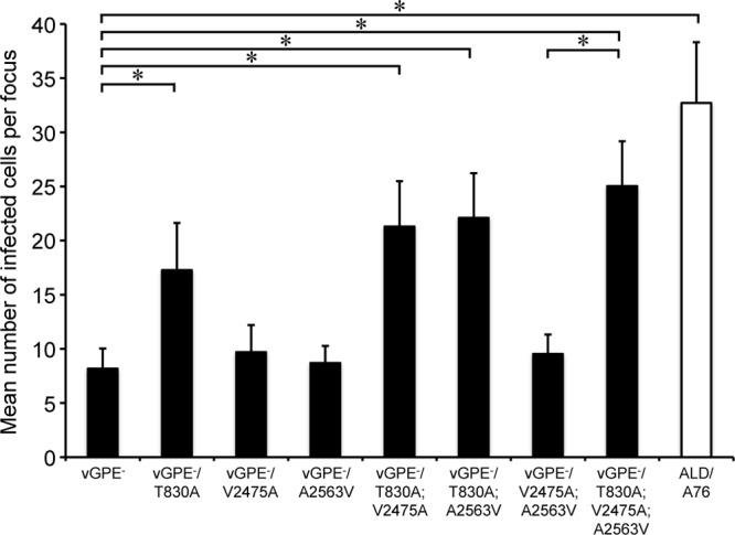
Focus formation by vGPE−, mutant viruses, and ALD/A76 in SK-L cell cultures. SK-L cells were inoculated at an MOI of 0.001 with the parent and mutant viruses as indicated. The cell monolayers were overlaid with MEM containing 1.0% Bacto agar and incubated at 37°C in the presence of 5% CO2. After 72 h of incubation, the cells were fixed with acetone and viral NS3 was detected by immunofluorescence using MAb 46/1. The number of infected cells per focus was counted for 30 independent foci (n = 30). Error bars represent the standard deviations, and the significance of the differences was calculated with the t test. *, P < 0.05.
Effect of the mutations in NS4B on viral RNA replication.
Two independent approaches were used to determine the effect of the adaptive mutations in NS4B on viral RNA replication. With qPCR, the RNA replication efficiency of the vGPE−/V2475A and vGPE−/V2475A; A2563V viruses was significantly higher than that of the parental vGPE− virus at 7 h postinfection (Fig. 6). At 12 h postinfection, the RNA replication efficiencies of the 2 viruses carrying a single mutation were significantly higher than that of the parent vGPE− virus.
Fig 6.
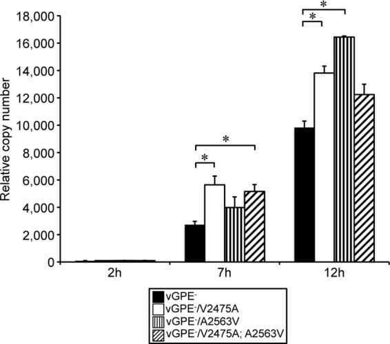
Effect of amino acid substitutions in NS4B on RNA replication efficiency. In order to assess the effect of the amino acid substitutions in NS4B on the viral RNA replication efficiency, confluent SK-L cell monolayers were inoculated at an MOI of 1.5 with the parent and mutant viruses carrying the indicated amino acid changes in NS4B. Parallel cultures of infected cells were incubated at 37°C in the presence of 5% CO2 for 2 h, 7 h, and 12 h. Total RNA was extracted from the infected cells at the indicated times. The relative RNA copy numbers compared with the copy number at 2 h postinoculation are shown. Each bar represents the mean value from 3 independent experiments, and the error bars show the standard deviations. Pairwise comparisons of the values were carried out using the t test. *, P < 0.05.
As an alternative approach to measure the activity of the viral RNA replicase complex, a bicistronic reporter replicon was constructed with the vGPE− genome. This replicon consisted of the 5′ and 3′ UTRs flanked by a chimeric Npro-firefly luciferase gene separated from the NS3 to NS5B genes of the replication complex by an EMCV IRES (Fig. 7A). The 2 adaptive mutations in NS4B were introduced separately or in combination in the reporter replicon backbone. The relative luciferase activities obtained with the parent and mutant replicons were determined in the swine CPK cell line. The replicon carrying the 2 adaptive mutations in NS4B resulted in 10-fold-higher luciferase activity than the parent rGPE−-Npro-Luc-IRES-NS3 replicon at 24 h after transfection (Fig. 7B). At this time point, luciferase activities significantly higher than those of the parent replicon were also observed with the replicons carrying a single amino acid substitution. These data indicate that the amino acid substitutions acquired in NS4B of the GPE− strain during adaptation in pigs contribute to enhanced virus replication in swine cells by increasing the activity of the viral replication complex.
Fig 7.
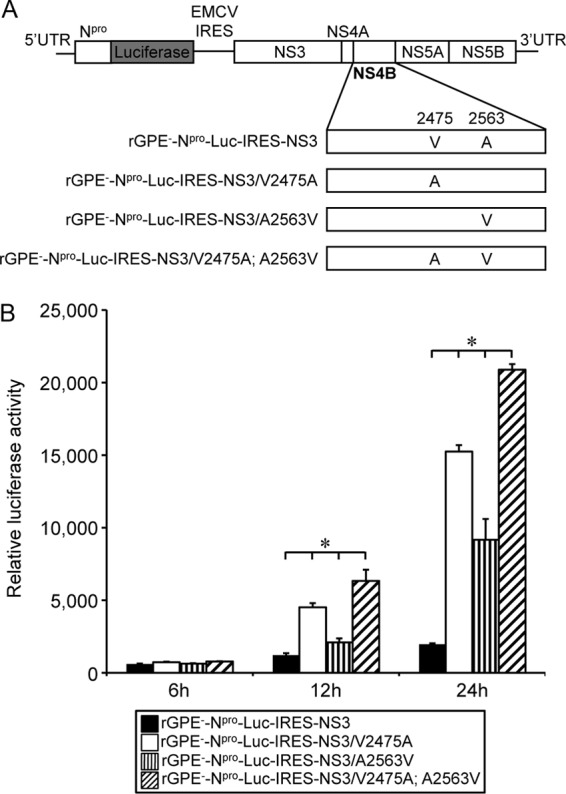
Effect of amino acid substitutions in NS4B on the viral RNA replication efficiency. (A) A GPE−-derived bicistronic replicon consisting of the 5′ and 3′ UTRs flanking a chimeric Npro-firefly luciferase gene separated from the NS3 to NS5B genes by the IRES of EMCV was constructed. Single amino acid substitutions at positions 2475 and 2563 were introduced by site-directed mutagenesis as indicated. (B) In order to analyze the effects of the amino acid substitutions on the viral RNA replication efficiency, 106.8 CPK cells were transfected with 1 μg of each replicon RNA and with 100 ng of pRL-TK (Int−) reporter plasmid for constitutive Renilla luciferase expression. After 6 h, 12 h, and 24 h of incubation at 37°C in the presence of 5% CO2, the firefly and Renilla luciferase activities expressed in relative light units were assayed in cell lysates. The firefly luciferase activity was normalized with the corresponding Renilla luciferase activity from the same lysate. Each bar represents the mean value from 3 independent experiments, with error bars showing the standard deviations. The values obtained with each construct were compared pairwise using the t test. *, P < 0.05.
DISCUSSION
The live attenuated vaccine strain GPE− was widely used in Japan to control CSF (57). The GPE− strain was generated from the virulent ALD strain through multiple passages and cloning in cells of swine, bovine, and guinea pig origin (48, 51). Field studies have proven that the GPE− vaccine virus is safe and efficacious. The virus is not able to spread, either horizontally or vertically, and reduces the prevalence of CSF by increasing herd immunity (34, 49). However, the molecular basis of the attenuation was unknown. Using an artificial virus transmission protocol in order to force readaptation to pigs of the live attenuated guinea pig-adapted GPE− virus, 3 amino acid residues that are critical for determining the pathogenic difference between the GPE− and the ALD strains were identified in E2 and NS4B. Importantly, mutation of these residues in the 2 genes had a synergistic effect on the pathogenicity in vivo and on virus spread and replication efficiency in cell culture. To the best of our knowledge, this is the first report showing that the pathogenicity of CSFV can be enhanced by the substitution of selected amino acids in the backbone of a low-virulent virus.
A total of 46 amino acid differences were found between the parent ALD virus and the GPE− vaccine strain (22). Since the pathogenicity of the triple mutant GPE− virus is not as high as the pathogenicity of the original ALD virus, additional changes are likely to determine the virulence of the ALD strain. Importantly, several virulent strains, such as CSFV Brescia, Eystrup, and Alfort/Tübingen, carry the same amino acid residues as those acquired by the GPE− virus in E2 and NS4B during passaging in pigs. However, these amino acids are also present in the CSFV C strain, a vaccine strain generated by adaptation in rabbits, indicating that other mutations are responsible for the attenuation of the C strain. Considering all of this, one can conclude that the specific residues identified in this study to be critical for determining virulence in pigs do not represent general virulence determinants of CSFV, suggesting that virulence of CSFV is a complex multigenic trait involving different genes and functions. This is reflected in numerous reports in which mutations of different functional domains in the structural proteins C, Erns, E1, and E2 and in the nonstructural proteins p7 and NS4B were shown to reduce the pathogenicity in pigs, indicating that these genes may be involved in determining CSFV virulence (7, 8, 9, 10, 11, 12, 13, 32, 36, 37, 38, 39, 40, 46, 54).
The fact that the amino acid change V2475A was found in NS4B after 1 passage in pigs suggests that the alanine at position 2475 was already present in the quasispecies population of the original inoculum. This hypothesis was indeed confirmed by deep sequencing analysis (Table 4). Additional direct sequencing data further indicate that the changes acquired during the passaging in pigs occurred from a shift of the quasispecies distribution during host-driven selection in vivo.
Table 4.
Quasispecies composition of the original GPE− virus at the codon sites at which adapted mutations were rescued
| Amino acid position and nucleotide sequencea | Rate (%) | Deduced amino acidb |
|---|---|---|
| E2 830 | ||
| ACA | >99.9 | T |
| NS4B 2475 | ||
| GUC | 97.5 | V |
| . C . | 2.3 | A |
| . G . | 0.2 | G |
| NS4B 2563 | ||
| GCA | 99.0 | A |
| . A . | 0.8 | E |
| . U . | 0.2 | V |
Periods indicate the same nucleotide as in the sequence above.
The amino acids found after adaptation in pigs are underlined.
In the present study, the T830A substitution in E2 acquired during passaging in pigs reduced the virus replication in guinea pig cells while enhancing the spreading and replication in pig cells, compared with those of the parent vGPE− virus. These results suggest that the residue at position 830 in E2 is involved in host tropism and plays a role in cell-to-cell spreading, probably at the level of viral attachment and entry, as the glycoproteins E2 and Erns of pestiviruses mediate viral attachment and entry into host cells (20, 41, 56). Unfortunately, there is only limited information available on the receptors and pathways involved in CSFV binding and entry. Further investigations are necessary to clarify the impact of the alanine at position 830 of E2 on virus infection of swine cells. E2 is exposed on the outer surface of the virus and is the most immunodominant protein inducing the major neutralizing antibodies in infected pigs (58). The amino acid at position 830 is located in the N-terminal region of E2, and this region is the minimal domain required for binding to pig antibodies generated during CSFV infection (29, 61). Recent studies support the importance of this domain for the pathogenicity of CSFV. In the Brescia strain, amino acid substitutions in this domain, including the amino acid at position 830, attenuated virulence in pigs (37).
In NS4B, the amino acid substitutions at positions 2475 and 2563 have not yet been reported to be involved in virulence. On the basis of the predicted topology of CSFV NS4B, the residues at positions 2475 and 2563 are located in the N-terminal and C-terminal transmembrane domains, respectively. A recent study on the biosynthesis of the CSFV nonstructural proteins showed that NS4B is mostly found attached to NS5A (27). The role of transmembrane domains of CSFV NS4B in the virus life cycle is not well understood, while these domains of hepatitis C virus (HCV) are reported to play a role in viral replication (15). Importantly, a study identified a putative Toll/interleukin-1 receptor-like domain in the C-terminal region of CSFV NS4B. Mutations in this domain of NS4B in the highly virulent CSFV Brescia backbone, immediately downstream of residue 2563 identified here, resulted in an attenuated phenotype along with enhanced activation of Toll-like receptor 7 (TLR-7)-induced genes (7). NS4B also harbors a nucleoside triphosphatase (NTPase) motif. The NTPase activity is an absolute requirement for CSFV replication, but there is no evidence yet that modulating the NTPase activity may affect virulence (10). The 2 amino acids of NS4B identified in the present study are not located within the NTPase motif but clearly determine the replication efficiency of the virus in vivo and in vitro. These substitutions may enhance the efficiency of viral RNA replication by influencing the stability of the replicase complex and/or the interaction of the replicase complex with host factors in swine. However, the function of NS4B in the replication complex of pestiviruses is not well understood. CSFV NS4B is relatively labile compared to other nonstructural proteins (27). Several studies show that NS4B of flaviviruses and HCV is an integral membrane protein of the endoplasmic reticulum and is involved in viral genome replication, interacting closely with other viral nonstructural proteins during the cycle of replication and in virus assembly and release (5, 14, 19, 24, 31).
With the adaptive mutations in E2 and NS4B combined in the GPE− backbone, the pathogenicity of the mutant viruses was clearly enhanced but did not equal the pathogenicity of the ALD/A76 strain, indicating that additional amino acid substitutions elsewhere in the genome may play a role in the pathogenicity of CSFV. Pestiviruses suppress innate immune defenses by preventing alpha/beta interferon (IFN-α/β) induction, a property attributed to the viral proteins Npro and Erns. Npro interferes with the induction of IFN-α/β by promoting the proteasomal degradation of interferon regulatory factor 3 (IRF3) (28, 42). During the generation of the live attenuated GPE− vaccine strain, Npro has acquired a mutation that abolishes its capacity to bind and degrade IRF3 and thus to interfere with the induction of IFN-α/β (43). In the parental ALD strain, Npro is functional in terms of inhibition of IFN-α/β induction (42, 55). Interestingly, during the readaptation of the GPE− vaccine in pigs, the IRF3-degrading function of Npro was not recovered. Whether restoring the Npro function in the triple mutant GPE− virus would further enhance its pathogenicity is under current investigation. Previous data suggest that the IRF3-degrading function of Npro may support the pathogenicity of low- and moderately virulent strains but has only little influence on the pathogenicity of highly virulent viruses: mutations to knock out the Npro-mediated IRF3 degradation did not alter the pathogenicity of the highly virulent CSFV strain Eystrup and resulted in only slight attenuation of the moderately virulent vA187-1 virus (43). Erns forms disulfide-linked homodimers and possesses RNase activity (45, 50). Erns interacts with extracellular double-stranded RNA (dsRNA), thereby targeting a major viral IFN-inducing signal (21). Knocking out the RNase activity in the CSFV Alfort/Tübingen backbone resulted in attenuation in pigs (32). Amino acid substitution at the glycosylation sites of Erns of the Brescia strain did not affect RNase activity but attenuated the virus (46). The amino acids of Erns reported to determine virulence of CSFV are found in virulent strains and in the GPE− strain as well. Therefore, it is not clear whether Erns may also be involved in determining the pathogenicity of the ALD strain.
In summary, this is the first study showing that amino acid substitutions in E2 and NS4B can enhance the pathogenicity of CSFV in pigs by an additive effect enhancing viral spread and genome replication. The acquisition of pathogenicity is related to the selection of a pig-adapted mutant virus using a vaccine strain that had been depathogenized through adaptation in a nonnatural host. Nevertheless, by demonstrating gain of virulence and increased virus replication after site-directed mutagenesis, these data show that a structural protein and a nonstructural protein of CSFV can carry virulence determinants that can function in a synergistic manner to determine the pathogenicity of the virus in pigs. Improved understanding of the molecular basis of CSFV pathogenicity contributes to understanding the molecular mechanisms involved in CSF pathogenesis, which is important for the implementation of efficient disease control and vaccine strategies.
ACKNOWLEDGMENTS
We thank H. Aoki (Department of Basic Science, School of Veterinary Nursing and Technology, Faculty of Veterinary Science, Nippon Veterinary and Life Science University, Tokyo, Japan) and I. Leifer (Institute of Virology and Immunoprophylaxis, Mittelhäusern, Switzerland) for their helpful advice and cooperation with the present study. We also thank A. Ishii and A. Ohuma (Research Center for Zoonosis Control, Hokkaido University, Sapporo, Japan) for technical support and help with deep sequencing. We thank N. Isoda (Research Center for Zoonosis Control, Hokkaido University) for advice on the statistical analysis of the experimental data. We also thank Y. Nomoto and A. Sawata (Kitasato Daiichi Sankyo Vaccine Co., Ltd., Saitama, Japan) for technical help with the GPK cells, Y. Sugita, H. Yoshida, M. Endo, N. Nomura, N. Nagashima, and K. Mitsuhashi for excellent technical support, and the laboratory staff members for continuous assistance.
Footnotes
Published ahead of print 6 June 2012
REFERENCES
- 1. Behrens SE, Grassmann CW, Thiel HJ, Meyers G, Tautz N. 1998. Characterization of an autonomous subgenomic pestivirus RNA replicon. J. Virol. 72:2364–2372 [DOI] [PMC free article] [PubMed] [Google Scholar]
- 2. Bintintan I, Meyers G. 2010. A new type of signal peptidase cleavage site identified in an RNA virus polyprotein. J. Biol. Chem. 285:8572–8584 [DOI] [PMC free article] [PubMed] [Google Scholar]
- 3. Collett MS, Larson R, Belzer SK, Retzel E. 1988. Proteins encoded by bovine viral diarrhea virus: the genomic organization of a pestivirus. Virology 165:200–208 [DOI] [PubMed] [Google Scholar]
- 4. Collett MS, Moennig V, Horzinek MC. 1989. Recent advances in pestivirus research. J. Gen. Virol. 70:253–266 [DOI] [PubMed] [Google Scholar]
- 5. Egger D, et al. 2002. Expression of hepatitis C virus proteins induces distinct membrane alterations including a candidate viral replication complex. J. Virol. 76:5974–5984 [DOI] [PMC free article] [PubMed] [Google Scholar]
- 6. Elbers K, et al. 1996. Processing in the pestivirus E2-NS2 region: identification of proteins p7 and E2p7. J. Virol. 70:4131–4135 [DOI] [PMC free article] [PubMed] [Google Scholar]
- 7. Fernandez-Sainz I, et al. 2010. Mutations in classical swine fever virus NS4B affect virulence in swine. J. Virol. 84:1536–1549 [DOI] [PMC free article] [PubMed] [Google Scholar]
- 8. Fernandez-Sainz I, et al. 2009. Alteration of the N-linked glycosylation condition in E1 glycoprotein of classical swine fever virus strain Brescia alters virulence in swine. Virology 386:210–216 [DOI] [PubMed] [Google Scholar]
- 9. Fernández-Sainz I, et al. 2011. Substitution of specific cysteine residues in the E1 glycoprotein of classical swine fever virus strain Brescia affects formation of E1-E2 heterodimers and alters virulence in swine. J. Virol. 85:7264–7272 [DOI] [PMC free article] [PubMed] [Google Scholar]
- 10. Gladue DP, et al. 2011. Identification of an NTPase motif in classical swine fever virus NS4B protein. Virology 411:41–49 [DOI] [PubMed] [Google Scholar]
- 11. Gladue DP, et al. 2010. Effects of the interactions of classical swine fever virus core protein with proteins of the SUMOylation pathway on virulence in swine. Virology 407:129–136 [DOI] [PubMed] [Google Scholar]
- 12. Gladue DP, et al. 2011. Interaction between core protein of classical swine fever virus with cellular IQGAP1 protein appears essential for virulence in swine. Virology 412:68–74 [DOI] [PubMed] [Google Scholar]
- 13. Gladue DP, et al. 2012. Classical Swine Fever Virus p7 protein is a viroporin involved in virulence in swine. J. Virol. 86:6778–6791 [DOI] [PMC free article] [PubMed] [Google Scholar]
- 14. Grant D, et al. 2011. A single amino acid in nonstructural protein NS4B confers virulence to dengue virus in AG129 mice through enhancement of viral RNA synthesis. J. Virol. 85:7775–7787 [DOI] [PMC free article] [PubMed] [Google Scholar]
- 15. Han Q, et al. 2011. Conserved GXXXG- and S/T-like motifs in the transmembrane domains of NS4B protein are required for hepatitis C virus replication. J. Virol. 85:6464–6479 [DOI] [PMC free article] [PubMed] [Google Scholar]
- 16. Harada T, Tautz N, Thiel HJ. 2000. E2-p7 region of the bovine viral diarrhea virus polyprotein: processing and functional studies. J. Virol. 74:9498–9506 [DOI] [PMC free article] [PubMed] [Google Scholar]
- 17. Heimann M, Roman-Sosa G, Martoglio B, Thiel HJ, Rümenapf T. 2006. Core protein of pestiviruses is processed at the C terminus by signal peptide peptidase. J. Virol. 80:1915–1921 [DOI] [PMC free article] [PubMed] [Google Scholar]
- 18. Hoffmann B, Beer M, Schelp C, Schirrmeier H, Depner K. 2005. Validation of a real-time RT-PCR assay for sensitive and specific detection of classical swine fever. J. Virol. Methods 130:36–44 [DOI] [PubMed] [Google Scholar]
- 19. Hügle T, et al. 2001. The hepatitis C virus nonstructural protein 4B is an integral endoplasmic reticulum membrane protein. Virology 284:70–81 [DOI] [PubMed] [Google Scholar]
- 20. Hulst MM, Moormann RJ. 1997. Inhibition of pestivirus infection in cell culture by envelope proteins Erns and E2 of classical swine fever virus: Erns and E2 interact with different receptors. J. Gen. Virol. 78:2779–2787 [DOI] [PubMed] [Google Scholar]
- 21. Iqbal M, Poole E, Goodbourn S, McCauley JW. 2004. Role for bovine viral diarrhea virus Erns glycoprotein in the control of activation of beta interferon by double-stranded RNA. J. Virol. 78:136–145 [DOI] [PMC free article] [PubMed] [Google Scholar]
- 22. Ishikawa K, et al. 1995. Comparison of the entire nucleotide and deduced amino acid sequences of the attenuated hog cholera vaccine strain GPE− and the wild-type parental strain ALD. Arch. Virol. 140:1385–1391 [DOI] [PubMed] [Google Scholar]
- 23. Kameyama K, et al. 2006. Development of an immunochromatographic test kit for rapid detection of bovine viral diarrhea virus antigen. J. Virol. Methods 138:140–146 [DOI] [PubMed] [Google Scholar]
- 24. Kim M, Mackenzie JM, Westaway EG. 2004. Comparisons of physical separation methods of Kunjin virus-induced membranes. J. Virol. Methods 120:179–187 [DOI] [PubMed] [Google Scholar]
- 25. Komaniwa H, Fukusho A, Shimizu Y. 1981. Micro method for performing titration and neutralization test of hog cholera virus using established porcine kidney cell strain. Natl. Inst. Anim. Health Q. (Tokyo) 21:153–158 [PubMed] [Google Scholar]
- 26. Lackner T, Müller A, König M, Thiel HJ, Tautz N. 2005. Persistence of bovine viral diarrhea virus is determined by a cellular cofactor of a viral autoprotease. J. Virol. 79:9746–9755 [DOI] [PMC free article] [PubMed] [Google Scholar]
- 27. Lamp B, et al. 2011. Biosynthesis of classical swine fever virus nonstructural proteins. J. Virol. 85:3607–3620 [DOI] [PMC free article] [PubMed] [Google Scholar]
- 28. La Rocca SA, et al. 2005. Loss of interferon regulatory factor 3 in cells infected with classical swine fever virus involves the N-terminal protease, Npro. J. Virol. 79:7239–7247 [DOI] [PMC free article] [PubMed] [Google Scholar]
- 29. Lin M, Lin F, Mallory M, Clavijo A. 2000. Deletions of structural glycoprotein E2 of classical swine fever virus strain Alfort/187 resolve a linear epitope of monoclonal antibody WH303 and the minimal N-terminal domain essential for binding immunoglobulin G antibodies of a pig hyperimmune serum. J. Virol. 74:11619–11625 [DOI] [PMC free article] [PubMed] [Google Scholar]
- 30. Lindenbach BD, Thiel HJ, Rice CM. 2007. Flaviviridae: the viruses and their replication, p 1101–1152 In Knipe DM, Howley PM. (ed), Fields virology, 5th ed, vol 1 Lippincott-Raven Publishers, Philadelphia, PA [Google Scholar]
- 31. Lindström H, Lundin M, Häggström S, Persson MA. 2006. Mutations of the hepatitis C virus protein NS4B on either side of the ER membrane affect the efficiency of subgenomic replicons. Virus Res. 121:169–178 [DOI] [PubMed] [Google Scholar]
- 32. Meyers G, Saalmüller A, Büttner M. 1999. Mutations abrogating the RNase activity in glycoprotein Erns of the pestivirus classical swine fever virus lead to virus attenuation. J. Virol. 73:10224–10235 [DOI] [PMC free article] [PubMed] [Google Scholar]
- 33. Moser C, Stettler P, Tratschin JD, Hofmann MA. 1999. Cytopathogenic and noncytopathogenic RNA replicons of classical swine fever virus. J. Virol. 73:7787–7794 [DOI] [PMC free article] [PubMed] [Google Scholar]
- 34. Okaniwa A, Nakagawa M, Shimizu Y, Furuuchi S. 1969. Lesions in swine inoculated with attenuated hog cholera viruses. Natl. Inst. Anim. Health Q. (Tokyo) 9:92–103 [PubMed] [Google Scholar]
- 35. Reed L, Muench H. 1938. A simple method of estimating fifty per cent endpoints. Am. J. Hyg. (Lond.) 27:493–497 [Google Scholar]
- 36. Risatti GR, et al. 2005. The E2 glycoprotein of classical swine fever virus is a virulence determinant in swine. J. Virol. 79:3787–3796 [DOI] [PMC free article] [PubMed] [Google Scholar]
- 37. Risatti GR, et al. 2006. Identification of a novel virulence determinant within the E2 structural glycoprotein of classical swine fever virus. Virology 355:94–101 [DOI] [PubMed] [Google Scholar]
- 38. Risatti GR, et al. 2007. Mutations in the carboxyl terminal region of E2 glycoprotein of classical swine fever virus are responsible for viral attenuation in swine. Virology 364:371–382 [DOI] [PubMed] [Google Scholar]
- 39. Risatti GR, et al. 2007. N-linked glycosylation status of classical swine fever virus strain Brescia E2 glycoprotein influences virulence in swine. J. Virol. 81:924–933 [DOI] [PMC free article] [PubMed] [Google Scholar]
- 40. Risatti GR, et al. 2005. Mutation of E1 glycoprotein of classical swine fever virus affects viral virulence in swine. Virology 343:116–127 [DOI] [PubMed] [Google Scholar]
- 41. Ronecker S, Zimmer G, Herrler G, Greiser-Wilke I, Grummer B. 2008. Formation of bovine viral diarrhea virus E1-E2 heterodimers is essential for virus entry and depends on charged residues in the transmembrane domains. J. Gen. Virol. 89:2114–2121 [DOI] [PubMed] [Google Scholar]
- 42. Ruggli N, et al. 2005. Npro of classical swine fever virus is an antagonist of double-stranded RNA-mediated apoptosis and IFN-alpha/beta induction. Virology 340:265–276 [DOI] [PubMed] [Google Scholar]
- 43. Ruggli N, et al. 2009. Classical swine fever virus can remain virulent after specific elimination of the interferon regulatory factor 3-degrading function of Npro. J. Virol. 83:817–829 [DOI] [PMC free article] [PubMed] [Google Scholar]
- 44. Ruggli N, Tratschin JD, Mittelholzer C, Hofmann MA. 1996. Nucleotide sequence of classical swine fever virus strain Alfort/187 and transcription of infectious RNA from stably cloned full-length cDNA. J. Virol. 70:3478–3487 [DOI] [PMC free article] [PubMed] [Google Scholar]
- 45. Rümenapf T, Unger G, Strauss JH, Thiel HJ. 1993. Processing of the envelope glycoproteins of pestiviruses. J. Virol. 67:3288–3294 [DOI] [PMC free article] [PubMed] [Google Scholar]
- 46. Sainz IF, Holinka LG, Lu Z, Risatti GR, Borca MV. 2008. Removal of a N-linked glycosylation site of classical swine fever virus strain Brescia Erns glycoprotein affects virulence in swine. Virology 370:122–129 [DOI] [PubMed] [Google Scholar]
- 47. Sakoda Y, Hikawa M, Tamura T, Fukusho A. 1998. Establishment of a serum-free culture cell line, CPK-NS, which is useful for assays of classical swine fever virus. J. Virol. Methods 75:59–68 [DOI] [PubMed] [Google Scholar]
- 48. Sasahara J. 1970. Hog cholera: diagnosis and prophylaxis. Natl. Inst. Anim. Health Q. (Tokyo) 10:57–81 [Google Scholar]
- 49. Sasahara J, Kumagai T, Shimizu Y, Furuuchi S. 1969. Field experiments of hog cholera live vaccine prepared in guinea-pig kidney cell culture. Natl. Inst. Anim. Health Q. (Tokyo) 9:83–91 [PubMed] [Google Scholar]
- 50. Schneider R, Unger G, Stark R, Schneider-Scherzer E, Thiel HJ. 1993. Identification of a structural glycoprotein of an RNA virus as a ribonuclease. Science 261:1169–1171 [DOI] [PubMed] [Google Scholar]
- 51. Shimizu Y, Furuuchi S, Kumagai T, Sasahara J. 1970. A mutant of hog cholera virus inducing interference in swine testicle cell cultures. Am. J. Vet. Res. 31:1787–1794 [PubMed] [Google Scholar]
- 52. Suter R, et al. 2011. Immunogenic and replicative properties of classical swine fever virus replicon particles modified to induce IFN-α/β and carry foreign genes. Vaccine 29:1491–1503 [DOI] [PubMed] [Google Scholar]
- 53. Tautz N, Elbers K, Stoll D, Meyers G, Thiel HJ. 1997. Serine protease of pestiviruses: determination of cleavage sites. J. Virol. 71:5415–5422 [DOI] [PMC free article] [PubMed] [Google Scholar]
- 54. Tews BA, Schürmann EM, Meyers G. 2009. Mutation of cysteine 171 of pestivirus Erns RNase prevents homodimer formation and leads to attenuation of classical swine fever virus. J. Virol. 83:4823–4834 [DOI] [PMC free article] [PubMed] [Google Scholar]
- 55. Toba M, Matumoto M. 1969. Role of interferon in enhanced replication of Newcastle disease virus in swine cells infected with hog cholera virus. Jpn. J. Microbiol. 13:303–305 [DOI] [PubMed] [Google Scholar]
- 56. Tscherne DM, Evans MJ, Macdonald MR, Rice CM. 2008. Transdominant inhibition of bovine viral diarrhea virus entry. J. Virol. 82:2427–2436 [DOI] [PMC free article] [PubMed] [Google Scholar]
- 57. van Oirschot JT. 2003. Vaccinology of classical swine fever: from lab to field. Vet. Microbiol. 96:367–384 [DOI] [PubMed] [Google Scholar]
- 58. Weiland E, et al. 1990. Pestivirus glycoprotein which induces neutralizing antibodies forms part of a disulfide-linked heterodimer. J. Virol. 64:3563–3569 [DOI] [PMC free article] [PubMed] [Google Scholar]
- 59. Wiskerchen M, Belzer SK, Collett MS. 1991. Pestivirus gene expression: the first protein product of the bovine viral diarrhea virus large open reading frame, p20, possesses proteolytic activity. J. Virol. 65:4508–4514 [DOI] [PMC free article] [PubMed] [Google Scholar]
- 60. Xu J, et al. 1997. Bovine viral diarrhea virus NS3 serine proteinase: polyprotein cleavage sites, cofactor requirements, and molecular model of an enzyme essential for pestivirus replication. J. Virol. 71:5312–5322 [DOI] [PMC free article] [PubMed] [Google Scholar]
- 61. Zhang F, et al. 2006. Characterization of epitopes for neutralizing monoclonal antibodies to classical swine fever virus E2 and Erns using phage-displayed random peptide library. Arch. Virol. 151:37–54 [DOI] [PubMed] [Google Scholar]


