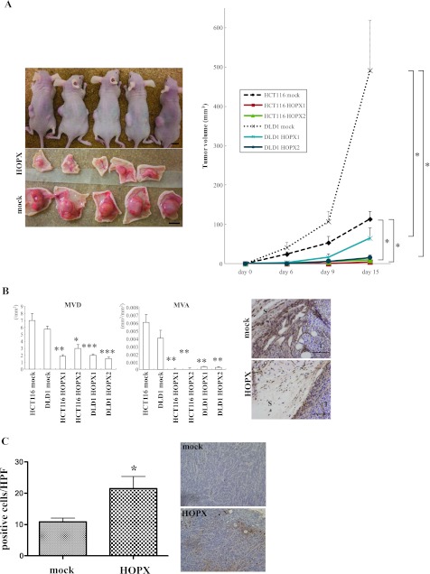
Figure 4.
Effects of HOPX on tumorigenicity and angiogenesis in vivo. (A) Mock cells were injected into the left flank and HOPX-expressing cells into the right flank of each mouse. Fifteen days after injection, mice were killed, and pictures were taken (left). Time course of tumor growth (right). *P < .05. Error bars, SEM. (B) Left: Stromal angiogenesis on day 15. P values were calculated by comparison with a mock group of each cell line. *P < .05. **P < .01. ***P < .001. Error bars, SEM. Right: Immunohistochemical staining with CD31 was performed to detect endothelial cells. Angiogenesis was significantly suppressed in mice injected with HOPX transfectants. S indicates stroma; T, tumor. Bars, 100 µm. (C) TUNEL assay was performed in tumor derived from HOPX or mock-transfected DLD1 (original magnification, x100). Apoptotic cells were counted in four different sections, four fields in each. Necrotic areas were excluded. *P < .05. Error bars, SEM.

