Abstract
Objective:
Iron deficiency is the most common form of malnutrition in developing countries. Iron containing supplements have been used effectively to solve this problem. In children, because of teeth staining after taking iron drops, parents have the idea that iron drops are the cause of tooth decay; therefore, they limit this vital supplement in their children’s diet. Hereby, we evaluate the histologic effect of iron containing supplements on tooth caries in rice rats with cariogenic or non-cariogenic diet.
Materials and Methods:
Twelve rats were selected and divided into four groups for this interventional experimental study. Four different types of dietary regimens were used for four months; group A, cariogenic diet with iron containing supplements; group B, cariogenic diet without iron containing supplements; group C, non-cariogenic diet with iron containing supplements; group D, non-cariogenic diet without iron containing supplements. After sacrificing the rats, 20-micron histological sections of their posterior teeth were prepared using the Ground Section method, then they were studied under polarized light microscopy. In order to compare the progression of caries in different samples, the depth of the lesions in the enamel was measured as three grades I, II and III.
Results:
The mean grade value of A, B, C and D groups were 1.61, 2.61, 1.37 and 1.80, respectively. Statistical analysis revealed that significantly fewer caries were seen in the group which had received iron containing supplements and cariogenic diet compared with cariogenic diet without iron supplements (p<0.05).
Conclusion:
Ferrous sulfate reduces the progression of dental caries in the cariogenic dietary regimen.
Keywords: Iron, Dental Caries, Dietary Regimen, Rat
INTRODUCTION
Iron deficiency is the most widely distributed nutrient deficiency state, affecting more than 2 billion people worldwide [1,2]. In some instances, this deficiency is alleviated by supplementing various foods with iron salts [3]. On the other hand, iron supplementation for children under 5-years old is recommended on the basis of anemia prevalence [1].
Caries is a diet-dependent disease, the prevalence of which may be reduced by avoiding frequent ingestion of sugars [4]. An alternative strategy would be to reduce the cariogenicity of sucrose through appropriate supplementation [5].
In countries where iron deficiency is apparent, the prevalence of dental caries is high, even though the two phenomena are not necessarily directly related [3].
Results from studies conducted in vitro, in situ and in vivo, both in humans and animals, support the belief that iron may have cariostatic properties [6].
Pecharki et al suggested that iron ions may reduce in situ the cariogenic potential of sucrose and the effect seems to be related to the reduction of Streptococci mutans in the dental biofilm formed [5].
Miguel et al in two studies done in desalivated rats found that iron added to sucrose, alone or in combination with other ions has a great effect on the reduction of the carigenic potential of the sugar [3,7].
In addition Devulapalle and Mooser showed the inhibition of the activity of glucosyltransferase by ferrous sulfate in vitro [8]. Recently, it has been shown that iron can inhibit acid demineralization by directly affecting mineral dissolution [9,10–13].
Polarized light microscopy has been used to explore the histologic progression of caries lesions.
Microscopic inspection is based on optical changes in the tissues. For the enamel, this is fairly straightforward, since a reduced mineral content is readily observed as opacity [14].
Knowing the fact that pediatricians prescribe iron supplements for children routinely, this study was designed to histologically evaluate the effect of an iron containing supplement (ferrous sulfate oral drops, Daropakhsh, Iran) on tooth caries in rice rats either with cariogenic or non-cariogenic diet.
MATERIALS AND METHODS
In this study, twelve 21-day-old weaning rice rats of both genders were selected. The animals were maintained under standard laboratory conditions and divided into four groups according to their diet (A, B, C and D). As each rat has twelve posterior teeth, 36 teeth were studied in each group. The dietary regimen was specified for each group as:
Group A: Cariogenic diet with iron containing supplement. Group B: Cariogenic diet without iron containing supplement. Group C: Non-cariogenic diet with iron containing supplement and Group D: Non-cariogenic diet without iron containing supplement.
The difference between cariogenic and non-cariogenic diet in this study was the sucrose sweetened water (5% w/v) added to a commercial laboratory diet (non-cariogenic).
The iron containing supplement “ferrous sulfate oral drop which contains 125 mg/ml iron” (Daroopakhsh, Tehran, Iran) was administrated as five drops each time, three times a week. All groups were infected with Streptococcus sobrinus which had been passaged through a desalivated rat to enhance virulence to obtain the desirable cariogenic condition [3].
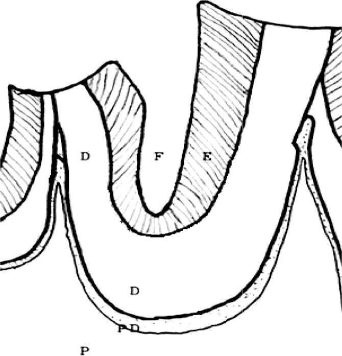
After four months, the rats were sacrificed by chloroform solution and the mandible and maxilla were removed, washed in running water and placed in 10% formaldehyde for 24 hours. The pieces were washed, dried with gauze and dissected. Each hemi jaw was sectioned along the sagittal plane. Ground Section Method was used for preparing histological sections. Longitudinal sections of 20 microns were prepared in a manner that the central grooves were perfectly exposed (Fig 1). After preparing, the samples were studied by one pathologist using polarized light microscopy. As cariogenesis in enamel causes a disruption in the arrangement of hydroxyapatite crystals, discolored areas with irregularities in the lateral joints to the central grooves were observed under polarized light. In order to compare these areas in different samples, based on the depth of caries in the enamel, an arbitrary qualitative classification in three different grades was established; grade I: A discolored area extending from the surface to the first one-third of the distance from the central groove surface to the dentin surface; grade II: A discolored area extending from the surface to the first two-thirds of the distance from the central groove surface to the dentine surface; grade III: A discolored area extending from the surface over the first two-thirds of the distance from the central groove surface to the dentine surface.
Fig 1.
A schematic presentation of the main central fissure of a rat’s mandibular second molar. Abbreviations: D, dentin; E, enamel; F, fissure; P, pulp tissue; PD, predentin layer
To examine the reliability of measurements, 20 samples were chosen randomly and caries grading was performed a second time after a long time interval, and a high correlation of repeated measurements was found (r = 0.91). The teeth in a rat mouth could not be analyzed independently, so the mean value obtained from one rat (three mean values for each group) were analyzed as the data using ANOVA test. SPSS software (version 11.5) was used and the significance limit was set at 5%.
RESULTS
All animals maintained good health during the experimental period.
But in the preparation of the sections of the teeth some of them were missed. It is necessary to mention that due to the rather sustained exposure to cariogenic bacteria, no sample was observed without any decay.
There was no dentinal lesion among the groups and for the enamel lesion the following results were obtained: group A, which had received iron supplement with cariogenic diets, provided a total number of 17 teeth.
Microscopic examinations revealed that 10 caries were grade I, four were grade II, and the decay in three samples was of grade III (mean grade value, 1.61).
Group B, which had received cariogenic diet yielded 17 teeth out of which only one sample was grade I, while four were grade II and 12 samples had caries of grade III (mean grade value, 2.61).
In group C, which had received iron containing supplement with non-cariogenic diets, of the total 24 teeth which were maintained sound in preparation, 15 samples were grade I, nine were grade II and none showed grade III (mean grade value, 1.37).
Group D, which had received only non-cariogenic diet without any supplement, provided 21 teeth for the study, six samples were grade I, 11 were grade 2 and four were grade III (mean grade value, 1.80) (Table 1).
Table 1.
Distribution of Caries in Four Groups with Different Diets
| Group | Grade I | Grade II | Grade III | Mean Grade value |
|---|---|---|---|---|
|
| ||||
| Coronal one third of enamel | Coronal two third of enamel | (Whole enamel or beyond it) | ||
| A | 10 | 4 | 3 | 1.61 |
| B | 1 | 4 | 12 | 2.61 |
| C | 15 | 9 | 0 | 1.37 |
| D | 6 | 11 | 4 | 1.80 |
Groups: A, Cariogenic diet with iron supplement B, Cariogenic diet without any supplement C, non-Cariogenic diet with iron supplement D, Non-cariogenic diet without any supplement
Statistical analysis revealed that significantly fewer caries were only seen in groups which had received iron containing supplement and cariogenic diet compared with cariogenic diet without iron supplement (p<0.05).
DISCUSSION
The purpose of this study was to evaluate the effect of iron containing supplements on rats’ dental caries progression and we found that such supplements might have an inhibitory role on progression of dental caries when using the cariogenic regimen. Even though using a non-cariogenic diet could reduce the depth of the enamel lesion, it could not prevent caries occurrence. Iron containing supplements could not significantly inhibit progression of caries in the non-cariogenic type of regimen.
Our results confirmed the results of the previous studies and agreed with the inhibitory effect of iron ion in the progression of dental caries.
Although missing teeth in the section preparation procedure was a limitation of our study, the total number of samples compromises it. The relevance of data obtained from numbers of teeth in one rat’s mouth was the other limitation.
Several investigations have previously offered the inhibitory effect of iron on caries development. Some studies evaluated this effect by using iron in sucrose and experimental diet suggesting the inhibitory role of iron in dental caries progression [3,5,7,15]. Results from our study confirmed these findings only in the cariogenic type of regimen, but in the non-cariogenic regimen, the inhibitory effect of iron was not significant. Maybe more specimens were needed to make it. Some others studied the effect of iron on acid demineralization of the enamel in vitro. They also found that a specific concentration of iron could reduce acid demineralization of the enamel [6,10–13].
Another study traced iron ions in human and rice rats’ teeth using X-ray fluorescent. They observed that teeth with fewer caries had higher pigment concentration indicating that they received more iron [16].
Although the results of these studies indicated the inhibitory effect of iron absorption on cariogenesis, none of them had a controlled diet or used histological observations.
The present study obtained the same results, but in a solid manner in which the specific dietary regimen used in different groups during the investigation period and samples was histologically evaluated under polarized light microscope (a more accurate method), leading to a higher level of reliability for results. Based on our results, it may be concluded that although oral iron supplements may cause teeth discoloration due to the formation of iron salts on teeth surfaces [15,17], it plays an inhibitory role in progression of dental caries. This inhibitory role seems to be due to increased enamel resistance, resulting from the protection by iron salts against acidic productions of oral bacteria [3,6,10–13] and the reduction in the population of Streptococci mutans in the dental biofilm formed [5]. However, the inactivation of oral bacterial glucosyltransferase by metallic ions such as iron should be mentioned [3,8].
CONCLUSION
In conclusion, it is suggested that when iron containing supplements are used, despite some side effects such as teeth discoloration, reduction of dental caries progression is expected when the cariogenic dietary regimen is used.
REFERENCES
- 1.Schuman K, Ettle T, Szegner B, Elsenhans B, Solmons NW. On risks and benefits of iron supplementation recommendations for iron intake revisited. J Trace Elem Med Biol. 2007;21(3):147–68. doi: 10.1016/j.jtemb.2007.06.002. [DOI] [PubMed] [Google Scholar]
- 2.Domellöf M. Benefits and harms of iron supplementation in iron-deficient and iron-sufficient children. Nestle Nutr Workshop Ser Pediatr Program. 2010;65:153–625. doi: 10.1159/000281159. [DOI] [PubMed] [Google Scholar]
- 3.Miguel JC, Bowen WH, Pearson SK. Influence of iron alone or with fluoride on caries development in desalivated and intact rats. Caries Res. 1997;31(3):244–8. doi: 10.1159/000262407. [DOI] [PubMed] [Google Scholar]
- 4.Ismail AI, Tanzer JM, Dingle JL. Current trends of sugar consumption in developing societies. Community Dent Oral Epidemiol. 1997 Dec;25(6):438–43. doi: 10.1111/j.1600-0528.1997.tb01735.x. [DOI] [PubMed] [Google Scholar]
- 5.Pecharki GD, Cury JA, Paes Leme AF, Tabchoury CP, Del Bel Cury AA, Rosalen PL, Bowen WH. Effect of sucrose containing iron (II) on dental biofilm and enamel demineralization in situ. Caries Res. 2005 Mar-Apr;39(2):123–9. doi: 10.1159/000083157. [DOI] [PubMed] [Google Scholar]
- 6.Martinhon CC, Italiani F de M, Padilha Pde M, Bijella MF, Delbem AC, Buzalaf MA. enamel and on the composition of the dental biofilm formed in “si-tu”. Arch Oral Biol. 2006 Jun;51(6):471–5. doi: 10.1016/j.archoralbio.2005.10.003. [DOI] [PubMed] [Google Scholar]
- 7.Miguel JC, Bowen WH, Pearson SK. Effects of frequency of exposure to iron-sucrose on the incidence of dental caries in desalivated rats. Caries Res. 1997;31(3):238–43. doi: 10.1159/000262406. [DOI] [PubMed] [Google Scholar]
- 8.Devulapalle KS, Mooser G. Glucosyltransferase inactivation reduces dental caries. J Dent Res. 2001 Feb;80(2):466–9. doi: 10.1177/00220345010800021301. [DOI] [PubMed] [Google Scholar]
- 9.Buzalaf MA, de Moraes Italiani F, Kato MT, Martinhon CC, Magalhães AC. Effect of iron on inhibition of acid demineralization of bovine dental enamel in vitro. Arch Oral Biol. 2006 Oct;51(10):844–8. doi: 10.1016/j.archoralbio.2006.04.007. [DOI] [PubMed] [Google Scholar]
- 10.Kato MT, Sales-Peres SH, Buzalaf MA. Effect of iron on acid demineralization of bovine enamel blocks by a soft drink. Arch Oral Biol. 2007;52(11):1109–11. doi: 10.1016/j.archoralbio.2007.04.012. [DOI] [PubMed] [Google Scholar]
- 11.Kato MT, Maria AG, Sales-Peres SH, Buzalaf MA. Effect of iron on the dissolution of bovine enamel powder in vitro by carbonated beverages. Arch Oral Biol. 2007 Jul;52(7):614–7. doi: 10.1016/j.archoralbio.2006.12.006. Epub 2007 Jan 22. [DOI] [PubMed] [Google Scholar]
- 12.Bueno MG, Marsicano JA, Sales-Peres SH. Preventive effect of iron gel with or without fluoride on bovine enamel erosion in vitro. Aust Dent J. 2010 Jun;55(2):177–80. doi: 10.1111/j.1834-7819.2010.01224.x. [DOI] [PubMed] [Google Scholar]
- 13.Alves KM, Franco KS, Sassaki KT, Buzalaf MA, Delbem AC. Effect of iron on enamel demineralization and remineralization in vitro. Arch Oral Biol. 2011 Nov;56(11):1192–8. doi: 10.1016/j.archoralbio.2011.04.011. [DOI] [PubMed] [Google Scholar]
- 14.Huysmans MC, Longbottom C. The challenges of validating diagnostic methods and selecting appropriate gold standards. J Dent Res. 2004;83:C48–52. doi: 10.1177/154405910408301s10. Spec No C: [DOI] [PubMed] [Google Scholar]
- 15.Sintes JL, Miller SA. Influence of dietary iron on the dental caries incidence and growth of rats fed an experimental diet. Arch Latinoam Nutr. 1983 Jun;33(2):322–38. [PubMed] [Google Scholar]
- 16.Torell P. Iron and dental caries. Swed Dent J. 1988;12(3):113–24. [PubMed] [Google Scholar]
- 17.Miguel JC, Bowen WH, Pearson SK. Effects of iron salts in sucrose on dental caries and plaque in rats. Arch Oral Biol. 1997 May;42(5):377–83. doi: 10.1016/s0003-9969(97)00018-6. [DOI] [PubMed] [Google Scholar]


