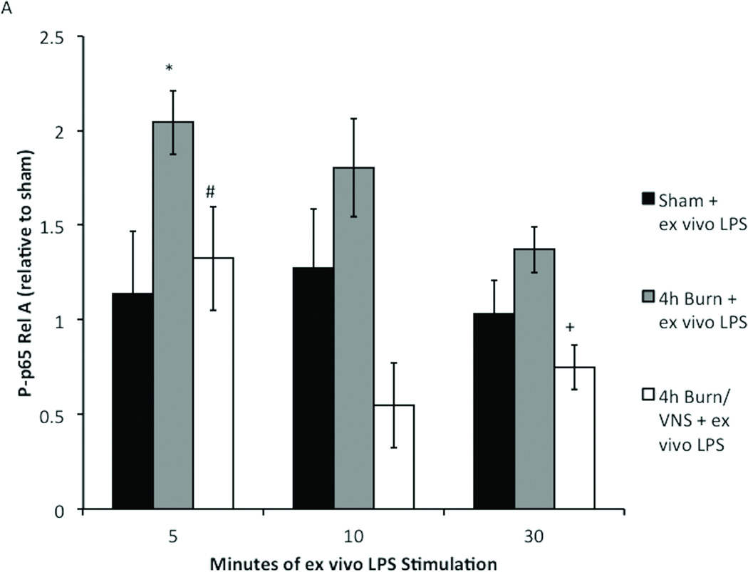
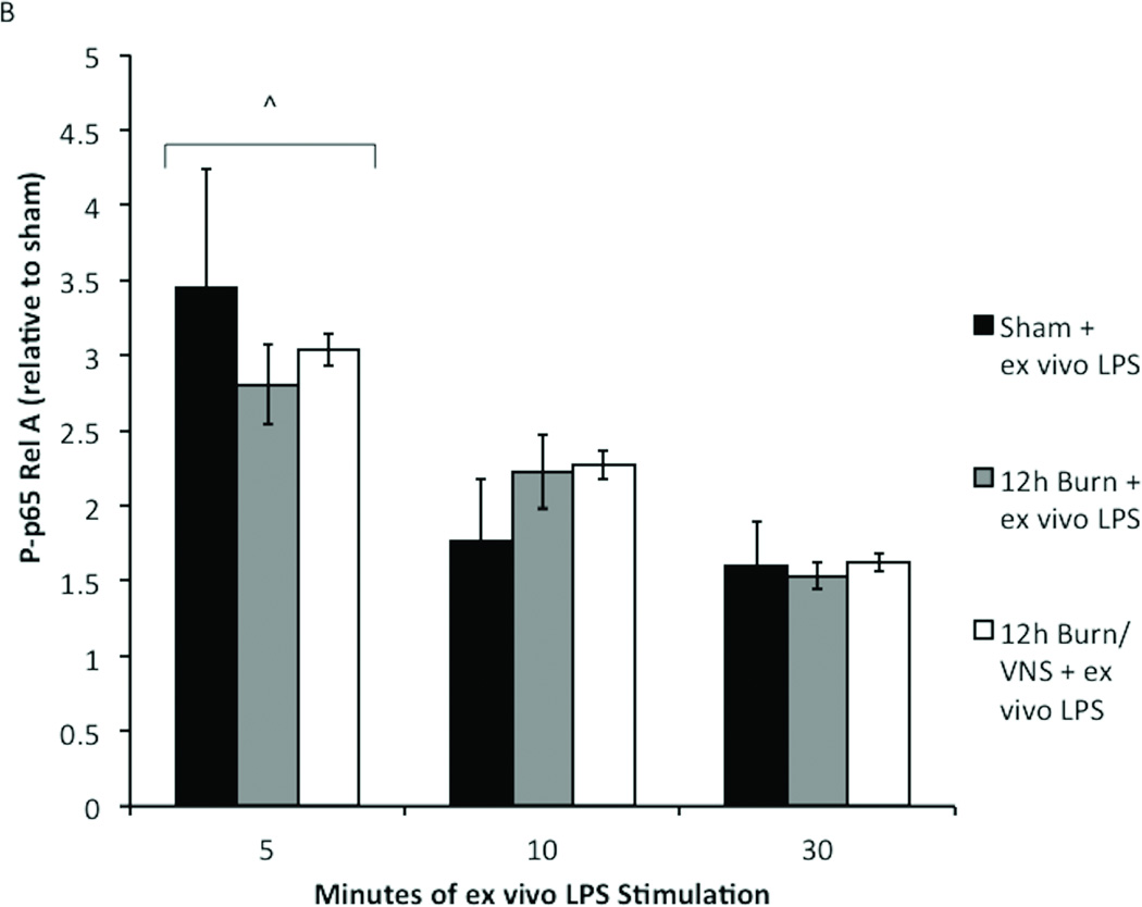
Figure 5. Kinetics of p-65 Rel A phosphorylation are unchanged with VNS and increased time after burn injury.
To confirm that our findings were not due to altered kinetics of phosphorylation, macrophages from sham, burn and burn/VNS animals were stimulated with LPS for 5, 10 and 30 minutes. (A) At the 4 hour time point only 5 minute LPS stimulation caused a significant increase in P-p65 in 4h burn macrophages. P-p65 in the 4h burn/VNS group remained lower than both sham and 4h burn after LPS stimulation for 5, 10 and 30 minutes, and was significantly lower than burn at stimulation times of 5 and 30 minutes. (B) Additionally, kinetics of phosphorylation were similar at the 12 hour time point; maximal increase in P-p65 was at 5 minutes. There was no difference in P-p65 Rel A between the groups regardless of LPS exposure time. This suggests that neither macrophage activation following burn injury, nor its inhibition by VNS results from differential kinetics of phosphorylation of p-65 Rel A. Error bars represent SEM. *p<0.05 vs. sham + ex vivo LPS (5 minute); #p<0.05 vs. 4h burn + ex vivo LPS (5 minute); + p=0.03 vs. 4h burn + ex vivo LPS (30 minute); ^p<0.001 vs. 10 and 30 minutes.


