Abstract
Myxomatous tumours can arise from different cardiac structures. They have a special predilection for the left atrium and are an exceedingly uncommon finding in cardiac valves. We report the case of a 28-year old man who presented with a stroke and was found to have a mass arising from his aortic valve. The patient underwent a successful surgical excision of the aortic valve with the implantation of a mechanical prosthesis. The histopathological examination of the aortic valve confirmed the diagnosis of myxoma. Some aspects related to the diagnosis and management of this entity are discussed in this article.
Keywords: Myxoma, Aortic valve
INTRODUCTION
Myxomas are the most common cardiac tumours in adults. They derive from multipotential mesenchymal cells located within the endocardium and can originate from any chamber or cardiac structure [1]. Myxomas arising from heart valves are very infrequent [2], and to the best of our knowledge, only eight cases of isolated aortic valve myxomas have been published previously [2–10]. We report the case of a patient with a stroke who was found to have a mass implanted on his aortic valve. After surgical resection, the histopathological examination confirmed the diagnosis of myxoma.
CASE REPORT
A 28-year old male was admitted to the emergency department with a sudden onset of left hemiplegia and dysarthria. An urgent cerebral CT scan demonstrated an ischaemic stroke involving the territory of the middle cerebral artery. His medical history included epilepsy of unknown aetiology since the age of 18, but the patient had been free from seizures during the last 5 years.
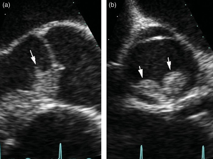
On physical examination, peripheral pulses were present and normal. An early diastolic murmur could be heard in the aortic area. The ECG showed a normal sinus rhythm with signs of mild left ventricular hypertrophy. Transoesophageal echocardiography revealed a grade II/IV aortic incompetence and a mobile mass measuring 15 × 7 mm originating from the ventricular surface of the aortic valve was identified (Fig. 1).
Figure 1:
Preoperative transoesophageal echocardiogram. (a) Longitudinal view. The tumour is attached to the ventricular aspect of the aortic valve (arrow). (b) Transversal view. The tumour arises from the right and left aortic leaflets (arrows).
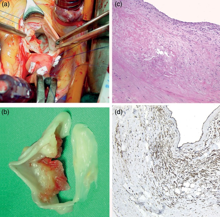
A doppler ultrasound of the carotid arteries was normal. Blood cultures, inflammatory markers and immunological tests were all negative. The patient remained afebrile during the whole period. Early physiotherapy was initiated with satisfactory progress of the patient's neurological status. Weekly follow-up transthoracic echocardiographic examinations were performed. Four weeks after admission, the mobile mass that was identified in relation to the aortic valve remained unchanged; therefore, surgical excision was scheduled after discussing the risk of new perioperative thromboembolic complications with the patient. An intraoperative inspection of the aortic valve revealed a 15 × 7 mm sessile mass attached to the ventricular surface of the right and left coronary leaflets. The mass was gelatinous soft, friable and yellowish in colour, with multiple haemorrhagic areas (Fig. 2). The patient underwent a standard aortic valve replacement with mechanical prosthesis.
Figure 2:
(a) Intraoperative image of the aortic valve showing a mass on the ventricular surface of the right and left cusps of the aortic valve. (b) Macroscopic view of the excised valve. (c) Histological section through the tumour (haematoxylin and eosin ×20). (d) Vascular channels are demonstrated using CD34 immunostaining (×10).
The surgical specimen was fixed in 10% neutral buffered formalin. Routine 5 µm-thick sections were prepared from paraffin-embedded tissue and stained with haematoxylin–eosin. An immunohistochemical stain was performed using EnVision Plus method (Dako Glostrup, Denmark). The antibody used was directed again CD34 (clone QBEND/10, BioGenex; 1:20). Histopathological examination of the mass confirmed the diagnosis of myxoma. There was a sparse population of round and stellate cells mostly concentrating beneath the surface, with some cells forming solid cords and vascular channels (demonstrated with CD34 antibody). Cells were surrounded by abundant myxoid stroma. Mitosis, pleomorphism and necrosis were all absent (Fig. 2).
DISCUSSION
Although myxomas can originate from any cavity or cardiac structure, they originate most frequently from the left atrium, followed to a lesser extent by the right atrium, then the left and right ventricles [1]. Myxomas of the cardiac valves are very unusual, especially those of the aortic valve [2]. To our knowledge, only eight cases of aortic valve myxomas have been reported. The first was described as a post mortem finding [2], while the clinical presentations of the other seven cases included: acute embolic stroke [3, 9], acute embolic myocardial infarction [8], acute embolic lower limb ischaemia [4], aortic stenosis [5] and accidental finding during a routine echocardiogram [6, 7]. This type of tumour has been described as arising from both the ventricular aspect [3, 6, 7] and the margin [8] of the valve cusps. The right [3, 6], left [5, 7] and non-coronary [8] leaflets may be affected either together or individually. In one of the reported cases [4], the right and left cusps were simultaneously affected (as in our patient).
Differential diagnosis of aortic valve myxoma includes vegetations, papillary fibroelastoma and Lambl's excrescences [1, 3, 4, 7]. Microscopic and immunohistochemical characteristics allow the distinction between these entities. As we have observed, aortic vale myxomas are a potential source of emboli; therefore, surgical removal should be indicated as soon as the diagnosis is confirmed. Surgical excision should include not only the tumour but also the implantation site to minimize the risk of local recurrence. Tumour resection with conservation of the native valve should be intended [7, 8], but sometimes due to a big tumour size and/or structural valve degeneration, replacement of the aortic valve may become necessary [4, 6]. Follow-up of these patients is highly recommended since distal tumour growth at the site of previous embolization as well as local recurrence of the tumour have been described [1] in previous reports.
Conflict of interest: none declared.
REFERENCES
- 1.Reardon MJ, Smythe WR. Cardiac neoplasms. In: Cohn LH, Edmunds LH, editors. Cardiac Surgery in the Adult. New York: McGraw-Hill; 2003. pp. 1373–400. [Google Scholar]
- 2.Wold LE, Lie JT. Cardiac myxomas: a clinico-pathologic profile. Am J Pathol. 1980;101:219–33. [PMC free article] [PubMed] [Google Scholar]
- 3.Ramsheyi A, Deleuze P, D'Attelis N, Bical O, Lefort JF. Aortic valve myxoma. J Card Surg. 1998;13:491–3. doi: 10.1111/j.1540-8191.1998.tb01089.x. doi:10.1111/j.1540-8191.1998.tb01089.x. [DOI] [PubMed] [Google Scholar]
- 4.Kennedy P, Parry AJ, Parums D, Pillai R. Myxoma of the aortic valve. Ann Thorac Surg. 1995;59:1221–3. doi: 10.1016/0003-4975(94)00969-e. doi:10.1016/0003-4975(94)00969-E. [DOI] [PubMed] [Google Scholar]
- 5.Gorlach G, Hagel KJ, Mulch J, Scheld HH, Moosdorf R, Fitz H, et al. Myxoma of the aortic valve in a child. J Cardiovasc Surg (Torino) 1986;27:679–80. [PubMed] [Google Scholar]
- 6.Watarida S, Katsuyama K, Yasuda R, Magara T, Onoe M, Nojima T, et al. Myxoma of the aortic valve. Ann Thorac Surg. 1997;63:234–6. doi: 10.1016/s0003-4975(96)00771-0. doi:10.1016/S0003-4975(96)00771-0. [DOI] [PubMed] [Google Scholar]
- 7.Okamoto T, Doi H, Kazui T, Suzuki M, Koshima R, Yamashita T, et al. Aortic valve myxoma mimicking vegetation: report of a case. Surg Today. 2006;36:927–9. doi: 10.1007/s00595-006-3273-y. doi:10.1007/s00595-006-3273-y. [DOI] [PubMed] [Google Scholar]
- 8.Dyk W, Konka M. Unusual complication of aortic valve grape-like myxoma. Ann Thorac Surg. 2009;88:1022. doi: 10.1016/j.athoracsur.2008.12.051. doi:10.1016/j.athoracsur.2008.12.051. [DOI] [PubMed] [Google Scholar]
- 9.Koyalakonda SP, Mediratta NK, Ball J, Royle M. A rare case of aortic valve myxoma: an unusual cause of embolic stroke. Cardiology. 2011;118:101–3. doi: 10.1159/000327081. doi:10.1159/000327081. [DOI] [PubMed] [Google Scholar]
- 10.Perek B, Misterski M, Stefaniak S, Ligowski M, Puslecki M, Jemielity M. Early and long-term outcome of surgery for cardiac myxoma: experience of a single cardiac surgical centre. Kardiol Pol. 2011;69:558–64. [PubMed] [Google Scholar]


