Summary
Sweat glands are abundant glands of our body and essential for thermoregulation. Like mammary glands, they originate from epidermal progenitors. However, they display few signs of cellular turnover, and whether they have stem cells and tissue regenerative capacity remain largely unexplored. Here we address these issues. Using lineage-tracing, we identify multipotent progenitors in sweat duct that transition to unipotency after developing the sweat gland. In characterizing four adult stem cell populations of glandular skin, we show that they display distinct regenerative capabilities and remain unipotent when healing epidermal, myoepithelial-specific and luminal-specific injuries. We devise purification schemes, isolate and transcriptionally profile progenitors. Exploiting molecular differences between sweat and mammary glands, we show that only some progenitors regain multipotency to produce de novo ductal and glandular structures, but that these can retain their identity even within certain foreign microenvironments. Our findings provide new concepts about glandular stem cells and sweat gland biology.
Introduction
Failure to regulate body temperature in hot climates or during exercise can lead to hyperthermia, stroke and death. Eccrine sweat glands perform this vital function. In response to thermal (trunk skin) and/or emotional stimuli (palmoplantar skin), they produce and secrete salt-enriched sweat fluids (water, sodium, potassium, chloride, urea, lactate and creatine) into small luminal orifices that extend to the skin surface (Lobitz and Dobson, 1961). Reaching densities of 200–700/ cm2 of skin surface, they are by far the most abundant glandular structures of the human body (Sato, 1993). In mice, sweat glands are restricted to foot pads, and much of what is known about them stems from observations on human sweat gland morphogenesis and from limited parallels drawn with their distant mammary gland relatives.
In mouse, sweat buds emerge just before birth (embryonic day E17.5), while mammary buds appear at E13.5. Both bud types are initiated by Ectodysplasin-A receptor-mediated signaling, and begin as morphologically similar, undifferentiated invaginations from epidermis (Cui and Schlessinger, 2006; Mustonen et al., 2004). However, while the nascent mammary duct is relatively short and undergoes elaborate branching morphogenesis during development, sweat buds generate a single long duct that extends deep within the dermis.
As the straight duct matures, it forms a coiled gland from its tip (Figure 1A). Like the mammary gland, the secretory coil consists of an outer basal layer of keratin 5 and 14 (K5/K14)+, smooth muscle actin (SMA)+ myoepithelial cells and an inner suprabasal layer of K8/K18/K19+ luminal cells (Langbein et al., 2005; Moll and Moll, 1992; Schon et al., 1999). However, the coiled architecture of the sweat gland is highly divergent from the lobular structure of the mammary gland. Additionally, sweat glands have small lumens and show little or no signs of homeostatic change, while mammary glands have large lumens and undergo dramatic tissue expansion during pregnancy (Richert et al., 2000). These natural differences in cellular proliferation and turnover may also explain why cancers are so rare in sweat glands, but frequent in mammary tissue.
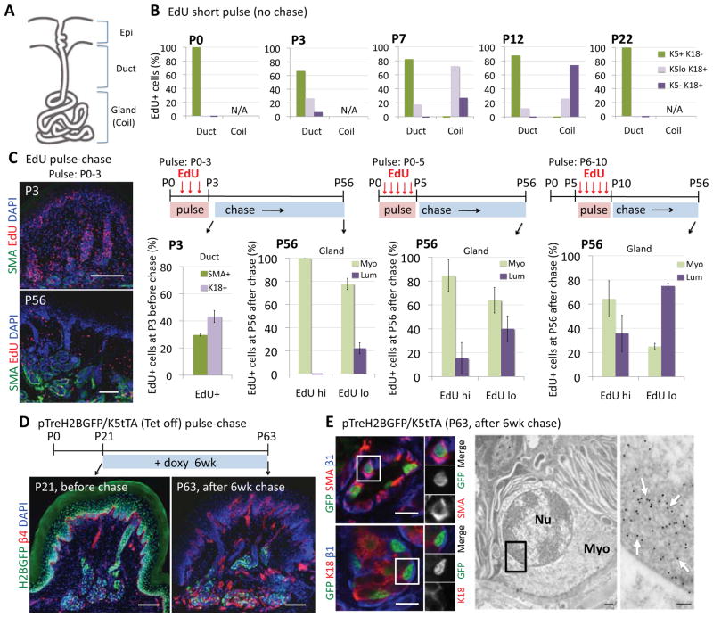
Figure 1. Dramatic changes in proliferation occur within developing paw skin during sweat gland morphogenesis.
(A) Diagram showing orifice and intraepidermal portion of a sweat duct, which extends from epidermis (Epi) into dermis, and terminates in a coiled, secretory gland.
(B) Mice constitutively expressing H2BGFP in K5 promoter-active skin epithelium were injected with EdU 4 hr prior to analyses at postnatal ages indicated. EdU+ (S-phase) cells were classified according to keratin and location (within ducts or glands) (n=100–250 EdU+ cells from 2–3 mice/stage). Overall gland but not ductal proliferation waned by maturity (P22).
(C) EdU was injected daily for times indicated. Paw pads were analyzed following the last EdU injection and again at P56. Quantifications of EdU label show %myoepithelial (SMA+) and luminal (K18+) cells in EdUhi and EdUlo populations (n=3 mice, >40 pads). Representative immunofluorescence images for pulse P0–3 pawskin are shown on the left. Note: label-retaining cells were confined to glands. Scale bars, 50 μm.
(D,E) H2BGFP, expressed in K5+ cells since embryogenesis, was silenced by doxy in Tet-off pTreH2BGFP/K5tTA mice as shown. (D) H2BGFP retention occurred only in glands. Scale bar, 50 μm. (E) (Left) Confocal images. Note H2BGFP label retention in SMA+ but not K18+ sweat gland cells (split channels are for boxed areas). Scale bars, 10 μm. (Right) Ultrathin sections of GFP immunogold labeled pawskin. Boxed area is magnified to see that myoepithelial cells were labeled (white arrows). Nu, nucleus. Myo, myoepithelial cells. White arrows indicate positive immunogold labeling. Scale bars, 1 μm.
Proliferation and tissue generation are features fueled by adult stem cells. Given the enormous tissue regenerative capabilities of the mammary gland, it is not surprising that their glandular stem cells have been a long-standing topic of intense study, and are now well-characterized. As suggested by earlier studies (Kordon and Smith, 1998) and then documented with purified (K14+α6-integrinhiCD24+) cells, individual myoepithelial progenitors can reconstitute functional mammary glands when introduced into a cleared fat pad (Shackleton et al., 2006; Stingl et al., 2006). Based upon these results, it seemed likely that myoepithelial stem cells might give rise to luminal cells within the mammary gland. However, recent lineage tracings show that during puberty and homeostasis, expansion and maintenance of the mammary gland are fueled largely if not solely by unipotent luminal and myoepithelial progenitors (Van Keymeulen et al., 2011). The experimental basis for this discrepancy is unknown. It has been speculated that the purified myoepithelial transplantation assays may reflect a wound healing state that does not occur in normal homeostasis.
For the sweat gland, the paucity of massive hormonally-induced fluctuations in tissue production raises the question as to whether sweat stem cells exist at all, and if they do, where they are and how they behave in morphogenesis, homeostasis and wound repair. Moreover, the prominence of the straight duct in this epidermal appendage adds an additional level of complexity that has been largely ignored in the mammary stem cell field.
Much of what is known about proliferative potential within the sweat duct and gland comes from histological studies dating back to the early 1950’s. When researchers performed deep dermal injury to the backskin of adult human volunteers, signs of regenerative activity were observed in the damaged glands. In initial experiments, injury-provoked mitoses seemed largely confined to basal cells of the straight duct (Lobitz et al., 1954). However in subsequent experiments, occasional mitoses were observed in several morphologically distinct epithelial cells residing within closer proximity to the secretory coils, prompting speculation that perhaps these cells might be able to dedifferentiate to repair sweat glands, ducts and epidermis (Lobitz et al., 1956).
Despite the tantalizing aspects of this hypothesis put forth so long ago, only a few follow-up reports in the past 65 years substantiate regenerative potential for sweat gland cells. Several of these were documented proliferation of dissociated sweat gland cells in culture; most of the in vivo studies focused on the contribution of sweat gland cells to epidermal repair after superficial skin injuries (Biedermann et al., 2010; Miller et al., 1998). Even in these limited studies, the precise cells involved were not identified, nor was the overall regenerative capacity of the sweat gland explored.
Since mammary gland stem cells are the only glandular stem cells studied in detail, it remains unknown to what extent their features might establish a paradigm for glandular stem cells. Similarly, until other glandular stem cells are isolated and profiled, it remains unresolved whether stem cells from different adult glands are analogous and merely differentiate distinctly in response to unique microenvironments or whether defining molecular characteristics are established during morphogenesis. Given the paucity of knowledge about the most abundant gland of our body and the intrigue that sweat glands are much less dynamic than mammary glands, we’ve now used the sweat gland as a model to address these fundamentally important and fascinating questions about glandular stem cells. In addition, we also explore the constituents of the adult sweat duct, and test for their stemness.
Here, we report the identification, characterization and regenerative potential of three types of adult progenitors within the sweat gland and its duct. Remarkably, each of these three populations responds distinctly to different types of skin injuries and to engraftments in a variety of foreign environments. Our findings provide important new general concepts about glandular stem cell biology, and they also reveal fundamental differences in the homeostatic dynamics and the molecular biology of stem cell lineages of glandular and ductal tissues and of other types of epidermal appendages. Moreover, our knowledge gained on how sweat glands form, are maintained and differ from mammary glands and hair follicles provides a valuable framework for understanding the roots of some genetic disorders in which sweat glands, and not other epidermal appendages, are specifically affected.
Results
Sweat Gland Development: Emergence of the Straight Duct and Secretory Coil
To gain insights into how the sweat duct and gland develop, we began by monitoring proliferative activity by administering a short pulse (4hr) of nucleotide analogue EdU just prior to analysis. We used mice which express histone H2B-green fluorescent protein (H2BGFP) in all K5-expressing cells and analyzed EdU incorporation in pawskin during postnatal development. Since the K5 promoter is silenced as basal progenitors differentiate, we used fluorescence intensity as a predictor of potential precursor-product relation between basal and suprabasal populations.
Representative immunofluorescence images and quantifications of the data are provided in Figures S1A and 1B. At P0, emerging sweat buds were uniformly GFPhi (K5-expressing). By P3, each bud had grown downward, producing a nascent straight duct. The developing duct was bilayered, with a K5+ GFPhi basal layer and a K5lo GFPdim suprabasal layer. Suprabasal cells within the lower nascent duct also expressed K18 and accounted for ~30% of all EdU+ cells at this stage.
Ductal proliferation and K18 expression was accompanied by the emergence of a nascent sweat gland from the tip (Figure S1A). K5+/K18(−) basal cells within developing glands were distinguished from their ductal counterparts by SMA expression and a paucity of EdU+ cells. By contrast, whether from the lower duct or the gland, K18+ cells still incorporated EdU over a two week developmental window. Once mature, proliferation was largely if not solely confined to K5+/K18(−) basal cells of the sweat duct and was not detected in the sweat gland (Figure 1B, P22). Moreover, whether in the sweat duct or gland, cells were either K5+ or K18+ at this stage, underscoring the transient nature of double-positive, proliferative suprabasal progenitors during morphogenesis. These results also highlighted early molecular and architectural similarities between proliferative tips of sweat ducts and emerging glands.
Fates of Proliferating Cells During Early Gland Development: Emergence of Infrequently Cycling Myoepithelial cells
To further evaluate cycling behavior within developing sweat ducts and glands, we conducted a series of pulse-chase experiments (Figure 1C). In the first experiment, EdU was administered from P0→P3 to mark proliferating nascent ductal cells and then analyzed at P3 and after 8 weeks of chase. Whether EdUhi or EdUlo, label-retaining cells (LRCs) were found exclusively within mature coiled secretory sweat glands and not straight ducts. Moreover, the majority of EdUhi and ~80% of Edulo cells were myoepithelial. With EdU labelings of P0→P5, and P6→P10, more luminal LRCs were detected (Figures 1C; S1B). That said, even at P6→P10, some myoepithelial LRCs were still seen, even though any that had formed by this time were not themselves proliferating (see Figure 1B).
To further explore relative proliferation rates within ductal and glandular populations, we turned to pTREH2BGFP/K5tTA (Tet-off) mice (Tumbar et al., 2004). This more sensitive method enabled us to express H2BGFP in all K5+ cells beginning in the embryo, and then silence H2BGFP transcription by administering doxycycline. At P21, all K5+ basal cells from untreated pawskin epidermis, ducts and sweat glands were marked by H2BGFP, but after 6 weeks, fluorescence was only retained within sweat gland coils (Figure 1D). Immunofluorescence and immunoelectron microscopy confirmed the myoepithelial identity of these H2BGFP-LRCs (Figure 1E). Together, our collective pulse and pulse-chase studies support the view that cells within the developing luminal compartment undergo several more rounds of proliferation than myoepithelial cells, but that the entire sweat gland is largely static once it reaches maturity. By contrast, adult basal cells of the sweat duct remain proliferative.
Emergence of Multiple Progenitor Populations During Pawskin Development: Multipotency Versus Unipotency
To test progenitor potential of cells within epidermis, sweat ducts and glands in vivo, we employed lineage tracing with RosaLacZ or RosaYFP reporter mice, in which a stop codon excisable by Cre-recombinase exists between a ubiquitously expressed Rosa26 promoter and the reporter gene. We mated these lines with transgenic strains driving constitutive or inducible expression of Cre recombinase in distinct cell populations.
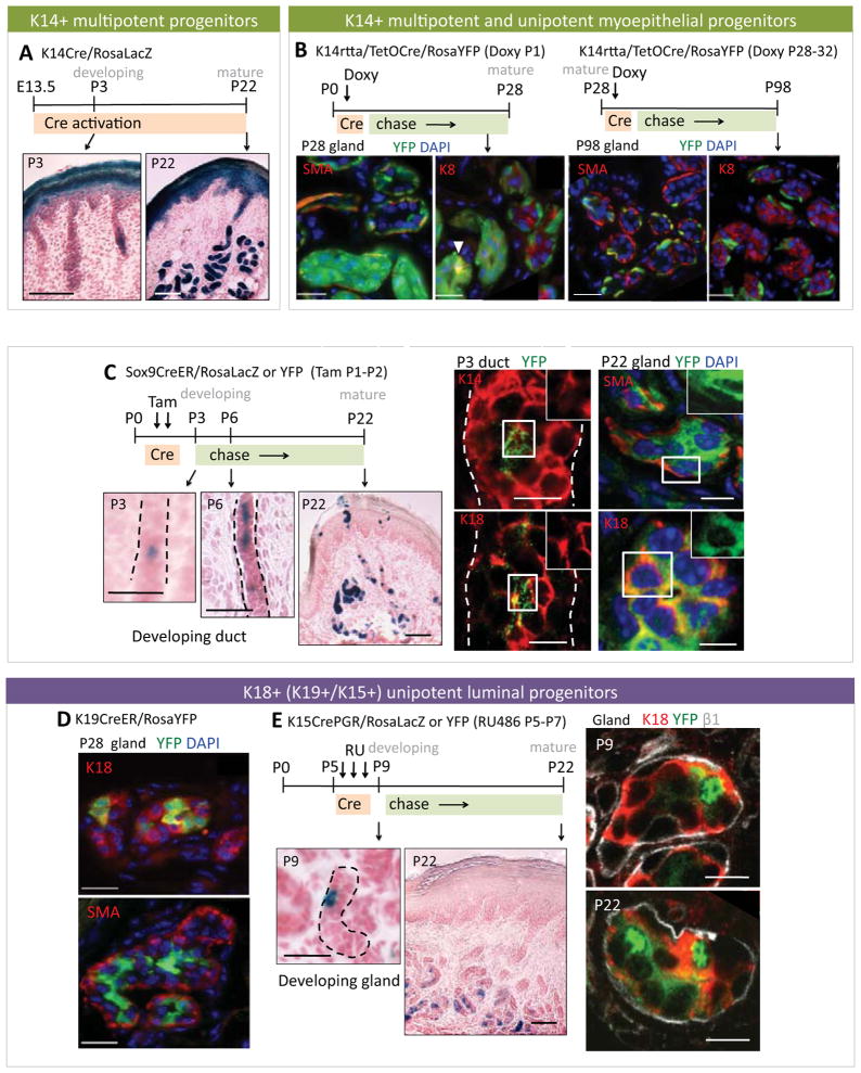
To assess the contribution of K5/K14-derived embryonic skin basal cells to sweat gland development, we used K14-Cre mice and induced β-galactosidase in K14+ cells at E13.5–E15.5 (Vasioukhin et al., 1999). Similar to previous findings on HFs (Vasioukhin et al., 1999) and mammary glands (Van Keymeulen et al., 2011), this tracing established that sweat duct and gland develop from K5/K14-expressing multipotent basal progenitors (Figure 2A).
Figure 2. Multipotent progenitors in developing sweat ducts give rise to both luminal and myoepithelial cells in sweat glands.
(A) Bright field images of paw pad sections from K14Cre/RosaLacZ mice to show emerging (P3) and mature (P22) sweat glands. Sections were stained with X-gal, which turns blue in LacZ+ cells. Counter staining was done with nuclear fast red.
(B) Immunofluorescence images of mature glands from K14rtta/TetOCre/RosaYFP mice induced at P1 and P28 respectively, and examined at times indicated. Some glands showed YFP-labeling of both myoepithelial (SMA+) and luminal (K8+) cells (arrow), but others only had YFP+ myoepithelial cells.
(C) Cre was induced at P1–P2 in Sox9CreER X RosaLacZ or RosaYFP mice and pawskins were analyzed at times indicated. (Left), X-gal stained bright field images show that suprabasal nascent ductal cells were Sox9-promoter active and contributed to gland formation. (Right), Confocal immunofluorescence. Note: YFP+ nascent ductal cells were double positive for K14/K18. At P22, both myoepithelial and luminal glandular cells were YFP-marked (i.e. ductal-derived). Single channels of boxed areas are shown in insets.
(D) Immunofluorescence images from glands of P28 K19CreER/RosaYFP mice Cre-induced at P1. Only luminal (K18+) cells were YFP-marked.
(E) Cre was induced at P5–7 in K15CrePGR X RosaLacZ or RosaYFP mice and paw pads were examined at P9 and P22. Note increase over time of YFP+ cells that co-labeled with K18, indicative of clonal expansion of marked luminal cells during gland maturation.
All dashed lines denote basement membrane. All scale bars are 10 μm.
We shifted to K14rtta/TetO and K5CreER mice, which enabled us to induce RosaYFP in basal cells just after birth, when sweat buds are initiating. Four weeks after induction, two patterns of YFP-marked sweat glands were found: one had only YFP+ myoepithelial cells; the other displayed YFP in both luminal and myoepithelial compartments. This was true for both of these Cre drivers (Figures 2B). One possibility consistent with these results is that multipotent K5/K14+ progenitors initiate sweat gland morphogenesis but as development proceeds, the basal lineage becomes largely unipotent. We tested this directly by analyzing myoepithelial cells in mature sweat glands. When activated at P28→P30, both K5 and K14 Cre drivers selectively marked myoepithelial cells, and after 8–12 weeks of chase, no signs of YFP+ luminal cells were detected (Figures 2B; S2C).
The existence of multipotent and unipotent basal progenitors led us to wonder whether there might be equivalent types of suprabasal progenitors, or whether they derive solely from basal progenitors, analogous to what happens in skin epidermis. In searching for additional Cre drivers that might allow us to resolve this issue, we discovered that Sox9CreER (Krahl and Sellheyer, 2010) is first activatable within suprabasal nascent ductal cells. Moreover, when induced from P1→P3 and examined at P6, signs of clonal expansion were evident (Figure 2C, RosaLacZ). Immunofluorescence microscopy of RosaYFP lineage-traced cells confirmed their suprabasal location within nascent ducts. These β1integrin(−), K18+ cells were also weakly positive for K14, in agreement with the temporal appearance of proliferative double-positive progenitors at this site. By P22, progeny of Sox9CreER-induced cells were found throughout sweat glands, straight ducts, and even sweat duct orifices. Within the gland, marked progeny were predominately luminal (K18+), although occasional SMA+ myoepithelial cells were marked (Figure 2C, RosaYFP).
To explore the potential of suprabasal progenitors, we conducted lineage tracings in K19CreER and K18CreER X RosaYFP mice. At P28, YFP-marked luminal cells were prevalent (Figure 2D). Myoepithelial cells were negative for YFP, and this did not change after 8–12 weeks of chase (Figures S1D–E). These findings suggested that K19 and K18 promoters had been activated selectively in unipotent luminal progenitors.
We confirmed the existence of unipotent luminal progenitors by using K15CrePGR mice, whose Cre is inducible by the progesterone analogue RU486 (Morris et al., 2004; Langbein et al., 2005). In pawskin, K15 promoter activity was not detectable until P5, and it was restricted to luminal cells of emerging secretory coils (Figure 2E). When lineage traced to P22, signs of clonal expansion were apparent, but again, only luminal cells were marked. The lineage-restricted unipotent behavior of these glandular progenitors was confirmed by FACS analyses (introduced later in the text). Similar signs of maturation-associated fate restrictions were seen within sweat ducts, where proliferative activity became restricted to basal cells (Figure S2A). Moreover, given the overall quiescence of the sweat gland, the lack of label-retention within adult ductal basal cells (Figures 1C–D) appeared to reflect frequent basal to suprabasal turnover within the duct, analogous to what happens in epidermis.
Taken together, our data pointed to the emergence of three distinct types of unipotent progenitors within mature sweat ducts and glands. Our data further showed that these unipotent progenitors all derive from a common multipotent K14+ sweat bud progenitor. This progenitor then generates a nascent duct replete with distinct K14+ basal and K18/K14-double positive suprabasal progenitors, both of which transiently maintain multipotency: they expand within their respective layers during sweat duct maturation and they also form the sweat gland. At this point, progenitors of gland and duct become restricted in potential.
Adult Sweat Duct But Not Gland Progenitors Contribute to Epidermal Wound Repair
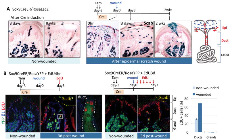
The relatively static state of mature sweat glands led us to wonder whether they have long-lived stem cells. To address this question, we focused on how cells within sweat duct and gland respond to injury. For this study, we used Sox9CreER/RosaLacZ mice, which when induced in adults, activated Cre throughout most of these cells (Figure 3A). 2d after induction, pawskin epidermis was scraped to induce re-epithelialization (t=0hr). Within 3d post-wounding, β-gal+ gland cells and/or their progeny appeared within regenerating epidermis under the scab. By 2 weeks, the narrow column of β-gal+ cells that surrounded sweat duct orifices at t=0 had broadened considerably (Figure 3A). However, many regenerated cells were unlabeled and hence appeared to come from neighboring epidermis.
Figure 3. Ductal but not glandular progenitors participate in epidermal wound repair.
(A) Cre was induced in adult Sox9CreER/RosaLacZ mice and pawskins were either examined for LacZ expression 3d and 8wks later (unwounded, left) or subjected to epidermal scratch-wounding and examined at times indicated (wounded, right). Note: in adult mice, Sox9Cre-induction resulted in LacZ expression (blue) in all cellular compartments within the sweat duct and gland. Note expansion of βgal+ interepidermal cells surrounding the ductal orifice after wounding. Blue dashed lines, removal of epidermis.
(B) Immunofluorescence images of non-wounded and 3d post-wound pawskin from Cre-induced Sox9CreER/RosaYFP mice injected with EdU for 4hr (left) or 3d (right) just prior to analysis as indicated. Magnification of boxed area is shown at its right. Quantifications are of EdU+ cells in ducts and glands (n=2 or 3 mice, >30 pads). Note that proliferation was restricted to ducts, while cells within the sweat gland remained quiescent, as depicted in the diagram in (A). Yellow dashed lines, wounded area covered by a scab. White dashed lines, basement membrane.
All scale bars are 50 μm.
To gain further insights, we repeated the assay, this time using a YFP reporter and administering EdU pulses prior to analyses (Figure 3B). As seen by a short (4 hr) EdU pulse (left). pawskin epidermis adjacent to the wound was highly proliferative. Sox9-YFP+ ductal cells were also proliferative. In a longer (3d) pulse, EdU+ ductal cells rose from ~30% to ~70% (right). Sweat glands, however, appeared to remain quiescent throughout wound healing. Their lack of contribution was unambiguously documented with wound studies on K15CrePGR/RosaLacZ mice (Figure S3).
Evidence for the Existence of Adult Sweat Gland Stem Cells
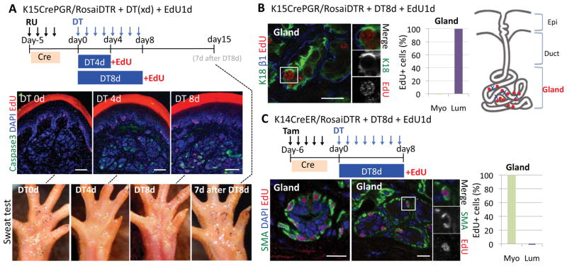
We next addressed whether adult sweat gland cells are refractory to wound repair, or whether their regenerative potential is only unleashed when wounds are in closer proximity. To generate more localized tissue damage, we mated K15CrePGR and K14CreER mice to RosaiDTR mice, and then induced diphtheria toxin receptor (DTR) expression specifically in either adult luminal (K15CrePGR) or myoepithelial (K14CreER) cells. After 4d DT, apoptosis (as determined by caspase 3 activation) was selectively induced within a fraction of DTR+ cells (Figure 4A). Few apoptotic cells were detected without DT.
Figure 4. Myoepithelial and luminal progenitors respond as unipotent stem cells to glandular injury.
(A, B) Cre was induced in K15CrePGR/RosaiDTR mice which were then treated with DT for times indicated to elicit DTR-mediated luminal cell death. EdU was given 1d before immunofluorescence analysis of pawskin, labeled with antibodies as shown (color coding is according to secondary antibodies. (A). Active caspase 3 (apoptosis) occurs only after DT. Scale bars, 50 μm. (Bottom), Sweat tests were performed on paw pads of K15CrePGR/RosaiDTR mice before, during, and after DT. Fine black dots reflect functional sweating pores, transiently reduced after glandular injury. (B) Images document luminal identity of EdU+ cells (split channels for boxed area are at right). Quantifications (n=2 mice, >80 EdU+ cells), reveal their selective proliferative response to luminal cell damage. Scale bar, 10 μm.
(C) Cre was induced in K14CreER/RosaiDTR mice which were then treated with DT for 8d to induce myoepithelial cell death. EdU was given 1d before analysis. Representative fluorescence images reveal the myoepithelial identity of EdU+ cells. Quantifications (n=2 mice, >120 EdU+ cells) reveal that only myoepithelial cells proliferate in response to myoepithelial apoptosis. Scale bars, 10 μm.
In K15CrePGR/iDTR DT-treated mice, myoepithelial cells were spared, not only validating targeting selectivity, but also demonstrating that cell death was not a global indirect effect, but rather a direct consequence of DT/DTR action on luminal epithelial cells. As evidenced by EdU pulses given prior to analysis, robust proliferation was induced selectively within K18+ luminal cells of secretory coils (Figures 4B). Ultrastructural analyses confirmed that apoptotic and mitotic cells were restricted to the luminal population, while neighboring myoepithelial cells remained unaffected (Figures S4). Further reflecting the selective damage were signs of lumen collapse within affected glands.
When similar experiments were performed with K14CreER/RosaiDTR mice, the reverse was observed. Although K14CreER-mediated targeting of DTR to adult myoepithelial cells was inefficient, it was selective. Moreover, the proliferative response that ensued after DT treatment appeared to be confined to myoepithelial and not luminal cells (Figure 4C).
To determine whether these proliferative responses were reflective of actual glandular tissue repair, we performed an iodine/starch-based sweat test on paw pads of our animals (Figure 4A, bottom). This procedure detects sweat, reflective of a functional gland. Prior to DT, paws of K15CrePGR/iDTR mice responded to the assay by displaying indigo-black dots, indicative of sweat production. After 4d of DT, indigo dots had decreased in numbers and intensity, suggesting that sweat production was impaired. Most importantly, 7d after DT treatment was terminated, dots resurfaced, indicating that sweat gland function had been restored.
Together, these results establish the existence of two distinct types of resident unipotent stem cells. Moreover, both luminal and myoepithelial stem cells of adult sweat glands possess regenerative potential that can be tapped in response to localized injury. Progenitor activation was reflected by their enhanced proliferation after injury; regenerative potential was reflected by subsequent restoration of secretory functions following activation and repair.
Purification and Culture of Distinct Progenitor Populations of the Adult Sweat Gland
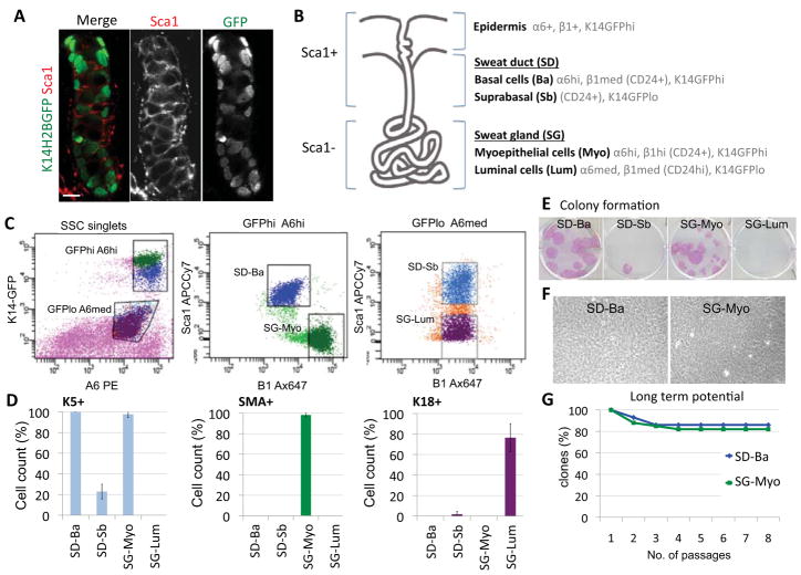
To delve more deeply into the molecular properties and differences of sweat gland progenitors, we devised methods to purify them. We first showed that a) similar to the HF infundibulum (Jensen et al., 2008), the sweat duct was marked by Sca1 (Figure 5A); b) CD29 (β1 integrin) labeled myoepithelial more prominently than straight duct basal cells; c) CD49f (α6 integrin) labeled all basal populations, and to lesser extent suprabasal glandular cells; and d) analogous to mammary gland luminal cells (Shackleton et al., 2006; Stingl et al., 2006), CD24 (sialoglycoprotein) strongly labeled sweat gland luminal cells.
Figure 5. Purification of distinct populations from adult sweat ducts and glands.
(A) Image of sweat duct segment from K14H2BGFP pawskin, showing that cells are positive for Sca1. Scale bar, 10 μm.
(B) Summary depicting localizations of markers used to isolate different populations from sweat glands and paw pad epidermis. SD, sweat duct. SG, sweat gland.
(C) FACS profiles illustrating the sorting strategy. Ba, basal cells. Sb, suprabasal cells. Myo, myoepithelial cells. Lum, luminal cells.
(D) Cytospin analyses for different populations isolated from the sweat ducts and glands. Percentages of K5+, SMA+, and K18+ cells in each population are shown. Quantifications were done with >150 cells from each of 3 independent sorts.
(E) Colony formation assay. Representative images of colonies formed from 3000 FACS-isolated cells after 16d in culture.
(F) Phase-contrast images of cells within the large holoclones formed from FACS-sorted sweat duct basal cells (SD-Ba, left) and sweat gland myoepithelial cells (SG-Myo, right). Note that both exhibit tightly packed and undifferentiated morphologies.
(G) Long-term potential of purified sweat duct basal cells and sweat gland myoepithelial cells, n=28 and 34 clones, respectively. Note that for both, >80% clones survived after 8 passages (>3 months) in culture.
Our purification scheme employed K14-H2BGFP transgenic mice, in which epidermis, sweat ducts and glands were brightly fluorescent. After separating pawskin epidermal and dermal (containing sweat ducts and glands) fractions, we used FACS on cells from the dermal fraction to isolate GFPhiα6hi and GFPloα6med populations, which were further subfractionated to generate straight duct cells (basal: GFPhiα6hiβ1medSca1pos; suprabasal: GFPloα6medβ1medSca1pos) and glandular cells (myoepithelial: GFPhiα6hiβ1hiSca1neg; luminal GFPloα6medβ1medSca1neg) (Figures 5B–C). We validated the sorting strategy by applying it to several transgenic lines expressing GFP or YFP in selective glandular compartments, which enabled us to match FACS with immunofluorescence (Figure S5). The identity and dramatic enrichment of our purified populations was further revealed by cytospin analysis (Figure 5D).
Interestingly, both myoepithelial cells and basal straight duct populations were enriched (1/200 - 1/750) for cells forming large colonies (holoclones) (Figures 5E–G). Cells within these colonies were small, tightly packed, and morphologically uniform, with little or no signs of terminal differentiation. Moreover, many colonies could be passaged long term. Notably, these are established features of epidermal and HF stem cells (Barrandon and Green, 1987; Blanpain et al., 2004). By contrast, suprabasal cells from gland and duct failed to grow. This could be due to either the particular culture conditions used, or to a difference in stemness. We return to this issue later.
Striking Molecular Differences Between Sweat Ducts, Sweat Glands and Mammary Glands
Equipped with our newfound ability to purify adult pawskin sweat duct and gland progenitors and GFPhiα6hiβ1hiSca1pos basal epidermal cells, we turned to transcriptionally profiling each population to dissect their distinguishing features. Purifications and microarray hybridizations (Affymetrix Mouse430A_2) were performed in duplicate. The raw and normalized microarray data will be made available at the Gene Expression Omnibus GSE37274 (http://www.ncbi.nlm.nih.gov/geo/).
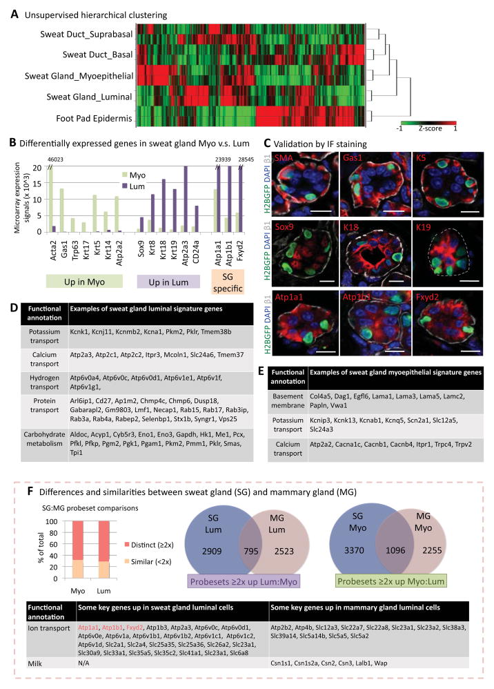
Principal component analysis demonstrated that duplicate data sets nicely clustered (data not shown). The relation of each cell population to the others was revealed by unsupervised hierarchical clustering algorithms. As shown by heatmap (Figure 6A), straight duct basal cells were most similar to their suprabasal counterparts, but differed markedly from glandular cells. Surprisingly, their most distant relation was pawskin epidermal cells.
Figure 6. Expression profiling of adult sweat duct/gland populations.
(A) Unsupervised Ward clustering and hierarchical analysis of the microarray profiles.
(B) Examples of differentially expressed genes in myoepithelial cells and luminal cells of the sweat gland. These genes are grouped into three categories: (left) upregulated in myoepithelial cells; (middle) up-regulated in luminal cells; (right) sweat gland-specific, compared to mammary gland (see below)
(C) Immunofluorescence microscopy to validate expression of sweat gland proteins whose RNA expression is up-regulated in either myoepithelial or luminal cells, or both. Sections are from K14H2BGFP adult pawskin. GFP marks myoepithelial cell nuclei. Dashed line, basement membrane. Scale bars, 10 μm.
(D) Functional annotations of some of the genes up-regulated ≥2X in sweat gland luminal cells.
(E) Functional annotations of some of the genes up-regulated ≥2X in sweat gland myoepithelial cells.
(F) Comparisons of sweat and mammary gland mRNA profiles. Graph at left shows percentage of total probesets that are similar (<2x) or distinct (≥2x) in arrays from myoepithelial cells and luminal cells of sweat and mammary glands. Venn diagrams showing marked differences between the sweat and mammary gland molecular signatures (genes up-regulated ≥2x) of both luminal (left) and myoepithelial (right) cells. Table highlights several of the key differentially-expressed genes in luminal cells of sweat and mammary glands, respectively.
Most intriguing were sweat gland features, where >400 genes scored as being upregulated by ≥2X in one compartment over the other. Other genes were upregulated specifically in either luminal or myoepithelial relative to sweat duct or pawskin epidermis. Myoepithelial and luminal signatures were generated as mRNAs upregulated by ≥2X relative to all other epithelial populations (Table S1). Besides proteins previously affiliated with sweat glands [(Cui et al., 2012; Sato and Sato, 2000; Schon et al., 1999; Song et al., 2002) and references therein], our data revealed a new array of genes (Figure 6B and Table S1). Representative expression-based validations are shown in Figure 6C.
Several functional annotations (grouped according to DAVID) featured prominently within the luminal signature (Figure 6D). Most notable were mRNAs encoding proteins involved in ion transport, such as Na+/K+ and/or Ca2+-ATPases. The luminal signature also featured transport-related categories, e.g. hydrogen and protein transport, that were not seen in the myoepithelial signature (compare Figures 6D and 6E). Another distinction was the preponderance of luminal genes involved in carbohydrate metabolism, a link made previously from pharmacologic studies (Sato and Dobson, 1973). More extensive lists for myoepithelial and luminal signature genes are shown in Table S1.
Given the structural similarities of mammary and sweat glands, we sought to compare our gene expression profiles with those published for mammary gland (Shackleton et al., 2006; Stingl et al., 2006). While inherent caveats are imposed by differences in purification schemes and transcriptional profiling platforms, the comparisons were nevertheless revealing (Figures 6F; Table S2). Whether myoepithelial or luminal, the overlap in molecular signatures of mammary and sweat gland counterparts was surprisingly modest. For myoepithelia, shared functional annotations included genes involved in smooth muscle contraction, cell adhesion/basement membrane and TGFβ, Wnt and hedgehog signaling pathways [see also (Kunisada et al., 2009)]. For luminal epithelia, this included cytoskeletal, metabolic and Notch pathway genes. These parallels aside, sweat and mammary signatures were otherwise strikingly divergent. Among notable luminal differences were mRNAs encoding ion transport proteins (preferentially expressed by sweat glands) and milk proteins (preferentially expressed by mammary glands).
Adult Myoepithelial and Sweat Duct Progenitors Can Regenerate De Novo Sweat Glands in Some But Not All Foreign Microenvironments
We next wondered whether foreign microenvironments might influence the identity and unipotency of adult sweat gland progenitors. To examine these issues, we challenged them to make sweat glands de novo when transplanted into different body sites. While kidney capsule implantations were not consistent and paw pads posed technical impracticalities, three distinct body locations were informative: mammary fat pads, shoulder fat pads and backskin.
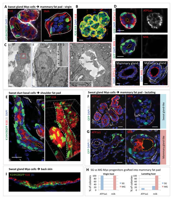
In the first experiment, we purified K14-H2BGFP-marked myoepithelial cells and transplanted them into the cleared mammary fat pad of virgin female Nu/Nu mice. Within 8 weeks, de novo green fluorescent glandular structures appeared (Figure 7A). Notably, K14-H2BGFP+ cells were SMA+ and oriented their integrin+ basal surface towards the gland periphery. Within the gland were K18+/K14(-) cells, many of which were suprabasal. Although lumens were small, their existence offered further signs of a polarized glandular structure. When administered during the regenerative process, EdU incorporated predominantly into K18+ cells, suggesting that the luminal compartment of these glands was still expanding at the time of analyses (Figure S6A).
Figure 7. Purified progenitors from sweat ducts and glands exhibit de novo tissue morphogenesis and maintain their identity when engrafted into some (but not all) foreign microenvironments.
(A) Sweat gland myoepithelial cells from K14H2BGFP mice were FACS purified and transplanted into mammary fat pads of virgin Nu/Nu mice. Engrafted tissues were analyzed at 10wks. K18, β1 and SMA immunolabelings of sections of K14H2BGFP gland-like structures reveal polarized architecture. Yellow dashed lines outline lumens. Scale bars, 10 μm.
(B) Image of a 4 mo. graft of transplanted FACS-purified myoepithelial cells as in (A) but from RosaYFP mice. The entire de novo glandular structure is YFP+, demonstrating that K18+ luminal cells are derived from the donor myoepithelial population and not host mammary luminal cells. Scale bars, 10μm.
(C) Ultrastructure analysis of de novo glandular structure within host mammary fat pad 10wk after transplanting donor K14H2BGFP+ myoepithelial cells. GFP+ glandular structures were identified by correlative immunofluorescence and transmission electron microscopy. I, Example of glandular structure. Cells are organized around an open luminal space. Boxed areas are enlarged in II and III. Scale bar, 10μm. II, Myoepithelial cell (Myo) beside a luminal cell (Lum). Boxed areas are magnified in insets. Boxed area (a), myoepithelial cell attachment to basement membrane (arrowheads). Boxed area (b) shows dense actin filament bundles within myoeptihelial cytoplasm. Scale bar, 500nm. III, Luminal cells within gland. Note numerous apical microvilli (Mv). Intercellular junctions are boxed, showing desmosome (De) and tight junction (Tj). Scale bar, 500nm.
(D) Immunofluorescence for ATP1a1 (sweat gland-specific marker) and milk protein (mammary gland-specific marker) in de novo glandular structures from transplanted FACS-purified K14H2BGFP sweat gland myoepithelial cells and from mammary gland control tissue. Note that sweat gland myoepithelial cells retain their character even when engrafted to a mammary microenvironment. Scale bar, 10 μm.
(E) Purified K14H2BGFP+ sweat duct basal cells were transplanted into mouse shoulder fat pad, and tissue was analyzed 8wks later. Left image shows a straight duct-like structure with K18+ suprabasal cells and K14H2BGFP+ basal cells. Yellow dashed lines outline lumens. Right image shows a 3D reconstruction from a Z-stack. White dashed lines demarcate the straight duct-like structure. Scale bar, 10 μm.
(F–G) Purified K14H2BGFP+ sweat gland myoepithelial cells were transplanted into mammary fat pad of recipient mouse that went through pregnancy after engraftment and was lactating when the graft was taken. (F) Representative images of some sweat gland-like colonies, which still express sweat gland markers and small lumens (yellow dashed lines). Note that some glands show slight branching morphology (left). (G) Representative images of some mammary gland-like colonies that show diminished expression of sweat gland marker (ATP1a1, left) and are positive for milk proteins (right). Note they exhibit clear branching morphology and enlarged lumens, not seen in grafts from virgin hosts. Scale bars, 10 μm.
(H) Graphs showing differences of sweat gland (SG) and mammary gland (MG) myoepithelial progenitors when FACS-purified from K14H2BGFP+ mice and engrafted into mammary fat pad of virgin and lactating hosts.
(I) Purified K14H2BGFP+ sweat gland myoepithelial cells were combined with dermal fibroblasts and transplanted into mouse back skin. Note epidermal differentiation of engrafted cells.
Since K14-H2BGFP is silenced in luminal cells, we repeated the transplantation experiments with FACS-purified myoepithelial cells from K14-Cre/RosaYFP mice (Figure 7B). The presence of YFP+/K18+ cells indicated that de novo glandular morphogenesis was driven by our FACS-purified RosaYFP+ donor cells and not a mixture of donor and host epithelial cells. De novo glands persisted for 4 months (the furthest time point we took), reflective of an ability to survive long-term in this foreign environment.
Ultrastructural analyses of de novo structures revealed classical glandular features (Figure 7C). An underlying basement membrane encased the well-polarized myoepithelial cells replete with bundles of actin filaments reflective of a rich actomyosin cytoskeleton. Inner luminal cells were also highly polarized. Desmosomes and adherens junctions denoted sealed intercellular membranes, culminating in tight junctions at the lumen boundary. By contrast, apical luminal surfaces were replete with microvilli (Mv) and lumens were small. As visualized by assembling Z-stacks of confocal images of thick tissue sections, de novo glands were coiled, and a continuous narrow lumen extended through the various coiled regions of the gland (Figure S6B and not shown).
To investigate glandular identity in further detail, we exploited our comparative gene expression data. Antibodies against sodium/potassium channel protein ATP1a1 labeled de novo K14-H2BGFP glandular structures but not mammary glands. Conversely, antibodies against milk proteins, a hallmark of mammary glands whether lactating or not, labeled only mammary glands and not de novo glands (Figure 7D).
Similar results were obtained when we engrafted sweat duct and gland cells into shoulder fat pads of recipient mice (Figure S6C). Ductal progenitors even showed hints of broader morphogenetic potential, as evidenced by a straight duct-like structure (see Figure 7E). Moreover, as visible in some sections and confirmed by three-dimensional reconstruction, both coiled gland and straight duct-like structures displayed lumen, which were small, as characteristic of sweat glands. Finally, neither mammary nor shoulder pad sites supported glandular growth by our purified luminal progenitors. Although dissecting the underlying bases for differences in progenitor behaviors ex vivo are beyond the scope of the present study, it is intriguing that these in vivo tests for stemness were in good agreement with those obtained from cell culture.
To further challenge the possible impact of microenvironment on progenitor behavior, we repeated our engraftment experiments, this time in cleared mammary fat pads of lactating mice. As before, de novo tissue structures arose with sweat gland-like features, including a small lumen and ATP1a1 expression (Figure 7F). Interestingly however, ~20% of de novo glands from sweat gland progenitors displayed an enlarged lumen and branching-like morphogenesis, accompanied by milk protein expression (Figure 7G). Additionally, some glands did not appear to express either ATP1a1 or milk protein, perhaps reflective of a transitory state. Although only partial, this fluctuation in glandular fate was not seen in engraftments into virgin females, nor was it observed in engraftments that we performed with mammary myoepithelial progenitors (Figure 7H). Moreover, since sweat gland progenitors in their native pawpad environment do not show such morphological or fate alterations during pregnancy, the changes appeared to be rooted in a hormonally induced change within the fat pad microenvironment which in turn affected the behavior of the de novo sweat gland-like structures.
A final example of the importance of microenvironment was revealed by transplanting basal sweat duct and gland progenitors to backskin. In this case, we saw no sign of de novo ducts or glands; rather, progenitors produced a stratified epidermis (Figure 7I). Moreover, the de novo epidermis resembled backskin and not pawskin. Collectively, our transplantation experiments into 4 distinct microenvironments underscored the existence of both intrinsic and microenvironmental factors that impact the behavior and fate specification of two types of stem cells from sweat ducts and glands.
Discussion
Two Populations of Progenitors in the Nascent Sweat Duct
Our studies provide long-awaited insights into the most abundant glandular epithelia of the human body, and ones that have endowed humans with the remarkable capacity to inhabit extreme climates of a range not possible for animals whose skin cannot sweat. We’ve found that like the mammary gland (Van Keymeulen et al., 2011), sweat gland development begins as an epidermal bud of multipotent K14+ progenitors. However, as downward growth proceeds, the nascent sweat duct never branches. Moreover, within its lower half, K14+ progenitors stratify to generate a transient but proliferative K14low/K18+ suprabasal layer of progenitors. Much of the morphogenetic potential to form sweat glands appears to stem from these K14+ basal and K14low/K18+ suprabasal ductal progenitors, which give rise to myoepithelial and luminal cells, respectively, of the sweat gland.
Four Populations of Stem Cells in Adult Pawskin: Multipotency Vs Unipotency
Our lineage tracing and EdU pulse-chase studies expanded upon early histological observations (Lobitz et al., 1956), and unveiled the existence of different progenitor populations within glandular skin. In adult pawskin, both sweat ducts and epidermis displayed considerable homeostatic turnover, and each followed distinct basal→suprabasal differentiation programs. Moreover, both ductal and epidermal progenitors participated in epidermal wound-repair, although ductal progenitors seemed focused on regenerating the sweat duct orifice, while epidermal progenitors adjacent to the wound showed more activity in epidermal repair. In contrast, sweat glands remained quiescent during epidermal wound repair and displayed little if any turnover during normal homeostasis.
The quiescence of sweat glands during normal homeostasis and after epidermal injury posed a challenge not faced for mammary gland. To discern the lineage relation between myoepithelial and luminal cells and test for stemness, we had to engineer mice and selectively target each sweat gland population for DT-induced cell death. In response, surviving glandular cells proliferated and sweat production was restored, thereby exposing their regenerative potential. Notably, myoepithelial progenitors only replaced damaged myoepithelial cells in this assay, while luminal progenitors only replaced damaged luminal cells.
Overall, our findings point to the existence of four resident progenitor populations, each of which supports primarily one lineage within glandular skin. Moreover, of the three K14+ basal progenitors, only two appear to contribute to a stratified program of differentiation.
Intrinsic Versus Extrinsic Features of Stemness and Multipotency
Our transplantation studies unleashed a diverse array of multipotent regenerative potentials not displayed by adult sweat gland progenitors in their normal tissue context. Previously, it was speculated that similar expanded potential displayed by adult mammary myoepithelia (Shackleton et al., 2006; Stingl et al., 2006) might be reflective of a wound response (Van Keymeulen et al., 2011). However, our studies show that at least for the sweat duct/gland, progenitors remain largely unipotent during wound-repair. By contrast, the de novo sweat ducts/glands generated by engraftment paralleled those generated in developmental glandular morphogenesis. Thus, purified adult ductal basal cells gave rise to glands and ducts, while purified myoepithelial counterparts formed sweat glands. That said, even this analogy isn’t sufficient to explain why adult myoepithelial progenitors generate a stratified epidermis when engrafted onto backskin. In skin development, such versatility is only exhibited by epidermal precursors of glandular tissue.
In transplantation assays, luminal cells did not show this diverse behavior. Moreover, in similar transplantation experiments performed with purified adult mammary myoepithelial cells, the frequency of luminal differentiation declined when the engraftment mixture was spiked with increasing numbers of unmarked luminal cells (Van Keymeulen et al., 2011). Overall, these findings suggest that even though luminal progenitors can replenish dying luminal cells in the context of normal glands, they are not able to support de novo morphogenesis either in normal development or when transplanted.
New Insights Into Glandular Biology
Despite their temporal lineage ties during morphogenesis, the relation between adult ductal and glandular progenitors has long remained unclear. Our studies now show that many distinguishing features of sweat glands, such as transcripts for secretory coil-specific sodium/potassium transporter proteins, are not found in sweat ducts. Moreover, ductal and glandular progenitors differ not only in normal homeostasis, but also in their responses to different types of tissue injury.
Our studies also offer new insights into distinctions between resident stem cells of different types of glands. Despite emerging paradigms between stem cells of sweat and mammary glands, their lineages remain molecularly and morphologically distinct. Moreover, the sweat gland-specific signature and morphology appear to some extent to be pre-programmed within adult myoepithelial progenitors. Thus, even when sweat gland myoepithelial progenitors were engrafted into virgin mammary or shoulder fat pad environments, these features persisted within sweat gland structures that formed de novo. The ability of glandular progenitors to faithfully maintain their identity even when placed within foreign microenvironments has hitherto not been recognized, and was surprising given prior reports that dissociated testis cells and cultured neural stem cells transdifferentiate into mammary glands when engrafted into cleared mammary pads (Booth et al., 2008; Boulanger et al., 2007).
Our results were also intriguing given the well-documented importance of embryonic mesenchyme in dictating sweat gland fate (Ferraris et al., 2000). The intrinsic differences displayed by adult mammary and sweat gland stem cells when placed in a common setting suggest that they are established during morphogenesis. In the future, it will be interesting to determine how reciprocal signaling between mesenchyme and epithelium differs spatially and temporally as mammary and sweat glands develop from epidermal precursors.
Given the ability of sweat gland myoepithelial cells to maintain their identity in the mammary fat pad of virgin females, it was intriguing to find that this fluctuated when the same engraftments were carried out in lactating animals. Since pregnancy does not induce milk production in pawskin sweat glands, the lactogenic hormones must specifically alter mammary stroma in a way that overpowers the preprogrammed features of adult glandular stem cells that otherwise govern identity. They further suggest that despite distinguishing features of adult myoepithelial stem cells of different glands, their relation is sufficiently close such that stromal changes may at least influence, if not fully override, their specified glandular behavior. This underscores the value of exploring different stem cells from related tissues, and has profound implications for regenerative medicine.
Experimental Procedures
Mouse manipulations
Unstimulated (no doxy) pTreH2BGFP X K5tTA (K5-Tet-off) mice were used for experiments in Fig. 1B. For experiments in Fig. 1D, E, H2BGFP was silenced by doxycycline (2 mg/kg). Ethynyldeoxyuridine (EdU), Cre inductions and scratch wounding are described in Supplemental Procedures. For sweat tests, an iodine-starch method was used [(Blecher, 1986) and references therein]: iodine/alcohol solution (2%) was applied to palmar surface of a restrained mouse. After the alcohol evaporated, a suspension of 1g starch /1ml castor oil was applied. Fine black dots appeared within 2 min on paw pads and at the toe tips of an actively sweating mouse.
Sweat gland cell preparation and FACS sorting and engraftment
Mouse paw pads were collected as biopsies and incubated in 5mM EDTA, 37°C, for 1hr. After manual dissection of epidermis, the dermal fraction containing sweat glands was placed in Hanks Balanced Salt Solution (Lonza) with 1000U/ml collagenase and 300U/ml hyaluronadase (Sigma), and digested for 1hr at 37°C. 0.25% Trypsin-EDTA (Gibco) was added for additional digestion for 10min. Digested tissues were suspended and washed with PBS containing 5% of fetal bovine serum (FBS), then filtered through 40μm cell strainers to make single cell suspensions. Samples were labeled with fluorescently-conjugated Abs: Sca1-APCCy7 (1:1500, eBioscience), α6-PE (1:1000, eBioscience), β1-Alexa647 (1:500, eBioscience) at 4°C for 15min and washed with PBS containing 0.1% BSA. DAPI was added for dead cell exclusion. Cell sorting was performed on FACS Aria system with FACS DiVa software (BD Bioscience). Sorted cells were mixed with Matrigel (1:1, BD Biosciences) and injected into cleared mammary or shoulder fat pads of female Nu/Nu mice.
Supplementary Material
Research Highlights.
4 populations of unipotent stem cells maintain adult homeostasis in glabrous skin
Depending on injury mode, each stem cell type responds differently but unipotently
Sweat duct basal cells and sweat gland myoepithelial cells exhibit multipotency when engrafted
Sweat gland and mammary gland stem cells differ in transcription and tissue regeneration
Acknowledgments
We thank D. Chu for sparking interest in sweat glands in the Fuchs lab. We thank Y.-C. Hsu, X. Wu, and B. Keyes for intellectual input and helpful suggestions. We thank Rockefeller’s Comparative Bioscience Center (AAALAC accredited) for care and breeding of mice in accordance with National Institutes of Health (NIH) guidelines; Bioimaging Center (A. North, director) for advice on image acquisition; Flow Cytometry facility (S. Mazel, director) for FACS sorting. E.F. is an Investigator of The Howard Hughes Medical Institute. C. L. is a Ruth Kirschstein Postdoctoral Fellow (NIH 5F32AR060654-02). This work was supported initially by pilot grants from Stem Cell Research and Starr Foundations (E.F.) and more recently NIH R01-AR050452 (E.F.).
Footnotes
Publisher's Disclaimer: This is a PDF file of an unedited manuscript that has been accepted for publication. As a service to our customers we are providing this early version of the manuscript. The manuscript will undergo copyediting, typesetting, and review of the resulting proof before it is published in its final citable form. Please note that during the production process errors may be discovered which could affect the content, and all legal disclaimers that apply to the journal pertain.
References
- Barrandon Y, Green H. Three clonal types of keratinocyte with different capacities for multiplication. Proc Natl Acad Sci U S A. 1987;84:2302–2306. doi: 10.1073/pnas.84.8.2302. [DOI] [PMC free article] [PubMed] [Google Scholar]
- Biedermann T, Pontiggia L, Bottcher-Haberzeth S, Tharakan S, Braziulis E, Schiestl C, Meuli M, Reichmann E. Human eccrine sweat gland cells can reconstitute a stratified epidermis. J Invest Dermatol. 2010;130:1996–2009. doi: 10.1038/jid.2010.83. [DOI] [PubMed] [Google Scholar]
- Blanpain C, Lowry WE, Geoghegan A, Polak L, Fuchs E. Self-renewal, multipotency, and the existence of two cell populations within an epithelial stem cell niche. Cell. 2004;118:635–648. doi: 10.1016/j.cell.2004.08.012. [DOI] [PubMed] [Google Scholar]
- Blecher SR. Anhidrosis and absence of sweat glands in mice hemizygous for the Tabby gene: supportive evidence for the hypothesis of homology between Tabby and human anhidrotic (hypohidrotic) ectodermal dysplasia (Christ-Siemens-Touraine syndrome) J Invest Dermatol. 1986;87:720–722. doi: 10.1111/1523-1747.ep12456718. [DOI] [PubMed] [Google Scholar]
- Booth BW, Mack DL, Androutsellis-Theotokis A, McKay RD, Boulanger CA, Smith GH. The mammary microenvironment alters the differentiation repertoire of neural stem cells. Proc Natl Acad Sci U S A. 2008;105:14891–14896. doi: 10.1073/pnas.0803214105. [DOI] [PMC free article] [PubMed] [Google Scholar]
- Boulanger CA, Mack DL, Booth BW, Smith GH. Interaction with the mammary microenvironment redirects spermatogenic cell fate in vivo. Proc Natl Acad Sci U S A. 2007;104:3871–3876. doi: 10.1073/pnas.0611637104. [DOI] [PMC free article] [PubMed] [Google Scholar]
- Cui CY, Childress V, Piao Y, Michel M, Johnson AA, Kunisada M, Ko MS, Kaestner KH, Marmorstein AD, Schlessinger D. Forkhead transcription factor FoxA1 regulates sweat secretion through Bestrophin 2 anion channel and Na-K-Cl cotransporter 1. Proc Natl Acad Sci U S A. 2012 doi: 10.1073/pnas.1117213109. [DOI] [PMC free article] [PubMed] [Google Scholar]
- Cui CY, Schlessinger D. EDA signaling and skin appendage development. Cell Cycle. 2006;5:2477–2483. doi: 10.4161/cc.5.21.3403. [DOI] [PMC free article] [PubMed] [Google Scholar]
- Ferraris C, Chevalier G, Favier B, Jahoda CA, Dhouailly D. Adult corneal epithelium basal cells possess the capacity to activate epidermal, pilosebaceous and sweat gland genetic programs in response to embryonic dermal stimuli. Development. 2000;127:5487–5495. doi: 10.1242/dev.127.24.5487. [DOI] [PubMed] [Google Scholar]
- Jensen UB, Yan X, Triel C, Woo SH, Christensen R, Owens DM. A distinct population of clonogenic and multipotent murine follicular keratinocytes residing in the upper isthmus. J Cell Sci. 2008;121:609–617. doi: 10.1242/jcs.025502. [DOI] [PMC free article] [PubMed] [Google Scholar]
- Kordon EC, Smith GH. An entire functional mammary gland may comprise the progeny from a single cell. Development. 1998;125:1921–1930. doi: 10.1242/dev.125.10.1921. [DOI] [PubMed] [Google Scholar]
- Krahl D, Sellheyer K. Sox9, more than a marker of the outer root sheath: spatiotemporal expression pattern during human cutaneous embryogenesis. J Cutan Pathol. 2010;37:350–356. doi: 10.1111/j.1600-0560.2009.01369.x. [DOI] [PubMed] [Google Scholar]
- Kunisada M, Cui CY, Piao Y, Ko MS, Schlessinger D. Requirement for Shh and Fox family genes at different stages in sweat gland development. Hum Mol Genet. 2009;18:1769–1778. doi: 10.1093/hmg/ddp089. [DOI] [PMC free article] [PubMed] [Google Scholar]
- Langbein L, Rogers MA, Praetzel S, Cribier B, Peltre B, Gassler N, Schweizer J. Characterization of a novel human type II epithelial keratin K1b, specifically expressed in eccrine sweat glands. J Invest Dermatol. 2005;125:428–444. doi: 10.1111/j.0022-202X.2005.23860.x. [DOI] [PubMed] [Google Scholar]
- Lobitz WC, Jr, Dobson RL. Dermatology: the eccrine sweat glands. Annu Rev Med. 1961;12:289–298. doi: 10.1146/annurev.me.12.020161.001445. [DOI] [PubMed] [Google Scholar]
- Lobitz WC, Jr, Holyoke JB, Brophy D. Response of the human eccrine sweat duct to dermal injury. J Invest Dermatol. 1956;26:247–259. doi: 10.1038/jid.1956.37. discussion, 259–262. [DOI] [PubMed] [Google Scholar]
- Lobitz WC, Jr, Holyoke JB, Montagna W. Responses of the human eccrine sweat duct to controlled injury: growth center of the epidermal sweat duct unit. J Invest Dermatol. 1954;23:329–344. doi: 10.1038/jid.1954.116. [DOI] [PubMed] [Google Scholar]
- Miller SJ, Burke EM, Rader MD, Coulombe PA, Lavker RM. Re-epithelialization of porcine skin by the sweat apparatus. J Invest Dermatol. 1998;110:13–19. doi: 10.1046/j.1523-1747.1998.00087.x. [DOI] [PubMed] [Google Scholar]
- Moll I, Moll R. Changes of expression of intermediate filament proteins during ontogenesis of eccrine sweat glands. J Invest Dermatol. 1992;98:777–785. doi: 10.1111/1523-1747.ep12499950. [DOI] [PubMed] [Google Scholar]
- Morris RJ, Liu Y, Marles L, Yang Z, Trempus C, Li S, Lin JS, Sawicki JA, Cotsarelis G. Capturing and profiling adult hair follicle stem cells. Nat Biotechnol. 2004;22:411–417. doi: 10.1038/nbt950. [DOI] [PubMed] [Google Scholar]
- Mustonen T, Ilmonen M, Pummila M, Kangas AT, Laurikkala J, Jaatinen R, Pispa J, Gaide O, Schneider P, Thesleff I, Mikkola ML. Ectodysplasin A1 promotes placodal cell fate during early morphogenesis of ectodermal appendages. Development. 2004;131:4907–4919. doi: 10.1242/dev.01377. [DOI] [PubMed] [Google Scholar]
- Richert MM, Schwertfeger KL, Ryder JW, Anderson SM. An atlas of mouse mammary gland development. J Mammary Gland Biol Neoplasia. 2000;5:227–241. doi: 10.1023/a:1026499523505. [DOI] [PubMed] [Google Scholar]
- Sato F, Sato K. cAMP-dependent Cl(-) channel protein (CFTR) and its mRNA are expressed in the secretory portion of human eccrine sweat gland. J Histochem Cytochem. 2000;48:345–354. doi: 10.1177/002215540004800304. [DOI] [PubMed] [Google Scholar]
- Sato K. Biology of the eccrine sweat gland. In: Fitzpatrick TB, Eisen AZ, Wolff K, Freedberg IM, Austen KF, editors. Dermatology in Medicine. McGraw-Hill; New York: 1993. pp. 221–241. [Google Scholar]
- Sato K, Dobson RL. Glucose metabolism of the isolated eccrine sweat gland. II. The relation between glucose metabolism and sodium transport. J Clin Invest. 1973;52:2166–2174. doi: 10.1172/JCI107401. [DOI] [PMC free article] [PubMed] [Google Scholar]
- Schon M, Benwood J, O’Connell-Willstaedt T, Rheinwald JG. Human sweat gland myoepithelial cells express a unique set of cytokeratins and reveal the potential for alternative epithelial and mesenchymal differentiation states in culture. J Cell Sci. 1999;112(Pt 12):1925–1936. doi: 10.1242/jcs.112.12.1925. [DOI] [PubMed] [Google Scholar]
- Shackleton M, Vaillant F, Simpson KJ, Stingl J, Smyth GK, Asselin-Labat ML, Wu L, Lindeman GJ, Visvader JE. Generation of a functional mammary gland from a single stem cell. Nature. 2006;439:84–88. doi: 10.1038/nature04372. [DOI] [PubMed] [Google Scholar]
- Song Y, Sonawane N, Verkman AS. Localization of aquaporin-5 in sweat glands and functional analysis using knockout mice. J Physiol. 2002;541:561–568. doi: 10.1113/jphysiol.2001.020180. [DOI] [PMC free article] [PubMed] [Google Scholar]
- Stingl J, Eirew P, Ricketson I, Shackleton M, Vaillant F, Choi D, Li HI, Eaves CJ. Purification and unique properties of mammary epithelial stem cells. Nature. 2006;439:993–997. doi: 10.1038/nature04496. [DOI] [PubMed] [Google Scholar]
- Tumbar T, Guasch G, Greco V, Blanpain C, Lowry WE, Rendl M, Fuchs E. Defining the epithelial stem cell niche in skin. Science. 2004;303:359–363. doi: 10.1126/science.1092436. [DOI] [PMC free article] [PubMed] [Google Scholar]
- Van Keymeulen A, Rocha AS, Ousset M, Beck B, Bouvencourt G, Rock J, Sharma N, Dekoninck S, Blanpain C. Distinct stem cells contribute to mammary gland development and maintenance. Nature. 2011;479:189–193. doi: 10.1038/nature10573. [DOI] [PubMed] [Google Scholar]
- Vasioukhin V, Degenstein L, Wise B, Fuchs E. The magical touch: genome targeting in epidermal stem cells induced by tamoxifen application to mouse skin. Proc Natl Acad Sci U S A. 1999;96:8551–8556. doi: 10.1073/pnas.96.15.8551. [DOI] [PMC free article] [PubMed] [Google Scholar]
Associated Data
This section collects any data citations, data availability statements, or supplementary materials included in this article.


