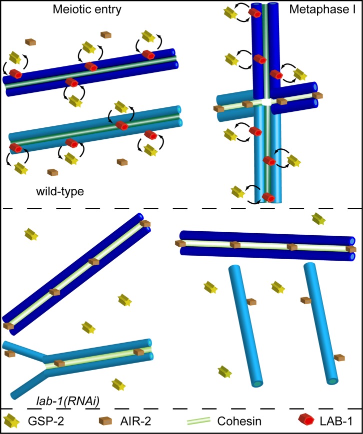
At the onset of the first meiotic division, the protein LAB-1 recruits the PP1 phosphatase to cohesion complexes, preventing Aurora B kinase from targeting cohesins for degradation prematurely and thereby ensuring proper progression of meiotic events in C. elegans.
Abstract
Successful execution of the meiotic program depends on the timely establishment and removal of sister chromatid cohesion. LAB-1 has been proposed to act in the latter by preventing the premature removal of the meiosis-specific cohesin REC-8 at metaphase I in C. elegans, yet the mechanism and scope of LAB-1 function remained unknown. Here we identify an unexpected earlier role for LAB-1 in promoting the establishment of sister chromatid cohesion in prophase I. LAB-1 and REC-8 are both required for the chromosomal association of the cohesin complex subunit SMC-3. Depletion of lab-1 results in partial loss of sister chromatid cohesion in rec-8 and coh-4 coh-3 mutants and further enhanced chromatid dissociation in worms where all three kleisins are mutated. Moreover, lab-1 depletion results in increased Aurora B kinase (AIR-2) signals in early prophase I nuclei, coupled with a parallel decrease in signals for the PP1 homolog, GSP-2. Finally, LAB-1 directly interacts with GSP-1 and GSP-2. We propose that LAB-1 targets the PP1 homologs to the chromatin at the onset of meiosis I, thereby antagonizing AIR-2 and cooperating with the cohesin complex to promote sister chromatid association and normal progression of the meiotic program.
Author Summary
A critical step for achieving successful cell division is the regulation of how the cohesin complexes that bind sister chromatids are initially deposited, then maintained, and finally removed to allow the chromatids to separate into daughter cells. This is particularly challenging during meiosis, when the sister chromatids must remain partially connected to each other through the first division. In organisms that have a single focal centromere on each chromosome, such as mammals and flies, cohesin is protected through the first meiotic division by the protein Shugoshin, which binds the PP2A phosphatase. PP2A counteracts phosphorylation by the Aurora B kinase; if certain cohesins are phosphorylated by Aurora B they become targeted for removal, which allows the chromatids to separate. In the nematode C. elegans, the chromosomes lack a localized centromere and the predicted Shugoshin homolog is not required for protection of cohesins; instead, this function is executed in metaphase of the first meiotic division by the protein LAB-1. But it is not completely understood what leads to the deposition of cohesin prior to entry into meiosis and to its maintenance throughout early meiosis I. In this study, we show that LAB-1 is also required for the loading and maintenance of the cohesin complex. LAB-1 ensures that the chromatids are not separated prematurely, and thus enables the proper progression of events through prophase I of meiosis. We propose that LAB-1 may act at the onset of meiosis in a manner akin to Shugoshin, by recruiting the PP1 phosphatase to counteract Aurora B kinase, thereby ensuring sister chromatid cohesion.
Introduction
Timely establishment and subsequent removal of Sister Chromatid Cohesion (SCC) between sister chromatids is necessary to facilitate faithful segregation of chromosomes in mitosis and meiosis. Failure to correctly segregate chromosomes in either mitosis or meiosis has been associated with tumorigenesis, miscarriages, and congenital defects [1],[2]. Premature loss of SCC prevents the correct bipolar attachment of sister kinetochores to the mitotic spindle, whereas a delay in removing SCC may prevent segregation and therefore result in mitotic arrest and aneuploidy (reviewed in [3],[4]). During meiosis, the control of SCC establishment and removal is even more intricate, and many meiotic processes fail when SCC is compromised. In this specialized cell division program, one cycle of chromosome replication is followed by two consecutive rounds of chromosome segregation, thus reducing the chromosome number by half to produce haploid sperm and oocytes. At the onset of meiosis I, homologous chromosomes pair and undergo synapsis mediated by the formation of a proteinaceous scaffold called the synaptonemal complex (SC). When the SC disassembles, homologs remain attached to each other through chiasmata as a result of earlier crossover recombination events underpinned by flanking SCC. A tight regulation of the establishment of SCC is required for the normal progression of these meiotic events. However, while studies from a number of organisms have revealed key insights into the regulation of SCC removal, far less is known about how the establishment of SCC is regulated.
Cohesin is a highly conserved multisubunit complex that establishes SCC by binding the newly formed chromatid as it is synthesized. The four core subunits of cohesin are the structural maintenance of chromosomes proteins Smc1 and Smc3, the Scc1/Rad21 kleisin, and the accessory protein Scc3 (reviewed in [5],[6]). During meiosis in monocentric organisms such as flies, vertebrates, and yeast, dissolution of chromosome cohesion occurs in a stepwise manner and involves phosphorylation and degradation of Rec8, a meiosis-specific Scc1 paralog. First, cohesin is removed at the chromosome arms during meiosis I, but actively maintained at the pericentromeric regions until anaphase II segregation when the remaining cohesin subset is degraded [7]–[9]. At the end of meiosis I, members of the Shugoshin protein family prevent cohesin removal from the centromeres by recruiting protein phosphatase 2A (PP2A), which counteracts the phosphorylation of Rec8 by Aurora B Kinase, thereby sparing cohesin from separase-mediated degradation [10]–[19].
In C. elegans, the holocentric homolog pairs undergo a single crossover event located at the terminal thirds of chromosomes [20],[21]. Upon chromosome condensation, this off-center crossover leads to the characteristic cruciform shape of the bivalents, which display long and short arms that play an important role for correct alignment on the metaphase I plate [22],[23]. As in monocentric organisms, cohesin is also removed in a two-step process in C. elegans. While REC-8 is lost on the short arm in meiosis I, REC-8 is preserved on the long arm past homolog segregation. This process coincides with the differential loading of the Aurora B Kinase homolog AIR-2, which during diakinesis is observed exclusively on the short arms where it is proposed to phosphorylate REC-8, thus licensing its cleavage by Separase [24],[25]. Recently, two other kleisin homologs, COH-3 and COH-4, were found to also play a part in SCC during C. elegans meiosis, but their specific roles and localization are still unknown [26].
In both mammals and fission yeast two Shugoshin paralogs function in cohesin protection as well as in the spindle assembly checkpoint [14],[19],[27]–[29], yet it was suggested that the latter is the ancestral role of Shugoshin, and that the protection of sister chromatid cohesion evolved as its consequence [27]. So far only a single sequence-predicted Shugoshin homolog was identified in C. elegans, SGO-1, but neither AIR-2 localization nor sister chromatid association are compromised during meiosis I in sgo-1 mutants [16],[30]. Instead, our studies suggested that the worm-specific LAB-1 (Long Arms of the Bivalent) protein participates in protecting REC-8 at the long arms during the metaphase I to anaphase I transition [30]. LAB-1 progressively forms continuous tracks throughout the full length of chromosomes starting at the onset of meiosis and co-localizes with the synaptonemal complex at pachytene [30]. During chromosome remodeling, LAB-1 becomes restricted to the long arms of the bivalents and, like Shugoshin in monocentric species, is finally removed from chromosomes in early anaphase I. lab-1 hypomorphic mutants show a spreading of AIR-2 signals to both arms of the bivalents, similar to mutants of the protein phosphatase 1 (PP1) homolog gsp-2. These results are consistent with a model in which LAB-1 impacts sister chromatid cohesion in late meiosis I by regulating cohesin phosphorylation. Based on these and other observations, as well as the lack of evidence for a direct role of Shugoshin in protecting meiotic cohesin in C. elegans, we have speculated that due to the holocentric nature of C. elegans chromosomes, Shugoshin maintained its roles in the spindle attachment checkpoint, but LAB-1 evolved as part of a process to protect cohesin during meiosis in this organism [30]. However, both how and when PP1 function is directed and regulated remained open questions.
Here we describe an earlier and distinct role for LAB-1 in the establishment of SCC via PP1 regulation. Moreover, we demonstrate how failing to properly establish SCC influences various downstream meiotic events. Depletion of lab-1 by RNAi reduces SCC and consequently impairs homolog pairing, alters the progression of meiotic recombination, and results in an increase in recombination intermediates (MSH-5 and ZHP-3 foci). We found that LAB-1, together with REC-8, is required for proper loading of SMC-3 and consequently for proper SC polymerization, which requires normal axis morphogenesis. While the different cohesin members and LAB-1 show some degree of interdependence with respect to either their initial localization or the subsequent maintenance of their localization on chromosomes, LAB-1 can promote partial SCC even in the absence of all three SCC-1 meiotic paralogs. Finally, underscoring a role in the regulation of phosphorylation, LAB-1 directly interacts with GSP-1 and its paralog GSP-2. Moreover, depletion of lab-1 results in reduced GSP-2 and increased AIR-2 signals in early meiotic nuclei. We propose that LAB-1 specifically targets PP1 to chromosomes in early meiotic stages to antagonize AIR-2 phosphorylation and to promote sister chromatid cohesion, thus supporting the normal progression of downstream meiotic events.
Results
LAB-1 Depletion Reduces Pairing and Sister Chromatid Cohesion
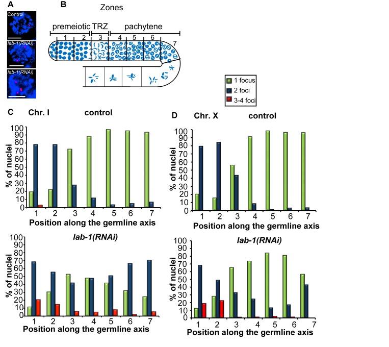
Our analysis of lab-1(RNAi) worms revealed the presence of >12 DAPI-stained bodies in 1.1% (n = 88) of oocytes at diakinesis, compared to the six DAPI-stained bodies corresponding to the six pairs of attached homologous chromosomes observed in wild type or the 7 to 12 univalents indicative of lack of chiasmata, suggesting instead a defect in sister chromatid cohesion (this study and [30]). To determine whether this defect results from a role for lab-1 in early meiosis, we examined the effects of lab-1 depletion on the various processes that take place earlier during prophase I (Figure S1). We first examined homologous chromosome pairing, a process that occurs upon entry into meiosis in most organisms (reviewed in [31]–[34]). To follow the progression of pairing in the germline, we used fluorescence in situ hybridization (FISH) with probes labeling the pairing center end of chromosome I. FISH signals either ≤0.75 µM or >0.75 µM apart represent paired and unpaired homologs, respectively (Figure 1A). In control gonads, homologous chromosomes are unpaired prior to meiotic entry and therefore only a few nuclei (n = 17/81) show a single FISH signal (Figure 1B and 1C, zones 1 and 2). Upon entry into meiotic prophase I (transition zone, which corresponds to the leptotene and zygotene stages; zone 3), the frequency of nuclei carrying a single focus increases (n = 52/72), and by pachytene, pairing is completed (Figure 1C, zones 4–7). In contrast, in lab-1-depleted gonads, pairing levels were reduced. Only 52% (n = 44/84) of nuclei in the transition zone carried paired homologs (Figure 1C, zones 3), and this was further decreased until only 24% (n = 9/37) of nuclei were observed with paired homologs at late pachytene (Figure 1C, zone 7, p<0.05, by the two-sided Fisher's Exact Test, 95% C.I.). Using a probe targeting the X chromosome pairing center revealed similar, albeit milder, results (Figure 1D). Although the reason for a stronger impairment of pairing on the autosomes compared to the X chromosome remains unclear, this has also been observed in other meiotic mutants [35]–[37]. Thus, depleting lab-1 reduces homolog pairing, suggesting a previously unknown function for LAB-1 during prophase I.
Figure 1. LAB-1 is required for homologous pairing.
(A) High-magnification images of DAPI stained mid-pachytene nuclei (blue) hybridized with FISH probes targeting the pairing center region on chromosome I (red). Nuclei with either one, two, or three signals (foci) are depicted. Bars, 3 µM. (B) Diagram of a C. elegans germline indicating the position of the zones scored in the analysis of the progression of homologous pairing. (C, D) Graphs depicting the percentage of nuclei showing one, two, and three to four foci within each zone in control and lab-1(RNAi) gonads hybridized with FISH probes recognizing the chromosome I pairing center (C) and the X chromosome pairing center (D).
In 20% (14/69) of premeiotic nuclei from lab-1-depleted gonads, we also noticed the presence of three or four FISH signals (Figure 1A), consistent with a defect in sister chromatid association (Figure 1C). At later stages, the frequency of nuclei with 3–4 signals decreased in lab-1(RNAi) gonads (5%, n = 2/37, at late pachytene). This temporal reduction could be explained either by residual LAB-1 or by a LAB-1-independent mechanism. Taken together, these observations suggest that LAB-1 may affect homologous pairing due to its role in the early establishment and/or maintenance of SCC.
Meiotic DNA Double-Strand Break Repair Progression Requires LAB-1
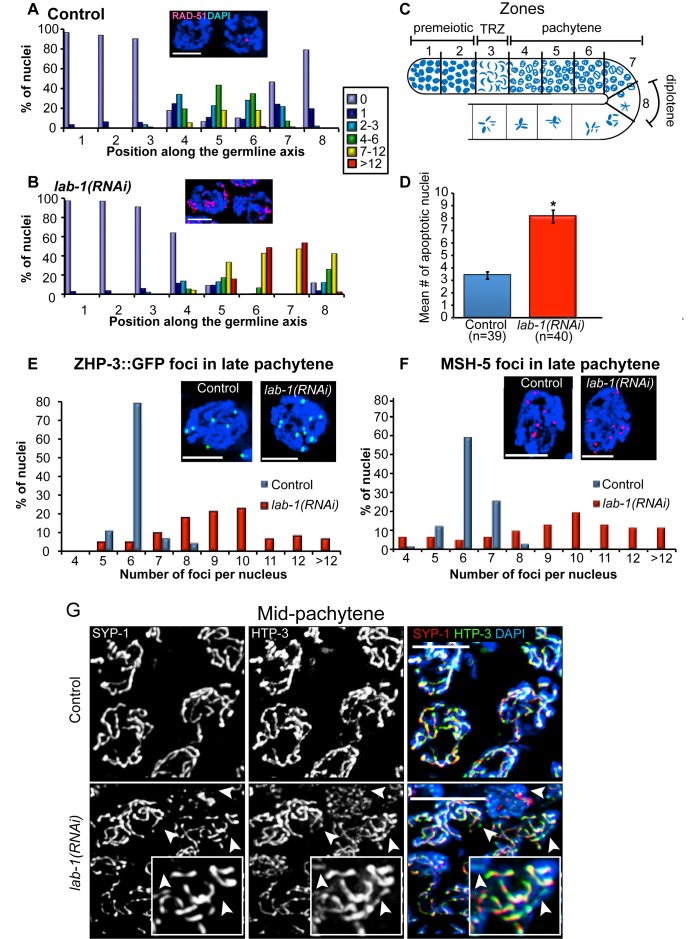
To examine whether lab-1 depletion affects the progression of meiotic DNA double-strand break repair (DSBR), we utilized an antibody against RAD-51, a protein involved in strand invasion/exchange during DSBR [38]. In control gonads, the levels of RAD-51 foci peaked in early/mid-pachytene (zone 5), and progressively decreased in later stages (Figure 2A and 2C). In contrast, levels of RAD-51 foci were elevated throughout mid to late pachytene (zones 5–7) and persisted in 88% (n = 95) of early diplotene (zone 8) nuclei in lab-1(RNAi) gonads compared to only 21% (n = 97) of nuclei in control gonads (Figure 2B and Figure S2). The elevated levels of RAD-51 foci observed in lab-1(RNAi) gonads depend on the formation of meiotic DSBs by SPO-11 and are therefore indicative of a meiosis-specific DSBR defect (Figure S3). These results can be explained by either a delay in meiotic DSBR or an increase in the levels of DSB formation upon lab-1 depletion. Consistent with the interpretation that nuclei in lab-1(RNAi) germlines contain unrepaired recombination intermediates, we observed a CEP-1/p53-dependent 2- to 3-fold increase in germ cell apoptosis in lab-1(RNAi) gonads compared to control, suggesting the activation of a late pachytene DNA damage checkpoint (p<0.0001 by the two-tailed Mann-Whitney test, 95% C.I.; Figure 2D and Figure S4). These results show that depletion of lab-1 perturbs normal meiotic DSBR.
Figure 2. LAB-1 is required for double-strand break repair progression, crossover control, and complete synapsis.
(A–B) Histograms depict the quantification of RAD-51 foci in (A) control and (B) lab-1(RNAi) germlines. The number of RAD-51 foci per nucleus is categorized according to the color code shown on the right. The percent of nuclei observed for each category (y-axis) is depicted for each zone along the germline axis (x-axis). Insets represent examples of late pachytene nuclei co-stained with DAPI (blue) and RAD-51 (red). Bars, 3 µM. (C) Germline diagram indicates the eight zones throughout which RAD-51 foci were scored for all nuclei. (D) Quantification of germline apoptosis. Error bars represent standard deviation of the mean. Asterisk indicates statistically significant increase in the number of apoptotic nuclei (p<0.0001, by the two-tailed Mann-Whitney test, 95% C.I.). n, number of gonad arms scored. (E–F) Histograms depict the quantification of (E) ZHP-3::GFP and (F) MSH-5 foci in late pachytene nuclei of control and lab-1(RNAi) germlines. Between 63 and 75 nuclei were scored from 5 to 7 gonads for each genotype. Insets depict representative nuclei (DAPI, blue; ZHP-3::GFP, green; and MSH-5, red). Bars, 3 µM. (G) Mid-pachytene nuclei in control and lab-1(RNAi) gonads co-stained with SYP-1 (red), HTP-3 (green), and DAPI (blue). Arrowheads indicate HTP-3-stained tracks that lack SYP-1 signal. Bars, 4 µM.
Lack of LAB-1 Alters the Number of ZHP-3- and MSH-5-Marked Crossover Recombination Sites
Meiotic crossovers are tightly regulated such that at least one crossover always forms between homologs while additional crossovers nearby are discouraged [39]. Due to the decreased levels of homologous pairing observed, we hypothesized that following lab-1 depletion, crossover levels would also be reduced. To highlight crossover precursor sites, we used a ZHP-3::GFP transgene [40],[41]. In C. elegans, six ZHP-3::GFP foci are observed in >78% of late pachytene nuclei, correlating with the expected one crossover event per bivalent (Figure 2E). Surprisingly, we observed a mean of 9.2 foci per nucleus in lab-1(RNAi) pachytene nuclei (n = 63, p<0.0001 by the two-tailed Mann-Whitney test, 95% C.I), and 4/63 had >12 foci (Figure 2E). To verify that these foci represent recombination events, we immunostained both control and lab-1(RNAi) gonads with an antibody that recognizes MSH-5, a conserved meiosis-specific protein required for crossover formation [42]–[45]. We found that the pattern of distribution of MSH-5 foci was almost identical to that of ZHP-3 (Figure 2F). These results suggest that lab-1 depletion may disrupt crossover control and lead to more crossover events. Utilizing a similar cytological approach to that in Rosu et al. [46] we did not observe any bivalents with two or more chiasmata in diakinesis oocytes from lab-1-depleted gonads (n = 0/88; Figure S5). This outcome, coupled with the significant reduction in homologous pairing observed in these gonads, suggests that some of these recombination events may represent crossovers between sister chromatids as opposed to between homologs.
Formation of the Central Region of the Synaptonemal Complex Is LAB-1-Dependent
The SC plays an essential role in promoting the maturation of DSBs into crossovers [31],[33],[34]. Therefore, we examined whether impaired chromosome synapsis might account for the altered DSBR progression observed in lab-1(RNAi) gonads. Specifically, we examined whether LAB-1 is required for the localization of SYP-1, a central region component of the SC. In wild type pachytene nuclei, SYP-1 is localized throughout the full length of thick DAPI-stained tracks representing paired and aligned homologous chromosomes (Figure 2G) [47]. In the lab-1-depleted gonads, SYP-1 signal was observed associating with chromosomes with wild type kinetics upon entry into meiosis. However, SYP-1 was not detected along the full length of DAPI-stained chromosomes in many nuclei at pachytene, as exemplified by co-staining with HTP-3, an axial element component [48] that is observed continuously throughout chromosome axes (Figure 2G). In contrast, LAB-1 is still observed localizing throughout chromosomes in syp-1 mutants (Figure S6B). This suggests that while assembly of lateral element components of the SC is apparently LAB-1-independent at this level of cytological observation, assembly of the central region components of the SC requires LAB-1 function.
LAB-1 Localization Depends on the Cohesin Complex
FISH analysis and the number of DAPI-stained bodies (>12) in diakinesis oocytes suggested that the impaired DSBR progression and chromosome synapsis observed in lab-1(RNAi) gonads may be due to an earlier role of LAB-1 in sister chromatid cohesion. To determine whether LAB-1 executes this role through interactions with the cohesin complex, we first examined if LAB-1 localization depends on cohesin. Depletion of the cohesin member smc-3 resulted in an overall decrease in LAB-1 signal throughout prophase I nuclei (Figure 3). In late pachytene nuclei, only short tracks of LAB-1 were observed on the chromosomes, and LAB-1 was detected on univalents at diakinesis (Figure 3). A similar pattern was observed following depletion of the cohesin complex subunit scc-3 (Figure S7).
Figure 3. LAB-1 localization is dependent on the cohesin complex.
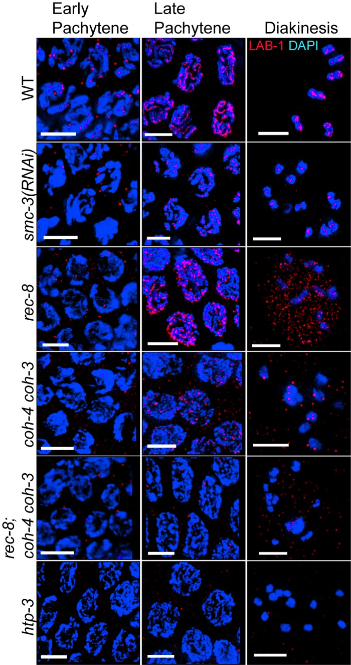
LAB-1 (red) and DAPI (blue) staining of early pachytene, late pachytene, and −1 oocytes at diakinesis in the germlines of the indicated genotypes. Bars, 4 µM.
We also examined whether meiosis-specific cohesin subunits were required for LAB-1 localization. In addition to REC-8, two other kleisins, COH-3 and COH-4, mediate meiotic sister chromatid cohesion in C. elegans [26],[49]. LAB-1 chromosomal localization was delayed in rec-8(ok978), but improved as nuclei proceeded through pachytene, and by late pachytene a mixture of both long and discontinuous tracks of LAB-1 were present throughout chromosomes (Figure 3). Interestingly, LAB-1 signals were no longer associated with chromosomes in 40% (n = 15/37) of −1 oocytes at diakinesis, and instead were distributed diffusely throughout the nuclei of rec-8 mutants (Figure 3). This is reminiscent of the early loss of chromosome-associated REC-8 signal observed in the lab-1 hypomorphs on metaphase I, suggesting some degree of interdependence between these proteins [30].
In coh-4(tm1857) coh-3(gk112) double mutants, LAB-1 associated with the chromosomes at early pachytene, but failed to form tracks (Figure 3). When all three meiotic kleisins were mutated, LAB-1 localization was further impaired as only very few and faint LAB-1 foci were detected on either pachytene or diakinesis chromosomes (Figure 3). This effect on LAB-1 localization was specific to members of the cohesin family, as we found no change in LAB-1 localization in mutants for either smc-5, which is a structural maintenance of chromosomes family member, but not part of the cohesin complex, or hcp-6, a gene encoding a member of the condensin II complex, which is structurally similar to cohesin (unpublished data) [50]. These results suggest that LAB-1 recruitment to the chromosomes depends on the cohesin complex, and that its association with meiotic chromosomes only completely fails when all three kleisins are absent.
LAB-1 Forms a Complex with Axial Element Proteins
To gain further insight into the function and regulation of LAB-1, we set out to identify the proteins interacting with LAB-1. Utilizing a transgenic line expressing GFP-LAB-1 and GFP antibodies for mass spectrometry analysis, we identified 19 proteins that were specifically co-immunoprecipitated from whole worm lysates with GFP-LAB-1, but not unrelated controls (Table S1). Prevalent among these were the axial element proteins HIM-3 (56.4%), HTP-1 (43.2% coverage), HTP-2 (38.9% coverage), and HTP-3 (15.8% coverage). All four proteins carry a HORMA domain that is also present in proteins involved in DSBR, synapsis, and mitotic spindle checkpoints from yeast to mammals [31],[51]–[53]. Importantly, HTP-1 is the only other known long arm-specific protein in C. elegans and HTP-3 was shown to be critical for meiotic sister chromatid cohesion, as multiple cohesin members fail to load onto chromosomes in htp-3(y428) mutants [26],[48],[54].
To assess the functional relevance of this set of interactions we examined the interdependency between the localization of LAB-1 and the HTP proteins. The localization of HTP-1, HTP-2, and HTP-3 in early prophase I was not altered in lab-1(RNAi) gonads compared to control (Figure 2G, Figure S6A, and Figure S6C). A reciprocal analysis revealed that LAB-1 localization is indistinguishable from wild type in htp-1 gonads (Figure S6B). In contrast, LAB-1 signals were not observed associated with chromosomes in htp-3 mutant germlines at any meiotic stage (Figure 3 and Figure S6D). Therefore, our analysis suggests that LAB-1 forms a complex with the HORMA domain proteins, and that HTP-3 is required for LAB-1 localization either directly or through its role in cohesin loading.
LAB-1 and REC-8 Cooperate to Load SMC-3 and Promote SC Formation
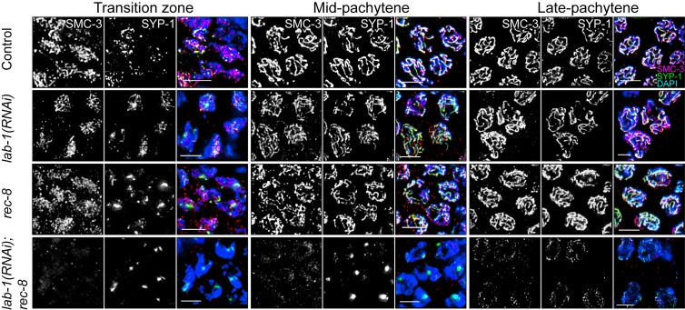
Our findings that lab-1 depletion results in reduced sister chromatid cohesion, and that the localization of both LAB-1 and REC-8 are partially co-dependent in late meiosis I, raise the question of whether LAB-1 could be involved in cohesin complex localization during early prophase I. Immunolocalization of either SMC-3 or REC-8 showed no differences between wild type and lab-1-depleted gonads (Figure 4, Figure S8, and [55]). Moreover, SMC-3 localization was indistinguishable from wild type in rec-8 mutants (Figure 4 and [26]). Therefore, we reasoned that as both REC-8 and LAB-1 are required for normal sister chromatid cohesion, yet seem to be dispensable for SMC-3 localization, they might work in parallel. Indeed, in lab-1(RNAi); rec-8 worms, SMC-3 loading was significantly impaired and, as expected due to abrogation of cohesin loading, the SC central region protein SYP-1 was restricted to mostly a single large aggregate per nucleus in mid-pachytene and few long tracks in late pachytene nuclei (Figure 4). Therefore, REC-8 and LAB-1 work in parallel to enable the loading of SMC-3 and facilitate SC formation.
Figure 4. Both LAB-1 and REC-8 are required for SMC-3 and SYP-1 localization onto chromosomes.
Transition zone, mid-pachytene, and late pachytene nuclei in the germlines of the indicated genotypes co-stained with SMC-3 (red), SYP-1 (green), and DAPI (blue). Bars, 4 µM.
LAB-1 Cooperates with REC-8, COH-4, and COH-3 to Ensure Sister Chromatid Cohesion
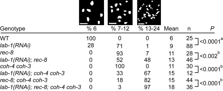
If LAB-1 and REC-8 cooperate in SMC-3 loading during meiosis, then lack of both should increase the premature loss of sister chromatid cohesion detected at diakinesis. Indeed, the number of diakinesis oocytes carrying 13–24 DAPI stained bodies is significantly increased in lab-1(RNAi); rec-8 gonads compared with either lab-1(RNAi) or rec-8 (48%, 1%, and 7%, n = 46, 88, and 28, respectively). Nevertheless, many chromatids were still held together in lab-1(RNAi); rec-8 as demonstrated by the 52% of oocytes that had 7–12 DAPI stained bodies (average 13.1±2.5; Figure 5). This could be explained by either residual LAB-1 that was not depleted or by other factors that contribute to sister chromatid cohesion independently. To examine whether the other two meiotic kleisins contribute to sister chromatid cohesion in parallel with lab-1, we looked at the effects of lab-1 depletion on coh-4 coh-3 double mutants. 7–12 DAPI stained bodies are observed in the diakinesis oocytes of coh-4 coh-3 double mutants (Figure 5 and [26]), suggesting that sister chromatids are still held together. However, when we depleted lab-1 in these worms, the average number of DAPI-stained bodies increased from 11±1 to 15±3 (Figure 5, n = 30 and 12, respectively, p<0.0001 by the two-tailed Mann-Whitney test, 95% C.I). These results suggest that LAB-1 affects both REC-8 and COH-3/COH-4 cohesin complexes. Moreover, the role of LAB-1 in SCC may be greater than suggested by these results, since this analysis relied on RNAi depletion and therefore residual LAB-1 activity cannot be ruled out.
Figure 5. lab-1 depletion weakens sister chromatid cohesion.
Number of DAPI-stained bodies in the −1 oocyte at diakinesis in the indicated genotypes. The p values were calculated by (a) the Wilcoxon signed-rank test and by (b) the two-tailed Mann-Whitney test, 95% C.I. Bars, 4 µM.
Two models for lab-1 contribution to sister chromatid cohesion can be envisioned from these results. In one, lab-1 ensures sister chromatid cohesion solely through rec-8, coh-4, and coh-3. Alternatively, lab-1 can contribute to sister chromatid cohesion even in their absence. To distinguish between these two possibilities, we examined the effect of lab-1 depletion when all three meiotic kleisins are mutated. Similar to previous observations [26], we found that most of the oocytes at diakinesis in the rec-8;coh-4 coh-3 worms had 13–24 DAPI stained bodies (Figure 5). However, the average number of bodies was only 15±3, indicating that sister chromatid cohesion was not completely lost. When lab-1 was depleted in these worms, the number of DAPI-stained bodies increased to 18±3, and 97% had 13–24 bodies (Figure 5, n = 44 and 36 for control and lab-1(RNAi), respectively, p<0.0001 by the two-tailed Mann-Whitney test, 95% C.I.). This result suggests that lab-1 acts to promote sister chromatid cohesion in parallel with rec-8, coh-4, and coh-3.
LAB-1 Directly Interacts with the PP1 Homologs
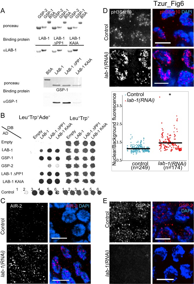
We have previously hypothesized that LAB-1 targets the PP1 homologs GSP-1 and GSP-2 to the long arms of the bivalents, thereby antagonizing AIR-2 localization to that region [30]. To test whether LAB-1 can directly interact with either PP1 homolog we utilized the far-western assay [56]. Bacterially expressed and purified GSP-1 and GSP-2 proteins transferred to nitrocellulose membranes bound purified LAB-1 (Figure 6A). Reciprocally, LAB-1 transferred to membranes bound GSP-1 (Figure 6A). These results suggest that LAB-1 binds to the PP1 homologs in vitro.
Figure 6. LAB-1 binds GSP-1 and GSP-2 and is required for correct AIR-2 and GSP-2 localization on early meiotic nuclei.
(A) Far-western analysis of in vitro binding of purified recombinant LAB-1, LAB-1 PP1 putative motif mutants, GSP-1, and GSP-2. Purified bacterially expressed proteins were transferred to membranes, incubated with the indicated binding protein, and probed with appropriate antibodies. BSA was used as control. (B) The yeast two-hybrid system was used to test the protein interactions between LAB-1, LAB-1 PP1 putative motif mutants, GSP-1, and GSP-2. Proteins were fused to either the DNA binding domain (DB) or the activation domain (AD) of GAL4. Interactions were scored by growth on SC-Leu-Trp-Ade plates. Growth on SC-Leu-Trp was used as a control. Negative (No. 1) and positive (No. 2–6) controls are shown. Positive interactions are shaded in gray. (C) Transition zone nuclei in control and lab-1(RNAi) gonads co-stained with AIR-2 (red) and DAPI (blue). (D) Transition zone nuclei in control and lab-1(RNAi) gonads co-stained with H3S10ph (red) and DAPI (blue). A quantification plot is presented below where each dot represents the ratio between the level of fluorescence detected for an individual nucleus and that of the adjacent background. Error bars represent standard deviation of the mean. Asterisk indicates statistically significant increase in fluorescence (p<0.0001, by the two-tailed Mann-Whitney test, 95% C.I.). (E) Transition zone nuclei in control and lab-1(RNAi) gonads co-stained with GSP-2 (red) and DAPI (blue). Bars, 4 µM.
A highly degenerate motif [(R/K) (V/I) X (F/W)] has been previously associated with the binding, localization, and function of PP1 phosphatases [57]–[59]. To test if the putative PP1 binding motif present in LAB-1 [30] is required for the in vitro interaction detected between LAB-1 and the PP1 homologs, we used purified LAB-1 protein either lacking the motif (ΔPP1) or carrying two alanine substitutions in this motif (KAIA). GSP-1 and GSP-2 proteins transferred to membranes were still able to bind both mutant LAB-1 proteins, and reciprocally, membrane-bound mutant LAB-1 proteins could bind GSP-1 (Figure 6A). Thus, the putative PP1 binding motif is probably not necessary for LAB-1 binding to GSP-1 and GSP-2.
To verify the interaction between LAB-1 and the PP1 homologs we utilized the yeast two-hybrid system. Full-length GSP-1 fused to the activation domain of GAL4 interacted with LAB-1 as well as with the LAB-1 PP1 mutants fused to the DNA binding domain of GAL4 (Figure 6B). Interestingly, GSP-2 could only weakly interact with the LAB-1 PP1 mutants but not with wild type LAB-1 (Figure 6B). It is possible that LAB-1 binding to GSP-2 is masked in yeast cells by the host's endogenous PP1 homologs, and only when these unrelated interactions are removed, LAB-1 and GSP-2 interaction can be detected. Taken together, these results support a direct interaction between LAB-1 and the PP1 homologs.
LAB-1 Is Required to Target GSP-2 to Early Meiotic Nuclei and Restrict AIR-2
The mislocalization of AIR-2 to the long arms of the bivalents during diakinesis in lab-1 hypomorph mutants [30] prompted us to test whether depletion of lab-1 by RNAi results in changes in AIR-2 localization during early meiotic stages as well. Indeed, unlike in most control gonads (n = 45/53), in which AIR-2 signal was not observed in transition zone nuclei, clear AIR-2 patches were observed in most transition zone nuclei upon lab-1 depletion (n = 16/30) (Figure 6C, p<0.0005, by the two-sided Fisher's Exact Test, 95% C.I.). Moreover, consistent with AIR-2 localization in early prophase I, increased histone H3 phosphorylation, a well-characterized chromosomal target for Aurora B was also observed at that stage (Figure 6D, mean relative fluorescence = 1.2, n = 284, and 1.5, n = 175, for control and lab-1(RNAi), respectively; p<0.0001 by the two-tailed Mann-Whitney test, 95% C.I.) [24],[60]. Since LAB-1 binds the PP1 homologs and was suggested to restrict AIR-2 through GSP-2 in metaphase I [30], we tested if depletion of lab-1 also changes GSP-2 localization in early meiotic nuclei. In control gonads, GSP-2 is found in foci throughout transition zone nuclei and the syncytial gonad (n = 12/12) (Figure 6E and Figure S9). However, in most lab-1 depleted gonads (n = 7/12), the level of nuclei-associated GSP-2 foci was reduced (Figure 6E and Figure S10, p<0.05, by the two-sided Fisher's Exact Test, 95% C.I.). These results suggest that LAB-1 directly targets GSP-2 to the chromatin during meiotic onset, thus restricting aberrant AIR-2 accumulation at that stage and promoting normal establishment and maintenance of sister chromatid cohesion.
Discussion
LAB-1 Is Required for Sister Chromatid Cohesion
The dynamic nature of chromosome interactions during the cell cycle requires the ability to establish, mobilize, and remove SCC. Since the discovery of the core components of the cohesin complex, a growing number of proteins have been found to take part in all aspects of cohesin function: proteins involved in loading cohesin, maintaining its binding, and removing it from chromosomes. The importance of SCC is highlighted in meiosis, due to the intricacy and complexity of this process. During prophase I, homologous chromosomes pair, synapse, undergo programmed meiotic DSBs and recombine [32]. Thus, while a small reduction in chromatid cohesion may still provide for normal mitotic segregation, it would be unable to support the structural requirements for the meiotic processes to proceed. For example, in the yeast smc3-42 temperature-sensitive mutant, both SC and crossover formation are perturbed and cells fail to undergo any meiotic division, even in the mitotic permissive temperature [61]. In C. elegans, lack or depletion of either cohesin members or their interactors have also been shown to alter pairing, synapsis, DSBR, and accurate chromosome segregation [26],[55],[62]–[64]. Another level of cohesin control must be employed in meiosis due to the requirement for protection of cohesin at specific chromosomal regions, namely at centromeres in the case of monocentric organisms and along the long arms of the bivalents in the case of the holocentric C. elegans chromosomes. Along the long arms, cohesin must be preserved during the first meiotic division, while in all other parts of the chromosomes it must be removed [6],[29],[65]. Without this protection, faithful chromatid segregation cannot take place during the second meiotic division. It is therefore not surprising that various different components are involved in regulating SCC. However, how these proteins modulate the way SCC is either enforced or relieved, and how these two processes are coordinated, is not completely understood.
Most of the factors implicated in SCC depend on the cohesin complex, yet some reports have also suggested cohesin-independent pathways [66]–[68]. Although LAB-1 can maintain some degree of SCC even in the absence of the meiotic kleisins, most of our data support a cohesin-dependent mechanism. Here we show that LAB-1 and different cohesin members are partially interdependent in their localization throughout prophase I and at metaphase I. The cooperation between LAB-1 and cohesin to ensure SCC can be observed cytologically through the number of DAPI-stained bodies at diakinesis when meiotic kleisins are mutated. Mutations in either rec-8 or coh-4 coh-3 do not result in precocious sister separation in most nuclei, yet depletion of lab-1 in those mutants significantly increases the frequency of unbound sister chromatids at diakinesis. These results are consistent with LAB-1 acting to maintain cohesion in cooperation with the cohesin complex. Yet it is possible that LAB-1 can promote SCC in a pathway that does not require the meiotic kleisins, since we observed a significant increase in sister chromatid separation when we depleted lab-1 in the rec-8, coh-3, and coh-4 triple mutant. The factors taking part in this pathway remain to be uncovered. Interestingly, our preliminary analysis did not reveal increased loss of sister chromatid cohesion following lab-1 depletion in either scc-1 or smc-3 depleted backgrounds (Y.B.T. and M.P.C. unpublished results). However, the lack of fully separated chromatids in the rec-8; coh-4 coh-3 triple mutant could be due to mechanisms involving yet other kleisins and/or the formation of tangles between sister chromatids.
Yan and colleagues have recently reported the finding of sisters on the loose (SOLO), a protein that together with stromalin on meiosis (snm) is required for centromere cohesion and SMC1 localization during Drosophila male meiosis [69]. We suggest that LAB-1 plays a similar role, in that it is required to both appropriately load and maintain the cohesin complexes on specific chromosomal subdomains during pre-meiotic S phase and prophase I, respectively, in C. elegans, thereby enabling the meiotic program. In this context (Figure S11), LAB-1 acts to properly load and maintain the association of cohesin complexes along the chromosome axes. Therefore, the meiotic defects observed in lab-1(RNAi) gonads are diagnostic of problems in SCC. First, lack of LAB-1 perturbs homolog paring. In this state, interhomolog repair is impaired, DSBR progression is altered and many nuclei undergo apoptosis. Second, nuclei that are not eliminated by apoptosis contain univalents as well as single chromatids at diakinesis. Finally, in the bivalents where SCC is not lost in early prophase I, reduced LAB-1 on the long arm is ultimately insufficient to prevent unchecked AIR-2 loading on all chromosome axes [30]. Upon metaphase I entry, REC-8 removal occurs at both short and long arms and accurate homolog segregation fails (Figure S11).
How does LAB-1 affect SCC? Our finding that LAB-1 and HTP-3 are present in the same complex raises the possibility that LAB-1 acts to maintain SCC via HTP-3-mediated control of cohesin loading [26]. Alternatively, HTP-3 may target LAB-1 either directly or indirectly to chromosome axes, and LAB-1 in turn recruits other factors that maintain SCC. The presence of other axial element proteins in LAB-1 immunoprecipitates as well as our finding that LAB-1 directly interacts with the PP1 homologs lead us to favor the latter.
We found that in the yeast two-hybrid system GSP-2 could only weakly interact with the LAB-1 protein carrying mutations in the putative PP1 binding motif, leading us to hypothesize that GSP-2 is a weaker binding partner of LAB-1 than GSP-1. Weak or transient binding is probably the reason why we did not detect GSP-1 and GSP-2 in our LAB-1 IPs, which were done using stringent conditions. Nevertheless, lab-1 depletion affected the localization of GSP-2, suggesting that LAB-1 indeed targets GSP-2 to early meiotic nuclei.
Early Versus Late Roles for LAB-1 During Meiosis I
LAB-1 localization in the germline is highly dynamic. We propose that at the onset of meiosis, the chromosomal association of LAB-1 opposes cohesin-removing proteins whose levels gradually increase during pachytene. Indeed, AIR-2 signal is only first observed in late pachytene in wild-type C. elegans [23], whereas it accumulates in transition zone nuclei following lab-1 depletion (this work). Given that AIR-2 promotes cohesin removal during mitotic prophase [24],[70]–[74], we suggest that a role for LAB-1 is to restrict AIR-2's ability to remove cohesin during early meiosis. According to this model, the system that protects cohesin during metaphase I, and involves LAB-1 antagonizing AIR-2 probably via the PP1s along the long arms of the bivalents, also operates throughout the early stages of meiosis to maintain SCC (Figure 7). This may be achieved by targeting the PP1s to chromosome axes and restricting AIR-2. In late prophase I, LAB-1 is lost from part of the chromosomes, permitting the removal of cohesin at these subdomains.
Figure 7. LAB-1 utilizes a similar mechanism to protect SCC during two different meiotic stages.
In wild type, LAB-1 starts associating with the chromosomes at the entry into meiosis and transiently targets GSP-2 to the chromosomes. This targeting antagonizes AIR-2 and maintains SCC. During diakinesis, LAB-1 localizes to the long arms, where it specifically antagonizes AIR-2 and protects REC-8 from premature removal. When lab-1 is depleted, AIR-2 associates with the chromosomes as early as transition zone, and SCC is perturbed. The weakened SCC prevents successful binding of homologs and many reach diakinesis as either univalents or detached chromatids.
In this article we show that LAB-1 plays an important role in protecting sister chromatid cohesion by localizing a phosphatase (PP1 by LAB-1 instead of PP2A by Shugoshin) and antagonizing Aurora B phosphorylation activity. Thus, our studies have revealed key conserved principles that guide proper regulation of meiotic sister chromatid cohesion. The use of PP1 to maintain and protect cohesin, while possibly the result of a necessary co-evolution with the holocentric nature of C. elegans chromosomes, may also be required for monocentric organisms. Similar to LAB-1, Sgo2 was found to maintain cohesion in mouse bivalents during late prophase I [75]. This raises the possibility that Shugoshin proteins in other metazoans may also play a role earlier in meiosis in establishing and/or maintaining sister chromatid cohesion. In support of this possibility, it was recently shown that PP2A has early meiotic roles, in addition to its role of protecting centromeric cohesin during metaphase I [76]. Therefore, the use of mouse meiotic conditional alleles may aid in assessing the effect of these proteins in early prophase I.
In conclusion, we have shown that LAB-1 has an earlier role in regulating the establishment and maintenance of SCC. Thus, LAB-1 emerges as a central protein in the regulation of SCC, exerting this role from the start of prophase I through homolog segregation at the metaphase I to anaphase I transition.
Materials and Methods
Strains and Alleles
The N2 Bristol strain was used as the wild-type background. C. elegans strains were cultured at 20 °C under standard conditions as described in [77]. The following mutations were used: LGI: htp-3(tm3655), cep-1(lg12501), lab-1(tm1791), LGIV: htp-1(gk174), rec-8(ok978), spo-11(ok79), LGV: coh-4(tm1857), coh-3(gk112), and syp-1(me17) ([26],[30],[35],[47],[78]–[80]). The GFP::LAB-1::HA line has been previously described in [30].
RNAi
Feeding RNAi experiments were performed at either 20 °C (for smc-3 and scc-3) or 25 °C (for lab-1) as described in [30],[63],[81]. Control RNAi was performed by feeding HT115 bacteria carrying the empty pL4440 vector. A feeding vector from the ORFeome RNAi collection [82] was used for smc-3 RNAi experiments. Successful depletions were verified at every single experiment by immunostaining with either LAB-1 or SMC-3 antibodies for lab-1(RNAi) and smc-3(RNAi), respectively, and by RT-PCR for scc-3(RNAi).
RT-PCR
cDNA was produced from single-worm RNA extracts using the Thermoscript RT-PCR system (Invitrogen). The effectiveness of scc-3 RNAi was determined by assaying the expression of the scc-3 transcript in at least four individual animals subjected to RNAi. Expression of gpd-1 (T09F3.3) was used as a control.
Immunostaining, DAPI Analysis, and FISH
Whole mount preparation of dissected gonads, DAPI staining, immunostaining, and analysis of germline nuclei were carried out as in [41],[83]. A rabbit polyclonal antibody against a C-terminal peptide of C. elegans GSP-2 (TPPRNAPAAQPKKGAKK) was generated by Sigma-Genosys. The antiserum was affinity-purified against the original peptide-antigen as described in [84]. Primary antibodies were used at the following dilutions: α-LAB-1 (1∶300; [30]), α-REC-8 (1∶50; Abcam), α-Histone H3 phospho-Ser10 (1∶300; Upstate Biotechnologies), α-HTP-3 (1∶100; [48]), α-HTP-1/2 (1∶200; [54]), α-SYP-1 (1∶200 [47]), α-RAD-51 (1∶100; [83]), α-SMC-3 (1∶500; Chemicom), α-MSH-5 (1∶100000; SDI), α-AIR-2 (1∶100 [30]), and α-GSP-2 (1∶100). The secondary antibodies used were: Cy3 α-rabbit, Cy3 α-rat, Cy3 α-mouse, FITC α-rabbit, and FITC α-guinea pig (Jackson Immunochemicals, 1∶200).
FISH was performed as in [37] utilizing a probe recognizing the left end of the X chromosome, derived from YAC Y51E2, and a probe recognizing the right end of chromosome I derived from pooled cosmids F32A7 and F14B11, prepared as in [36].
Imaging, Microscopy, and Mass Spectrometry
Immunofluorescence images were collected at 0.2 µm increments with an IX-70 microscope (Olympus) and a cooled CCD camera (model CH350; Roper Scientific) controlled by the DeltaVision system (Applied Precision). Images were subjected to deconvolution analysis using the SoftWorx 3.0 program (Applied Precision) as in [23]. For germ cell apoptosis, worms were transferred onto a drop of M9 on 1.5% agarose pads on slides and assayed using the Leica DM5000 B microscope (100× objective). Mass spectrometry analysis was done as described in [84]. Specificity was further supported by analysis of >7 other C. elegans proteins precipitated in the same approach (I. Cheeseman, personal communication and [85]–[87]).
Quantification of Immunofluorescence Signal
Control and lab-1(RNAi) worms were mounted on the same slides, but either the heads or tails were dissected to distinguish between genotypes. Immunostaining and imaging were performed as described above. Images were acquired from gonads still attached to carcasses. Fluorescence intensity was measured using ImageJ. Values were normalized by dividing the fluorescence intensity level detected in a rectangular area encompassing each nucleus with the intensity level detected within the same size area of the adjacent cytoplasm.
Far-Western and Yeast Two-Hybrid Assays
For far-westerns, 0.8 µg (as determined by Micro BCA protein assay kit, Pierce Biotechnology Rockford, IL) of each protein were resolved on 12.5% SDS-PAGE and transferred to nitrocellulose membranes. Membranes were stained with Ponceau S and then washed with PBS. The membranes were then blocked for 1 h in PBSTBM (PBS containing 1% Tween 20, 5% dry milk, and 1% BSA), and incubated overnight with 2 µg/ml of the appropriate binding protein diluted in PBSTBM at 4°C. After four washes with PBSTBM, the membranes were incubated for 1 h at RT with primary antibody diluted 1∶100,000 in PBSTMB for 1 h, washed four times with PBST, and incubated for 1 h with peroxidase-conjugated secondary antibodies diluted 1∶10,000 in PBST. Incubation with an N-terminus HIM-18 antibody was used as a negative control (Figure S12).
Yeast two-hybrid assays were performed as in [41].
Time Course Analysis for RAD-51 Foci
Quantitative analysis of RAD-51 foci was performed as in [83] except that eight instead of seven zones composing the germline were scored. The eighth additional zone included in this study consists of early diplotene nuclei. The average number of nuclei scored per zone (n) from six gonads each for control and lab-1(RNAi) were: zone 1 (n = 247), zone 2 (n = 311), zone 3 (n = 267), zone 4 (n = 204), zone 5 (n = 181), zone 6 (n = 160), zone 7 (n = 134), and zone 8 (n = 96).
Quantitative Analysis of Germ Cell Apoptosis
Germ cell corpses were scored in adult hermaphrodites 18 h post-L4 using acridine orange as described in [43]. A minimum of 35 gonads were scored for each genotype. Statistical analysis was performed using the two-tailed Mann-Whitney test, 95% C.I.
Supporting Information
lab-1 depletion by RNAi results in complete loss of LAB-1-specific immunofluorescence signal. Late pachytene nuclei in control and lab-1(RNAi) gonads co-stained with LAB-1 (red) and DAPI (blue). The weak residual signals observed in lab-1(RNAi) are unspecific and not associated with chromatin. Bars, 4 µM.
(TIF)
Depletion of lab-1 increases the mean number of RAD-51 foci during prophase I. Histograms depict the quantification of the mean number of RAD-51 foci observed per nucleus (y-axis) along the germline axis of both control and lab-1(RNAi) worms. Error bars represent standard deviation of the mean.
(TIF)
Levels of RAD-51 foci are elevated in a SPO-11-dependent manner in lab-1(RNAi) germlines. Histograms depict the quantification of RAD-51 foci in (A) control and (B) lab-1(RNAi) in spo-11 germlines. The number of RAD-51 foci per nucleus is categorized according to the color code shown on the right. The percent of nuclei observed for each category (y-axis) are depicted for each zone along the germline axis (x-axis).
(TIF)
Increased germ cell apoptosis in lab-1(RNAi) worms is CEP-1/p53-dependent, indicating activation of a DNA damage checkpoint. Quantification of germline apoptosis by scoring acridine orange positive nuclei in control, cep-1 control, lab-1(RNAi), and cep-1 lab-1(RNAi) worms. Error bars represent standard deviation of the mean. n, number of gonad arms scored.
(TIF)
Detection of chiasmata in lab-1-depleted gonads. Partial projection of a z stack of images collected from a diakinesis nucleus in a lab-1(RNAi) gonad co-stained with HTP-3 (green) and DAPI (blue). Arrowheads point towards bivalents in which a single chiasma is clearly detected by the cruciform organization of the axes highlighted by HTP-3. Bar, 4 µM.
(TIF)
Interdependency analysis of chromosomal localization of the HTP-1/-2/-3 and LAB-1 proteins. (A) Mid-pachytene nuclei in control and lab-1(RNAi) gonads co-stained with HTP-1/2 (red) and DAPI (blue). (B) Late-pachytene nuclei in wild type, htp-1, and syp-1 mutants co-stained with LAB-1 (red) and DAPI (blue). (C) Transition zone nuclei in control and lab-1(RNAi) gonads co-stained with HTP-3 (green) and DAPI (blue). (D) Transition zone nuclei in wild type, and htp-3 mutants co-stained with LAB-1 (red) and DAPI (blue). Bars, 4 µM.
(TIF)
LAB-1 localization is SCC-3-dependent. High-magnification images of early pachytene and late pachytene nuclei as well as −1 oocytes at diakinesis co-stained with LAB-1 (red) and DAPI (blue) in scc-3(RNAi) gonads. Bars, 4 µM.
(TIF)
REC-8 localization in early prophase I is not altered following lab-1 depletion. High-magnification images of transition zone, mid-pachytene, and late pachytene nuclei co-stained with REC-8 (green) and DAPI (blue) in control and lab-1(RNAi) germlines. Bars, 4 µM.
(TIF)
Specificity of GSP-2 antibodies. High-magnification images of pachytene nuclei co-stained with GSP-2 (red) and DAPI (blue) in wild-type and gsp-2 germlines. Bars, 4 µM.
(TIF)
GSP-2 signal associated with transition zone nuclei is LAB-1-dependent. Transition zone nuclei in control and lab-1(RNAi) gonads mildly squashed as in [83], and co-stained with GSP-2 (red) and DAPI (blue). Bars, 4 µm.
(TIF)
Depletion of lab-1 SCC reduction manifests as meiotic defects. We propose that LAB-1 assists in cohesin loading in the pre-meiotic region, and maintenance of the complex during early prophase I, whereas it protects REC-8 from premature removal at the long arms of the bivalents at metaphase I. When lab-1 is depleted, cohesin is not loaded correctly, potentially creating localized regions with either a lack or reduction of cohesin. The partial dissociation of sister chromatids reduces homologous pairing and impairs the repair of DSBs via interhomolog recombination, possibly due to the lack of a stable homologous template in close proximity. This results in either apoptosis or the use of alternative modes of meiotic DSB repair, such as intersister-based repair. Upon remodeling, the lack of both SCC and interhomolog crossovers leads to the formation of both univalents and single chromatids. Lack of LAB-1 in metaphase I results in the premature removal of REC-8 from the long arms and increased errors in chromosome segregation.
(TIF)
LAB-1 can specifically bind GSP-1 and GSP-2 in vitro. Far-western assay for in vitro binding of purified recombinant LAB-1 and N-HIM-18 (negative control) to GSP-1 and GSP-2 transferred to membranes.
(TIF)
LAB-1 interacting proteins. Immunoprecipitation (IP) from LAB-1::GFP whole worm extracts with an antibody against GFP was analyzed by mass spectrometry. Numbers indicate the total mass spectra collected in two samples.
(DOC)
Acknowledgments
We thank the Caenorhabditis Genetics Center, Anne Villeneuve, Verena Jantsch, and Barbara Meyer for kindly providing some strains. We thank Anne Villeneuve for the HTP-1/2 antibody and Monique Zetka for the HTP-3 antibody. We thank Hua Chen for advice on statistical analysis.
Abbreviations
- AD
activation domain
- DB
DNA binding domain
- DSB
double-strand break
- DSBR
double-strand break repair
- FISH
fluorescence in situ hybridization
- PP1
protein phosphatase 1
- PP2A
protein phosphatase 2A
- SC
synaptonemal complex
- SCC
sister chromatid cohesion
Funding Statement
This work was supported by US National Institutes of Health grant R01GM072551 and a John and Virginia Kaneb Fellowship to MPC; a fellowship from The Human Frontier Science Program to YBT; US NIH grant R01GM088313 to IMC; and National Science Foundation grant MCB-0747515 and US NIH grant R15 HD068996 to DSC. The funders had no role in study design, data collection and analysis, decision to publish, or preparation of the manuscript.
References
- 1. Hassold T, Hunt P (2001) To err (meiotically) is human: the genesis of human aneuploidy. Nat Rev Genet 2: 280–291. [DOI] [PubMed] [Google Scholar]
- 2. Kops G, Weaver B, Cleveland D (2005) On the road to cancer: aneuploidy and the mitotic checkpoint. Nat Rev Cancer 5: 773–785. [DOI] [PubMed] [Google Scholar]
- 3. Nasmyth K (2001) Disseminating the genome: joining, resolving, and separating sister chromatids during mitosis and meiosis. Annu Rev Genet 35: 673–745. [DOI] [PubMed] [Google Scholar]
- 4. Uhlmann F (2003) Chromosome cohesion and separation: from men and molecules. Curr Biol 13: R104–R114. [DOI] [PubMed] [Google Scholar]
- 5. Barbero JL (2009) Cohesins: chromatin architects in chromosome segregation, control of gene expression and much more. Cell Mol Life Sci 66: 2025–2035. [DOI] [PMC free article] [PubMed] [Google Scholar]
- 6. Onn I, Heidinger-Pauli JM, Guacci V, Unal E, Koshland DE (2008) Sister chromatid cohesion: a simple concept with a complex reality. Annu Rev Cell Dev Biol 24: 105–129. [DOI] [PubMed] [Google Scholar]
- 7. Clyne R, Katis V, Jessop L, Benjamin K, Herskowitz I, et al. (2003) Polo-like kinase Cdc5 promotes chiasmata formation and cosegregation of sister centromeres at meiosis I. Nat Cell Biol 5: 480–485. [DOI] [PubMed] [Google Scholar]
- 8. Lee B, Amon A (2003) Role of Polo-like kinase CDC5 in programming meiosis I chromosome segregation. Science 300: 482–486. [DOI] [PubMed] [Google Scholar]
- 9. Watanabe Y, Nurse P (1999) Cohesin Rec8 is required for reductional chromosome segregation at meiosis. Nature 400: 461–464. [DOI] [PubMed] [Google Scholar]
- 10. Hamant O, Golubovskaya I, Meeley R, Fiume E, Timofejeva L, et al. (2005) A REC8-dependent plant Shugoshin is required for maintenance of centromeric cohesion during meiosis and has no mitotic functions. Curr Biol 15: 948–954. [DOI] [PubMed] [Google Scholar]
- 11. Katis VL, Galova M, Rabitsch KP, Gregan J, Nasmyth K (2004) Maintenance of cohesin at centromeres after meiosis I in budding yeast requires a kinetochore-associated protein related to MEI-S332. Curr Biol 14: 560–572. [DOI] [PubMed] [Google Scholar]
- 12. Kerrebrock AW, Moore DP, Wu JS, Orr-Weaver TL (1995) Mei-S332, a Drosophila protein required for sister-chromatid cohesion, can localize to meiotic centromere regions. Cell 83: 247–256. [DOI] [PubMed] [Google Scholar]
- 13. Kitajima T, Sakuno T, Ishiguro K, Iemura S, Natsume T, et al. (2006) Shugoshin collaborates with protein phosphatase 2A to protect cohesin. Nature 441: 46–52. [DOI] [PubMed] [Google Scholar]
- 14. Kitajima TS, Kawashima SA, Watanabe Y (2004) The conserved kinetochore protein shugoshin protects centromeric cohesion during meiosis. Nature 427: 510–517. [DOI] [PubMed] [Google Scholar]
- 15. Marston AL, Tham WH, Shah H, Amon A (2004) A genome-wide screen identifies genes required for centromeric cohesion. Science 303: 1367–1370. [DOI] [PubMed] [Google Scholar]
- 16. Rabitsch KP, Gregan J, Schleiffer A, Javerzat JP, Eisenhaber F, et al. (2004) Two fission yeast homologs of Drosophila Mei-S332 are required for chromosome segregation during meiosis I and II. Curr Biol 14: 287–301. [DOI] [PubMed] [Google Scholar]
- 17. Riedel C, Katis V, Katou Y, Mori S, Itoh T, et al. (2006) Protein phosphatase 2A protects centromeric sister chromatid cohesion during meiosis I. Nature 441: 53–61. [DOI] [PubMed] [Google Scholar]
- 18. Tang Z, Shu H, Qi W, Mahmood N, Mumby M, et al. (2006) PP2A is required for centromeric localization of Sgo1 and proper chromosome segregation. Dev Cell 10: 575–585. [DOI] [PubMed] [Google Scholar]
- 19. Watanabe Y (2005) Shugoshin: guardian spirit at the centromere. Curr Opin Cell Biol 17: 590–595. [DOI] [PubMed] [Google Scholar]
- 20. Barnes TM, Kohara Y, Coulson A, Hekimi S (1995) Meiotic recombination, noncoding DNA and genomic organization in Caenorhabditis elegans. Genetics 141: 159–179. [DOI] [PMC free article] [PubMed] [Google Scholar]
- 21. Meneely PM, Farago AF, Kauffman TM (2002) Crossover distribution and high interference for both the X chromosome and an autosome during oogenesis and spermatogenesis in Caenorhabditis elegans. Genetics 162: 1169–1177. [DOI] [PMC free article] [PubMed] [Google Scholar]
- 22. Maddox PS, Oegema K, Desai A, Cheeseman IM (2004) “Holo”er than thou: chromosome segregation and kinetochore function in C. elegans. Chromosome Res 12: 641–653. [DOI] [PubMed] [Google Scholar]
- 23. Nabeshima K, Villeneuve AM, Colaiacovo MP (2005) Crossing over is coupled to late meiotic prophase bivalent differentiation through asymmetric disassembly of the SC. J Cell Biol 168: 683–689. [DOI] [PMC free article] [PubMed] [Google Scholar]
- 24. Kaitna S, Pasierbek P, Jantsch M, Loidl J, Glotzer M (2002) The aurora B kinase AIR-2 regulates kinetochores during mitosis and is required for separation of homologous Chromosomes during meiosis. Curr Biol 12: 798–812. [DOI] [PubMed] [Google Scholar]
- 25. Rogers E, Bishop J, Waddle J, Schumacher J, Lin R (2002) The aurora kinase AIR-2 functions in the release of chromosome cohesion in Caenorhabditis elegans meiosis. J Cell Biol 157: 219–229. [DOI] [PMC free article] [PubMed] [Google Scholar]
- 26. Severson AF, Ling L, van Zuylen V, Meyer BJ (2009) The axial element protein HTP-3 promotes cohesin loading and meiotic axis assembly in C. elegans to implement the meiotic program of chromosome segregation. Genes Dev 23: 1763–1778. [DOI] [PMC free article] [PubMed] [Google Scholar]
- 27. Kawashima SA, Tsukahara T, Langegger M, Hauf S, Kitajima TS, et al. (2007) Shugoshin enables tension-generating attachment of kinetochores by loading Aurora to centromeres. Genes Dev 21: 420–435. [DOI] [PMC free article] [PubMed] [Google Scholar]
- 28. Pouwels J, Kukkonen AM, Lan W, Daum JR, Gorbsky GJ, et al. (2007) Shugoshin 1 plays a central role in kinetochore assembly and is required for kinetochore targeting of Plk1. Cell Cycle 6: 1579–1585. [DOI] [PubMed] [Google Scholar]
- 29. Sakuno T, Watanabe Y (2009) Studies of meiosis disclose distinct roles of cohesion in the core centromere and pericentromeric regions. Chromosome Res 17: 239–249. [DOI] [PubMed] [Google Scholar]
- 30. de Carvalho CE, Zaaijer S, Smolikov S, Gu Y, Schumacher JM, et al. (2008) LAB-1 antagonizes the Aurora B kinase in C. elegans. Genes Dev 22: 2869–2885. [DOI] [PMC free article] [PubMed] [Google Scholar]
- 31. Colaiacovo M (2006) The many facets of SC function during C. elegans meiosis. Chromosoma 115: 195–211. [DOI] [PubMed] [Google Scholar]
- 32. Gerton JL, Hawley RS (2005) Homologous chromosome interactions in meiosis: diversity amidst conservation. Nat Rev Genet 6: 477–487. [DOI] [PubMed] [Google Scholar]
- 33. Page SL, Hawley RS (2004) The genetics and molecular biology of the synaptonemal complex. Annu Rev Cell Dev Biol 20: 525–558. [DOI] [PubMed] [Google Scholar]
- 34. Zickler D, Kleckner N (1999) Meiotic chromosomes: integrating structure and function. Annu Rev Genet 33: 603–754. [DOI] [PubMed] [Google Scholar]
- 35. Martinez-Perez E, Villeneuve AM (2005) HTP-1-dependent constraints coordinate homolog pairing and synapsis and promote chiasma formation during C. elegans meiosis. Genes Dev 19: 2727–2743. [DOI] [PMC free article] [PubMed] [Google Scholar]
- 36. Smolikov S, Eizinger A, Schild-Prufert K, Hurlburt A, McDonald K, et al. (2007) SYP-3 restricts synaptonemal complex assembly to bridge paired chromosome axes during meiosis in Caenorhabditis elegans. Genetics 176: 2015–2025. [DOI] [PMC free article] [PubMed] [Google Scholar]
- 37. Smolikov S, Schild-Prufert K, Colaiacovo MP (2008) CRA-1 uncovers a double-strand break-dependent pathway promoting the assembly of central region proteins on chromosome axes during C. elegans meiosis. PLoS Genet 4: e1000088 doi:10.1371/journal.pgen.1000088. [DOI] [PMC free article] [PubMed] [Google Scholar]
- 38. Sung P (1994) Catalysis of ATP-dependent homologous DNA pairing and strand exchange by yeast RAD51 protein. Science 265: 1241–1243. [DOI] [PubMed] [Google Scholar]
- 39. Martinez-Perez E, Colaiacovo MP (2009) Distribution of meiotic recombination events: talking to your neighbors. Curr Opin Genet Dev 19: 105–112. [DOI] [PMC free article] [PubMed] [Google Scholar]
- 40. Bhalla N, Wynne DJ, Jantsch V, Dernburg AF (2008) ZHP-3 acts at crossovers to couple meiotic recombination with synaptonemal complex disassembly and bivalent formation in C. elegans. PLoS Genet 4: e1000235 doi:10.1371/journal.pgen.1000235. [DOI] [PMC free article] [PubMed] [Google Scholar]
- 41. Saito TT, Youds JL, Boulton SJ, Colaiacovo MP (2009) Caenorhabditis elegans HIM-18/SLX-4 interacts with SLX-1 and XPF-1 and maintains genomic integrity in the germline by processing recombination intermediates. PLoS Genet 5: e1000735 doi:10.1371/journal.pgen.1000735. [DOI] [PMC free article] [PubMed] [Google Scholar]
- 42. Higgins JD, Vignard J, Mercier R, Pugh AG, Franklin FC, et al. (2008) AtMSH5 partners AtMSH4 in the class I meiotic crossover pathway in Arabidopsis thaliana, but is not required for synapsis. Plant J 55: 28–39. [DOI] [PubMed] [Google Scholar]
- 43. Kelly KO, Dernburg AF, Stanfield GM, Villeneuve AM (2000) Caenorhabditis elegans msh-5 is required for both normal and radiation-induced meiotic crossing over but not for completion of meiosis. Genetics 156: 617–630. [DOI] [PMC free article] [PubMed] [Google Scholar]
- 44. Lenzi ML, Smith J, Snowden T, Kim M, Fishel R, et al. (2005) Extreme heterogeneity in the molecular events leading to the establishment of chiasmata during meiosis i in human oocytes. Am J Hum Genet 76: 112–127. [DOI] [PMC free article] [PubMed] [Google Scholar]
- 45. Snowden T, Acharya S, Butz C, Berardini M, Fishel R (2004) hMSH4-hMSH5 recognizes Holliday Junctions and forms a meiosis-specific sliding clamp that embraces homologous chromosomes. Mol Cell 15: 437–451. [DOI] [PubMed] [Google Scholar]
- 46. Rosu S, Libuda DE, Villeneuve AM (2011) Robust crossover assurance and regulated interhomolog access maintain meiotic crossover number. Science 334: 1286–1289. [DOI] [PMC free article] [PubMed] [Google Scholar]
- 47. MacQueen AJ, Colaiacovo MP, McDonald K, Villeneuve AM (2002) Synapsis-dependent and -independent mechanisms stabilize homolog pairing during meiotic prophase in C. elegans. Genes Dev 16: 2428–2442. [DOI] [PMC free article] [PubMed] [Google Scholar]
- 48. Goodyer W, Kaitna S, Couteau F, Ward JD, Boulton SJ, et al. (2008) HTP-3 links DSB formation with homolog pairing and crossing over during C. elegans meiosis. Dev Cell 14: 263–274. [DOI] [PubMed] [Google Scholar]
- 49. Pasierbek P, Jantsch M, Melcher M, Schleiffer A, Schweizer D, et al. (2001) A Caenorhabditis elegans cohesion protein with functions in meiotic chromosome pairing and disjunction. Genes Dev 15: 1349–1360. [DOI] [PMC free article] [PubMed] [Google Scholar]
- 50. Chan RC, Severson AF, Meyer BJ (2004) Condensin restructures chromosomes in preparation for meiotic divisions. J Cell Biol 167: 613–625. [DOI] [PMC free article] [PubMed] [Google Scholar]
- 51. Aravind L, Koonin EV (1998) The HORMA domain: a common structural denominator in mitotic checkpoints, chromosome synapsis and DNA repair. Trends Biochem Sci 23: 284–286. [DOI] [PubMed] [Google Scholar]
- 52. Chen YT, Venditti CA, Theiler G, Stevenson BJ, Iseli C, et al. (2005) Identification of CT46/HORMAD1, an immunogenic cancer/testis antigen encoding a putative meiosis-related protein. Cancer Immun 5: 9. [PubMed] [Google Scholar]
- 53. Wojtasz L, Daniel K, Roig I, Bolcun-Filas E, Xu H, et al. (2009) Mouse HORMAD1 and HORMAD2, two conserved meiotic chromosomal proteins, are depleted from synapsed chromosome axes with the help of TRIP13 AAA-ATPase. PLoS Genet 5: e1000702 doi:10.1371/journal.pgen.1000702. [DOI] [PMC free article] [PubMed] [Google Scholar]
- 54. Martinez-Perez E, Schvarzstein M, Barroso C, Lightfoot J, Dernburg AF, et al. (2008) Crossovers trigger a remodeling of meiotic chromosome axis composition that is linked to two-step loss of sister chromatid cohesion. Genes Dev 22: 2886–2901. [DOI] [PMC free article] [PubMed] [Google Scholar]
- 55. Chan R, Chan A, Jeon M, Wu T, Pasqualone D, et al. (2003) Chromosome cohesion is regulated by a clock gene paralogue TIM-1. Nature 423: 1002–1009. [DOI] [PubMed] [Google Scholar]
- 56. Tzur YB, Margalit A, Melamed-Book N, Gruenbaum Y (2006) Matefin/SUN-1 is a nuclear envelope receptor for CED-4 during Caenorhabditis elegans apoptosis. Proc Natl Acad Sci U S A 103: 13397–13402. [DOI] [PMC free article] [PubMed] [Google Scholar]
- 57. Egloff MP, Johnson DF, Moorhead G, Cohen PT, Cohen P, et al. (1997) Structural basis for the recognition of regulatory subunits by the catalytic subunit of protein phosphatase 1. EMBO J 16: 1876–1887. [DOI] [PMC free article] [PubMed] [Google Scholar]
- 58. Wakula P, Beullens M, Ceulemans H, Stalmans W, Bollen M (2003) Degeneracy and function of the ubiquitous RVXF motif that mediates binding to protein phosphatase-1. J Biol Chem 278: 18817–18823. [DOI] [PubMed] [Google Scholar]
- 59. Zhao S, Lee EY (1997) A protein phosphatase-1-binding motif identified by the panning of a random peptide display library. J Biol Chem 272: 28368–28372. [DOI] [PubMed] [Google Scholar]
- 60. Hsu JY, Sun ZW, Li X, Reuben M, Tatchell K, et al. (2000) Mitotic phosphorylation of histone H3 is governed by Ipl1/aurora kinase and Glc7/PP1 phosphatase in budding yeast and nematodes. Cell 102: 279–291. [DOI] [PubMed] [Google Scholar]
- 61. Klein F, Mahr P, Galova M, Buonomo SB, Michaelis C, et al. (1999) A central role for cohesins in sister chromatid cohesion, formation of axial elements, and recombination during yeast meiosis. Cell 98: 91–103. [DOI] [PubMed] [Google Scholar]
- 62. Mito Y, Sugimoto A, Yamamoto M (2003) Distinct developmental function of two Caenorhabditis elegans homologs of the cohesin subunit Scc1/Rad21. Mol Biol Cell 14: 2399–2409. [DOI] [PMC free article] [PubMed] [Google Scholar]
- 63. Pasierbek P, Fodermayr M, Jantsch V, Jantsch M, Schweizer D, et al. (2003) The Caenorhabditis elegans SCC-3 homologue is required for meiotic synapsis and for proper chromosome disjunction in mitosis and meiosis. Exp Cell Res 289: 245–255. [DOI] [PubMed] [Google Scholar]
- 64. Wang F, Yoder J, Antoshechkin I, Han M (2003) Caenorhabditis elegans EVL-14/PDS-5 and SCC-3 are essential for sister chromatid cohesion in meiosis and mitosis. Mol Cell Biol 23: 7698–7707. [DOI] [PMC free article] [PubMed] [Google Scholar]
- 65. Rivera T, Losada A (2006) Shugoshin and PP2A, shared duties at the centromere. Bioessays 28: 775–779. [DOI] [PubMed] [Google Scholar]
- 66. Lam WW, Peterson EA, Yeung M, Lavoie BD (2006) Condensin is required for chromosome arm cohesion during mitosis. Genes Dev 20: 2973–2984. [DOI] [PMC free article] [PubMed] [Google Scholar]
- 67. Shimada K, Gasser SM (2007) The origin recognition complex functions in sister-chromatid cohesion in Saccharomyces cerevisiae. Cell 128: 85–99. [DOI] [PubMed] [Google Scholar]
- 68. Suter B, Tong A, Chang M, Yu L, Brown GW, et al. (2004) The origin recognition complex links replication, sister chromatid cohesion and transcriptional silencing in Saccharomyces cerevisiae. Genetics 167: 579–591. [DOI] [PMC free article] [PubMed] [Google Scholar]
- 69. Yan R, Thomas SE, Tsai JH, Yamada Y, McKee BD (2010) SOLO: a meiotic protein required for centromere cohesion, coorientation, and SMC1 localization in Drosophila melanogaster. J Cell Biol 188: 335–349. [DOI] [PMC free article] [PubMed] [Google Scholar]
- 70. Han Z, Riefler GM, Saam JR, Mango SE, Schumacher JM (2005) The C. elegans Tousled-like kinase contributes to chromosome segregation as a substrate and regulator of the Aurora B kinase. Curr Biol 15: 894–904. [DOI] [PMC free article] [PubMed] [Google Scholar]
- 71. Speliotes EK, Uren A, Vaux D, Horvitz HR (2000) The survivin-like C. elegans BIR-1 protein acts with the Aurora-like kinase AIR-2 to affect chromosomes and the spindle midzone. Mol Cell 6: 211–223. [DOI] [PubMed] [Google Scholar]
- 72. Gimenez-Abian JF, Sumara I, Hirota T, Hauf S, Gerlich D, et al. (2004) Regulation of sister chromatid cohesion between chromosome arms. Curr Biol 14: 1187–1193. [DOI] [PubMed] [Google Scholar]
- 73. Hauf S, Roitinger E, Koch B, Dittrich C, Mechtler K, et al. (2005) Dissociation of cohesin from chromosome arms and loss of arm cohesion during early mitosis depends on phosphorylation of SA2. PLoS Biol 3: e69 doi:10.1371/journal.pbio.0030069. [DOI] [PMC free article] [PubMed] [Google Scholar]
- 74. Losada A, Hirano M, Hirano T (2002) Cohesin release is required for sister chromatid resolution, but not for condensin-mediated compaction, at the onset of mitosis. Genes Dev 16: 3004–3016. [DOI] [PMC free article] [PubMed] [Google Scholar]
- 75. Lee J, Kitajima TS, Tanno Y, Yoshida K, Morita T, et al. (2008) Unified mode of centromeric protection by shugoshin in mammalian oocytes and somatic cells. Nat Cell Biol 10: 42–52. [DOI] [PubMed] [Google Scholar]
- 76. Nolt JK, Rice LM, Gallo-Ebert C, Bisher ME, Nickels JT (2011) PP2A (Cdc55) is required for multiple events during meiosis I. Cell Cycle 10. [DOI] [PubMed] [Google Scholar]
- 77. Brenner S (1974) The genetics of Caenorhabditis elegans. Genetics 77: 71–94. [DOI] [PMC free article] [PubMed] [Google Scholar]
- 78. Dernburg AF, McDonald K, Moulder G, Barstead R, Dresser M, et al. (1998) Meiotic recombination in C. elegans initiates by a conserved mechanism and is dispensable for homologous chromosome synapsis. Cell 94: 387–398. [DOI] [PubMed] [Google Scholar]
- 79. Hayashi M, Chin GM, Villeneuve AM (2007) C. elegans germ cells switch between distinct modes of double-strand break repair during meiotic prophase progression. PLoS Genet 3: e191 doi:10.1371/journal.pgen.0030191. [DOI] [PMC free article] [PubMed] [Google Scholar]
- 80. Schumacher B, Hanazawa M, Lee MH, Nayak S, Volkmann K, et al. (2005) Translational repression of C. elegans p53 by GLD-1 regulates DNA damage-induced apoptosis. Cell 120: 357–368. [DOI] [PubMed] [Google Scholar]
- 81. Timmons L, Court DL, Fire A (2001) Ingestion of bacterially expressed dsRNAs can produce specific and potent genetic interference in Caenorhabditis elegans. Gene 263: 103–112. [DOI] [PubMed] [Google Scholar]
- 82. Rual JF, Ceron J, Koreth J, Hao T, Nicot AS, et al. (2004) Toward improving Caenorhabditis elegans phenome mapping with an ORFeome-based RNAi library. Genome Res 14: 2162–2168. [DOI] [PMC free article] [PubMed] [Google Scholar]
- 83. Colaiacovo MP, MacQueen AJ, Martinez-Perez E, McDonald K, Adamo A, et al. (2003) Synaptonemal complex assembly in C. elegans is dispensable for loading strand-exchange proteins but critical for proper completion of recombination. Dev Cell 5: 463–474. [DOI] [PubMed] [Google Scholar]
- 84. Chase DL, Patikoglou GA, Koelle MR (2001) Two RGS proteins that inhibit Galpha(o) and Galpha(q) signaling in C. elegans neurons require a Gbeta(5)-like subunit for function. Curr Biol 11: 222–231. [DOI] [PubMed] [Google Scholar]
- 85. Cheeseman IM, Niessen S, Anderson S, Hyndman F, Yates JR 3rd, et al. (2004) A conserved protein network controls assembly of the outer kinetochore and its ability to sustain tension. Genes Dev 18: 2255–2268. [DOI] [PMC free article] [PubMed] [Google Scholar]
- 86. Cheeseman IM, MacLeod I, Yates JR 3rd, Oegema K, Desai A (2005) The CENP-F-like proteins HCP-1 and HCP-2 target CLASP to kinetochores to mediate chromosome segregation. Curr Biol 15: 771–777. [DOI] [PubMed] [Google Scholar]
- 87. Gassmann R, Essex A, Hu JS, Maddox PS, Motegi F, et al. (2008) A new mechanism controlling kinetochore-microtubule interactions revealed by comparison of two dynein-targeting components: SPDL-1 and the Rod/Zwilch/Zw10 complex. Genes Dev 22: 2385–2399. [DOI] [PMC free article] [PubMed] [Google Scholar]
Associated Data
This section collects any data citations, data availability statements, or supplementary materials included in this article.
Supplementary Materials
lab-1 depletion by RNAi results in complete loss of LAB-1-specific immunofluorescence signal. Late pachytene nuclei in control and lab-1(RNAi) gonads co-stained with LAB-1 (red) and DAPI (blue). The weak residual signals observed in lab-1(RNAi) are unspecific and not associated with chromatin. Bars, 4 µM.
(TIF)
Depletion of lab-1 increases the mean number of RAD-51 foci during prophase I. Histograms depict the quantification of the mean number of RAD-51 foci observed per nucleus (y-axis) along the germline axis of both control and lab-1(RNAi) worms. Error bars represent standard deviation of the mean.
(TIF)
Levels of RAD-51 foci are elevated in a SPO-11-dependent manner in lab-1(RNAi) germlines. Histograms depict the quantification of RAD-51 foci in (A) control and (B) lab-1(RNAi) in spo-11 germlines. The number of RAD-51 foci per nucleus is categorized according to the color code shown on the right. The percent of nuclei observed for each category (y-axis) are depicted for each zone along the germline axis (x-axis).
(TIF)
Increased germ cell apoptosis in lab-1(RNAi) worms is CEP-1/p53-dependent, indicating activation of a DNA damage checkpoint. Quantification of germline apoptosis by scoring acridine orange positive nuclei in control, cep-1 control, lab-1(RNAi), and cep-1 lab-1(RNAi) worms. Error bars represent standard deviation of the mean. n, number of gonad arms scored.
(TIF)
Detection of chiasmata in lab-1-depleted gonads. Partial projection of a z stack of images collected from a diakinesis nucleus in a lab-1(RNAi) gonad co-stained with HTP-3 (green) and DAPI (blue). Arrowheads point towards bivalents in which a single chiasma is clearly detected by the cruciform organization of the axes highlighted by HTP-3. Bar, 4 µM.
(TIF)
Interdependency analysis of chromosomal localization of the HTP-1/-2/-3 and LAB-1 proteins. (A) Mid-pachytene nuclei in control and lab-1(RNAi) gonads co-stained with HTP-1/2 (red) and DAPI (blue). (B) Late-pachytene nuclei in wild type, htp-1, and syp-1 mutants co-stained with LAB-1 (red) and DAPI (blue). (C) Transition zone nuclei in control and lab-1(RNAi) gonads co-stained with HTP-3 (green) and DAPI (blue). (D) Transition zone nuclei in wild type, and htp-3 mutants co-stained with LAB-1 (red) and DAPI (blue). Bars, 4 µM.
(TIF)
LAB-1 localization is SCC-3-dependent. High-magnification images of early pachytene and late pachytene nuclei as well as −1 oocytes at diakinesis co-stained with LAB-1 (red) and DAPI (blue) in scc-3(RNAi) gonads. Bars, 4 µM.
(TIF)
REC-8 localization in early prophase I is not altered following lab-1 depletion. High-magnification images of transition zone, mid-pachytene, and late pachytene nuclei co-stained with REC-8 (green) and DAPI (blue) in control and lab-1(RNAi) germlines. Bars, 4 µM.
(TIF)
Specificity of GSP-2 antibodies. High-magnification images of pachytene nuclei co-stained with GSP-2 (red) and DAPI (blue) in wild-type and gsp-2 germlines. Bars, 4 µM.
(TIF)
GSP-2 signal associated with transition zone nuclei is LAB-1-dependent. Transition zone nuclei in control and lab-1(RNAi) gonads mildly squashed as in [83], and co-stained with GSP-2 (red) and DAPI (blue). Bars, 4 µm.
(TIF)
Depletion of lab-1 SCC reduction manifests as meiotic defects. We propose that LAB-1 assists in cohesin loading in the pre-meiotic region, and maintenance of the complex during early prophase I, whereas it protects REC-8 from premature removal at the long arms of the bivalents at metaphase I. When lab-1 is depleted, cohesin is not loaded correctly, potentially creating localized regions with either a lack or reduction of cohesin. The partial dissociation of sister chromatids reduces homologous pairing and impairs the repair of DSBs via interhomolog recombination, possibly due to the lack of a stable homologous template in close proximity. This results in either apoptosis or the use of alternative modes of meiotic DSB repair, such as intersister-based repair. Upon remodeling, the lack of both SCC and interhomolog crossovers leads to the formation of both univalents and single chromatids. Lack of LAB-1 in metaphase I results in the premature removal of REC-8 from the long arms and increased errors in chromosome segregation.
(TIF)
LAB-1 can specifically bind GSP-1 and GSP-2 in vitro. Far-western assay for in vitro binding of purified recombinant LAB-1 and N-HIM-18 (negative control) to GSP-1 and GSP-2 transferred to membranes.
(TIF)
LAB-1 interacting proteins. Immunoprecipitation (IP) from LAB-1::GFP whole worm extracts with an antibody against GFP was analyzed by mass spectrometry. Numbers indicate the total mass spectra collected in two samples.
(DOC)


