Abstract
The EEC syndrome is a genetic anomaly characterized by the triad: ectodermal dysplasia (development of anomalies of the structures derived from the embryonic ectodermal layer), ectrodactyly (extremities, hands and feet malformations) and cleft lip and/or palate; these malformations can be seen together or in isolation. The prenatal diagnosis can be made by two-dimensional ultrasonography (2DUS) that identifies the facial and/or limb anomalies, most characteristic being the “lobster-claw” hands. The three-dimensional ultrasonography (3DUS) provides a better analysis of the malformations than the 2DUS. A 25-year-old primigravida, had her first transvaginal ultrasonography that showed an unique fetus with crow-rump length of 47 mm with poorly defined hands and feet,. She was suspected of having sporadic form of EEC syndrome. The 2DUS performed at 19 weeks confirmed the EEC syndrome, showing a fetus with lobster-claw hands (absence of the 2nd and 3rd fingers), left foot with the absence of the 3rd toe and the right foot with syndactyly, and presence of cleft lip/palate. The 3DUS defined the anomalies much better than 2DUS including the lobster-claw hands.
Keywords: Ectodermal dysplasia, ectrodactyly, lobster claw hand, prenatal diagnosis, three-dimensional ultrasonography, two-dimensional ultrasonography
INTRODUCTION

The ectrodactyly-ectodermal dysplasia cleft lip/palate (EEC) syndrome is a rare autosomal dominant genetic syndrome characterized by various degrees of ectrodactyly and syndactyly (hands and feet), cleft lip and/or palate and ectodermal dysplasia.[1] Usually it is associated with renal malformations, deafness, mental retardation, and cloacal atresia.[2] It has also been associated with other malformations such as prune-belly.[3]
A great majority of the classic EEC syndrome cases are due to heterozygote mutations on the p63 gene.[4] The prenatal diagnosis is usually confirmed by two-dimensional ultrasonography (2DUS), that demonstrates the classic extremity malformations, such as the lobster-claw hands[5] and face (cleft lip and/or palate), frequently associated with urinary tract malformations[6] The three-dimensional ultrasonography (3DUS) have been used to analyze the ectrodactyly cases[7] nonetheless, the classic form of EEC syndrome has been reported only once in literature.[8]
We present a case of EEC syndrome diagnosed on the 19th week of pregnancy in a mother, a carrier of the classic form, in whom the 2DUS showed the characteristic extremity and face malformations. The 3DUS allowed a spatial analysis of the malformations, specially the lobster-claw hands.
CASE REPORT
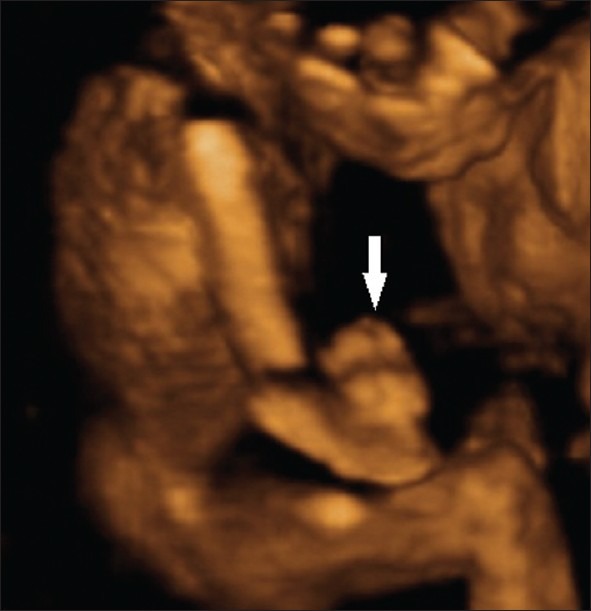
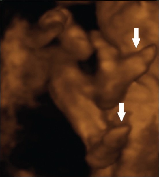
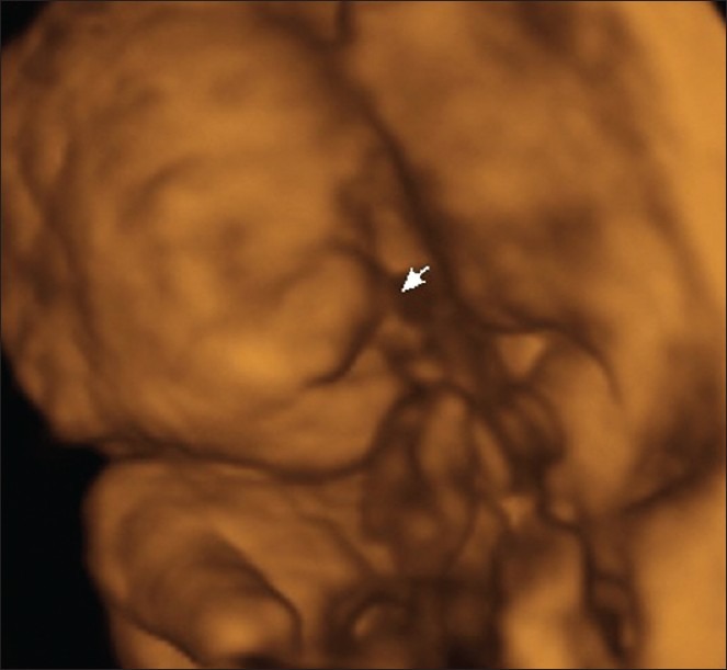
Primigravid woman, 25-year-old, had her first obstetric transvaginal ultrasonography, in which she was confirmed to have a singleton embryo with the crow-rump length (CRL) of 21 mm, compatible with the 9,0 weeks of gestation. Second ultrasonography, by transvaginal route, was performed on the 11th week that showed a fetus with CRL of 47.0 mm, nuchal translucency of 1.3 mm, hands and feet were poorly defined; at this time EEC syndrome was suspected. The second trimester 2DUS done at 19 weeks confirmed the EEC syndrome. 2DUS was able to demonstrate a fetus with lobster-claw hands (absence of the 2nd and 3rd fingers), left foot with the absence of the 3rd toes and a right foot with syndactyly on the 2nd and 3rd toes, and presence of a cleft lip/palate. The 3DUS, done with a convex volumetric transductor (RAB 4-8L) of Voluson 730 Pro machine (General Electric Medical Systems, Healthcare, Zipf, Austria) in rendering mode, showed the spatial relations of all malformations of the extremity and face, specially the lobster-claw hands [Figures 1, 2 and 3]. The 3DUS was not essential to the EEC syndrome diagnosis, however, it allowed a better depiction of the abnormalities and it was very helpful in explaining the abnormalities to the parents and for genetic counseling. The prenatal care was uneventful, the delivery was done by caesarian section on the 39th week, and the newborn female with weight of 3,150 g and Apgar score 9/9 was born.
Figure 1.

EEC syndrome. Three-dimensional ultrasound in rendering mode demonstrates the lobster-claw hands (white arrow).
Figure 2.

EEC syndrome. Three-dimensional ultrasound in rendering mode demonstrates the lobster-claw feet (white arrows).
Figure 3.

EEC syndrome. Three-dimensional ultrasound in rendering mode demonstrates the left cleft lip (white arrow).
DISCUSSION
The term EEC syndrome was first used by Rüdiger et al.,[1] to describe a girl with trimelic ectrodactyly, ectodermal dysplasia of hair, teeth, and nails, and also harelip facial cleft. The EEC syndrome has many phenotypic variability and may be associated with thin skin, with hyperkeratosis and nipple hypoplasia. The ophthalmologic abnormalities are also frequent, varying from blue iris, blepharitis, and dacryocystitis to corneal perforation.[9]
The EEC syndrome should be differentiated from the other syndromes that have ectodermal dysplasia and facial clefts such as: Rapp-Hodgkin syndrome, the Hay-Wells syndrome, ankyloblepharon ectodermal and harelip facial cleft; the split-hand and foot malformation syndrome (SHFM), syndactyly – median clefts of hands and feet – and aplasia/hypoplasia of the phalanges, metacarpals and metatarsals. Haratz-Rubinstein et al.,[10] described a case of prenatal diagnosis of SHFM at 18 weeks of pregnancy. In this case, the fetus presented with both hands with limited mobility than expected and the thumbs were fixed in an abducted position. The right hand had a median cleft with fusion of second and third fingers. The pregnant woman had pain and vaginal bleeding at 19 weeks, and she developed inevitable abortion. The post-mortem skeletal X-ray proved the anomalies detected by prenatal ultrasound.
The EEC syndrome has been linked to two genetic locations. The form of EEC syndrome designated EEC1 has been linked to chromosome 7q11.2-q21.3. The second and most common form of this disorder, designated EEC3, is caused by a mutation in the TP63 gene.[11] EEC 2 does not exist. The precise prenatal diagnosis is possible by DNA extraction by chorionic villus sample done between the 11th and 14th week of pregnancy. In our specific case, the prenatal diagnosis of EEC syndrome was based on characteristic sonographic features.
The prenatal diagnosis of EEC syndrome can be done early by the 2DUS, when the hands and feet malformations are associated with a cleft lip and/or palate deformities.[3,5] The 3DUS in the rendering mode has been used to better define the malformations, this mode is better to explain the malformations to the parents. There are evidences of benefits of 3DUS in isolated cases of ectrodactyly,[7] however there is only one image based report related to the EEC syndrome in the literaure.[8] Allen and Maestri[8] described a case of EEC syndrome, similar to ours, diagnosed by 2DUS on the 17th week of gestation in a high-risk pregnant woman. Her husband had EEC syndrome and their two children also had this syndrome. The 2DUS showed a cleft palate associated with bilateral ectrodactyly and “lobster-claw” anomaly of hands and feet. The 3DUS in the rendering mode allowed a proper view of facial anomalies and ectrodactyly of hands. In the absence of other anomalies such as ophthamolical or genito-urinary abnormalities, the postnatal prognosis is excellent, with the majority of the subjects presenting with normal intelligence and normal life style after the correction of the face and extremity anomalies.
CONCLUSION
In summary this is the second case of EEC syndrome diagnosed with 3DUS in prenatal period. 2DUS is sufficient for the diagnosis of EEC and 3DUS is not essential for its diagnosis. However 3DUS allowed better depiction of the abnormalities such “lobster claw hands” that were easy to explain to the parents.
Footnotes
Source of Support: Nil
Conflict of Interest: None declared.
Available FREE in open access from: http://www.clinicalimagingscience.org/text.asp?2012/2/1/40/99153
REFERENCES
- 1.Rüdiger RA, Haase W, Passarge E. Association of ectrodactyly, ectodermal dysplasia, and cleft lip-palate. Am J Dis Child. 1970;120:160–3. doi: 10.1001/archpedi.1970.02100070104016. [DOI] [PubMed] [Google Scholar]
- 2.Rodini ES, Richieri-Costa A. EEC syndrome: Report on 20 new patients, clinical and genetic considerations. Am J Med Genet. 1990;37:42–53. doi: 10.1002/ajmg.1320370112. [DOI] [PubMed] [Google Scholar]
- 3.Janssens S, Defoort P, Vandenbroecke C, Scheffer H, Mortier G. Prune belly anomaly on prenatal ultrasound as a presenting feature of ectrodactyly-ectodermal dysplasia-clefting syndrome (EEC) Genet Couns. 2008;19:433–7. [PubMed] [Google Scholar]
- 4.Brunner HG, Hamel BC, van Bokhoven H. The p63 gene in EEC and other syndromes. J Med Genet. 2002;39:377–81. doi: 10.1136/jmg.39.6.377. [DOI] [PMC free article] [PubMed] [Google Scholar]
- 5.Leung KY, MacLachlan NA, Sepulveda W. Prenatal diagnosis of ectrodactyly: The ‘lobster claw’ anomaly. Ultrasound Obstet Gynecol. 1995;6:443–6. doi: 10.1046/j.1469-0705.1995.06060443.x. [DOI] [PubMed] [Google Scholar]
- 6.Chuangsuwanich T, Sunsaneevithayakul P, Muangsomboon K, Limwongse C. Ectrodactyly-ectodermal dysplasia-clefting (EEC) syndrome presenting with a large nephrogenic cyst, severe oligohydramnios and hydrops fetalis: A case report and review of the literature. Prenat Diagn. 2005;25:210–5. doi: 10.1002/pd.1101. [DOI] [PubMed] [Google Scholar]
- 7.Koo BS, Baek SJ, Kim MR, Joo WD, Yoo HJ. Prenatally diagnosed ectrodactyly at 16 weeks’ gestation by 2- and 3-dimensional ultrasonography: A case report. Fetal Diagn Ther. 2008;24:161–4. doi: 10.1159/000151331. [DOI] [PubMed] [Google Scholar]
- 8.Allen LM, Maestri MJ. Three-dimensional sonographic findings associated with ectrodactyly ectodermal dysplasia clefting syndrome. J Ultrasound Med. 2008;27:149–54. doi: 10.7863/jum.2008.27.1.149. [DOI] [PubMed] [Google Scholar]
- 9.Kasmann B, Ruprecht KW. Ocular manifestations in a father and son with EEC syndrome. Graefes Arch Clin Exp Ophthalmol. 1997;235:512–6. doi: 10.1007/BF00947009. [DOI] [PubMed] [Google Scholar]
- 10.Haratz-Rubinstein N, Yeh MN, Timor-Tritsch IE, Monteagudo A. Prenatal diagnosis of the split hand anomaly: How early is early? Ultrasound Obstet Gynecol. 1996;8:57–61. doi: 10.1046/j.1469-0705.1996.08010057.x. [DOI] [PubMed] [Google Scholar]
- 11.Bethesda, MD: National Center for Biotechnology Information, National Library of Medicine; [Last accessed on 2012 May 10]. Online mendelian inheritance in man. Available from: http://ncbi.nlm.nih.gov/ omim . [Google Scholar]


