Abstract
Oral Myiasis is a rare disease that is mostly reported in developing countries. It is primarily caused by the invasion of the human body by fly larvae. The phenomenon is well-documented in the skin, especially among animals. This case report describes the presentation of Oral Myiasis caused by Musca Nebulo (common house fly), in a 28-year-old patient, with recent maxillofacial trauma. The patient was treated by manual removal of the larvae, after topical application of turpentine oil, followed by surgical debridement and oral therapy with Ivermectin.
Keywords: Ivermectin, Musca Nebulo, oral myiasis, turpentine oil
Introduction
Myiasis is derived from a Latin word ‘Muia,’ which means fly and ‘iasis,’ which means disease.[1] The term was coined by Hope in 1840,[2] and defined by Zumpt.[3] It is a pathological condition in which there is infestation of living mammals with the dipterous larvae, which, at least for a certain period feed on the host's dead or living tissue, and develop as parasites.[1,4]
Myiasis is a fairly common condition in rural areas, among animals such as cats and dogs, and has been frequently reported in humans residing in rural areas of developing countries.[5]
Clinically, myiasis is classified as:[1,6]
Depending upon the condition of the involved tissue, it is of two types:
Obligatory — require living tissue for larvae development
Facultative — require necrotic tissue for flies to lay eggs and incubate them
In humans the most common sites of Myiasis are the nose, eye, ear, vagina, skin, nasopharynx, and rarely, the oral cavity.[9,10]
Case Report
A 28-year-old male patient from a low socioeconomic status reported to the Department of Oral Pathology and Microbiology, Purvanchal Institute of Dental Sciences, Gorakhpur, with a chief complaint of pain and swelling in his upper lip since three weeks, which had increased over the past four days.
The patient's past dental history revealed that he had a road traffic accident, with no maxillofacial trauma of bones, three weeks back. An extraoral examination revealed several bruises and lacerations over the upper lip and left side of his face. He also had diffused swelling of the upper lip along with ulceration in the midline of upper lip, measuring approximately 0.5 × 2 cm. He was taken to a local hospital, where he received suturing of his laceration and medications.
An intraoral examination revealed mild bleeding from the gingival sulcus of the maxillary central incisors. The patient was having severe halitosis and poor oral hygiene, with severe periodontitis. Teeth numbers 11, 21, 31, 32, 41, and 42 showed grade III mobility. Radiographic examination showed no signs of bone trauma except for a slight widening of the periodontal ligament space in relation to 11 and 21, with generalized horizontal bone loss. Hematological investigations revealed that the Total Leukocyte Count (TLC) was slightly raised (TLC — 11,800cells / μL).
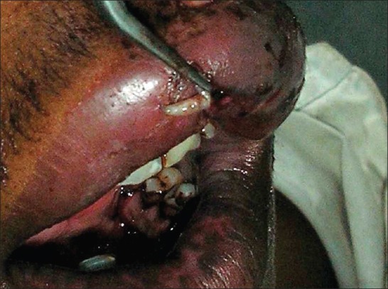
On clinical examination of the lesion, maggots were moving out [Figure 1]. Based on the patient's history, and the radiographic and hematological investigations, the provisional diagnosis was Oral Myiasis.
Figure 1.

Preoperative photograph showing maggots coming out of the lesion
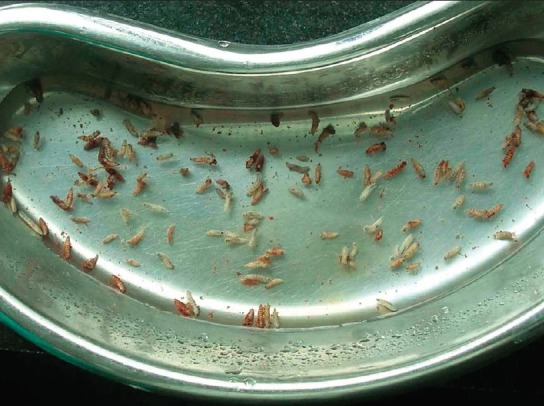
The patient was treated under local anesthesia. Cotton impregnated in turpentine oil (a topical irritant) was placed for 10 – 12 minutes and maggots were removed with a blunt tweezer. Around 45 – 50 maggots were removed [Figure 2]. The maggots were then sent for Entomological examination to a zoologist.
Figure 2.

Live maggots retrieved after application of turpentine oil
The wound was cleaned to remove the necrotic tissue, and periodontically weekend teeth with poor prognosis were extracted. The wound was thoroughly irrigated with betadine and normal saline. The necrotic tissue that was removed was sent for histopathological examination.
The histopathological examination of the Hematoxyline and Eosin (H and E)-stained section revealed connective tissue stroma consisting of collagen fibers along with fibroblasts and fibrocytes. Few areas showed numerous proliferating blood vessels along with red blood cells (RBCs). The tissue sections also showed numerous inflammatory cells; mainly plasma cells and lymphocytes. All these features were suggestive of a chronically inflamed granulation tissue.
The patient was put on Tab. Ivermectin 6 mg O.D. for the first three days along with Tab Metronidazole 400 mg thrice daily, for five days. The patient was advised to maintain proper oral hygiene and rinse the wound with 0.2% Chlorhexidine mouth wash, three to four times daily. The patient was asked to follow-up after five days, with a subsequent follow-up once a week. The wound was primarily closed and healed uneventfully in four weeks time.
An entomological examination of the removed maggots revealed that the larvae were of the house fly (Musca Nebulo).
Discussion
Musca Nebulo is the most common house fly in India. They are most active during the summer and rainy seasons.[11] The lifecycle of house fly starts with the egg stage, followed by the larva, pupa, and finally an adult fly. Open wound, ulcers, and open sores provide a favorable environment for their growth.[12]
Human Myiasis is a rare condition in today's world, especially Oral Myiasis.[13] It mainly occurs in rural areas.[14] A large number of predisposing factors favor the development of Oral Myiasis, such as, diabetes mellitus, psychiatric illness, leprosy, mental retardation,[15] and open neglected wounds. Apart from these predisposing factors, poor oral hygiene, low socioeconomic status, and maxillofacial trauma facilitate the disease.[16]
The best treatment modality is manual removal of maggots with a tweezer or tissue holding forceps, under local or general anesthesia.[17] Use of turpentine oil (a topical irritant) facilitates the removal of larvae;[2] other agents such as ether, Chloroform, Iodoform, and phenol mixtures can also be used.[18] This is followed by surgical debridement of the wound, to remove the necrotic tissue. Use of systemic Ivermectin gives good results in most cases.[19]
In the present case, maxillofacial trauma, poor oral hygiene, and poor periodontal status were the main factors.
Conclusion
Oral Myiasis is a rare disease in humans, but occurs with life-threatening results. Manual removal of maggots with application of turpentine oil, a topical irritant, is the treatment of choice, before a large amount of tissue necrosis takes place. As prevention is better than cure, prevention of this disease should be given utmost importance, to control the population of house flies.
Footnotes
Source of Support: Nil.
Conflict of Interest: None declared.
References
- 1.Sharma J, Mamatha GP, Acharya R. Primary oral myiasis: A case report. Med Oral Patol Oral Cir Bucal. 2008;13:E714–6. [PubMed] [Google Scholar]
- 2.Felices RR, Ogbureke KU. Oral myiasis: report of case and review of management. J Oral Maxillofac Surg. 1996;54:219–20. doi: 10.1016/s0278-2391(96)90452-8. [DOI] [PubMed] [Google Scholar]
- 3.Zumpt F. Myiasis in man and animals in the old world. In: Zumpt F, editor. A Textbook for Physicians, Veterinarians and Zoologists. London: Butterworth and Co. Ltd; 1965. p. 109. [Google Scholar]
- 4.Hope F. On insects and their larvae occasionally found in the human body. Trans R Soc Entomol. 1840;2:256–71. [Google Scholar]
- 5.Gabriel JG, Marinho SA, Verli FD, Krause RG, Yurgel LS, Cherubini K. Extensive myiasis infestation over a squamous cell carcinoma in the face. Case report. Med Oral Patol Oral Cir Bucal. 2008;13:E9–11. [PubMed] [Google Scholar]
- 6.Shinohara EH, Martini MZ, de Oliveira Neto HG, Takahashi A. Oral myiasis treated with ivermectin: case report. Braz Dent J. 2004;15:79–81. doi: 10.1590/s0103-64402004000100015. [DOI] [PubMed] [Google Scholar]
- 7.Gomez RS, Perdigao PF, Pimenta F, Rios Leite AC, Tanos de Lacerda JC, Custódio Neto AL. Oral myiasis by screwworm Cochlimyia hominivorax. Br J Oral Maxillofac Surg. 2003;41:115–6. doi: 10.1016/s0266-4356(02)00302-9. [DOI] [PubMed] [Google Scholar]
- 8.Sowani A, Joglekar D, Kulkarni P. Maggots. A neglected problem in palliative care. Indian J Palliat care. 2004;10:27–9. [Google Scholar]
- 9.Poon TS. Oral Myiasis in Hong Kong – A case Report. Hong Kong Pract. 2006;28:388–93. [Google Scholar]
- 10.Abdo EN, Sette-Dias AC, Comunian CR, Dutra CE, Aguiar EG. Oral myiasis: a case report. Med Oral Patol Oral Cir Bucal. 2006;11:E130–1. [PubMed] [Google Scholar]
- 11.Sandhu DB, Bhaskar H. Textbook of Invertebrate Zoology. In: Sandhu DB, Bhaskar H, editors. Musca: The Housefly. New Delhi: Campus; 2004. pp. 704–9. [Google Scholar]
- 12.Yazar S, Dik B, Yalcin S, Demirtas F, Yaman O, Ozturk M, et al. Nosocomial Oral Myiasis by Sarcophagi sp.in Turkey. Yonsei Med J. 2005;46:431–4. doi: 10.3349/ymj.2005.46.3.431. [DOI] [PMC free article] [PubMed] [Google Scholar]
- 13.Novelli MR, Haddock A, Eveson JW. Orofacial myiasis. Br J Oral Maxillofac Surg. 1993;31:36–7. doi: 10.1016/0266-4356(93)90095-e. [DOI] [PubMed] [Google Scholar]
- 14.Hakimi R, Yazdi I. Oral mucosa Myiasis caused by Oestrus ovis. Arch Iran Med. 2002;5:194–6. [Google Scholar]
- 15.Dandriyal R, Pant S. Oral myiasis in mentally challenged patient: a case report. J Clin Exp Dent. 2011;3:e155–7. [Google Scholar]
- 16.Godhi S, Goyal S, Pandit M. Oral Myiasis. A case report. J Maxillofac Surg. 2008;7:292–3. [Google Scholar]
- 17.Bhatt AP, Jayakrishna A. Oral myiasis.a case report. Int J Pediatric Dent. 2000;10:67–70. doi: 10.1046/j.1365-263x.2000.00162.x. [DOI] [PubMed] [Google Scholar]
- 18.Droma EB, Wilamowski A, Schnur H, Yarom N, Scheuer E, Schwartz E. Oral myiasis: a case report and literature review. Oral Surg Oral Med Oral Pathol Oral Radiol Endod. 2007;103:92–6. doi: 10.1016/j.tripleo.2005.10.075. [DOI] [PubMed] [Google Scholar]
- 19.Rossi-Schneider T, Cherubini K, Yurgel LS, Salum F, Figueiredo MA. Oral myiasis: a case report. J Oral Sci. 2007;49:85–8. doi: 10.2334/josnusd.49.85. [DOI] [PubMed] [Google Scholar]


