Abstract
Cutaneous metastases from internal malignancies are rare with a reported incidence between 0.7% and 10%. We report a case with distant skin and subcutaneous metastases in abdominal skin from breast cancer detected on 18F-fluoro-deoxyglucose–positron emission tomography/computed tomography imaging.
Keywords: Breast cancer, 18F-FDG–PET/CT, skin metastases
Introduction
Cutaneous metastases from internal malignancies are rare with a reported incidence between 0.7% and 10%. We report a case of distant subcutaneous metastases over the abdominal skin from breast cancer, detected on an 18F-fluoro-deoxyglucose-positron emission tomography/computed tomography (18F-FDG-PET/CT) scan. In addition to the detection of skin metastases, 18F FDG-PET/CT was also useful in defining the true extent of the disease.
Case Report
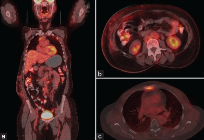
A 52-year-old woman diagnosed to have right-sided breast carcinoma was treated with total mastectomy and axillary lymph node clearance (TMAC) followed by chemoradiation. Five years after that she developed left-sided breast carcinoma, which was also treated with TMAC, followed by local irradiation of chest wall and chemotherapy. She was subjected to 18F-FDG-PET/CT [Figure 1] scan to restage the disease 2 months after chemotherapy. No abnormal FDG uptake was noted over chest wall and bilateral axillae. Mildly FDG avid skin thickening [standardized uptake value (SUVmax) = 3.4] and nodules in the subcutaneous fat over right lower abdominal wall were noted. Abnormal FDG avid sclerotic lesion (SUVmax = 6.2) was also noted in the sternum. FDG avid retroperitoneal, pelvic, and inguinal adenopathy was detected involving aortocaval, para-aortic, retrocrural, bilateral external iliac, and right inguinal nodal stations, indicating widespread metastatic disease. Hence, in addition to demonstration of skin metastases, 18F-FDG-PET/CT revealed widespread lymph node and bone metastases establishing the true extent of the disease. Subsequently, the patient underwent fine-needle aspiration cytology of skin nodules, which revealed metastatic deposits from breast cancer.
Figure 1.

Coronal fused PET/CT (a) and transaxial images (b) showing fluoro-deoxyglucose (FDG) uptake in multiple skin and subcutaneous nodules in abdominal wall on the right side. Transaxial image (c) showing FDG uptake in sternum
Discussion
Differential diagnosis of the skin lesions and subcutaneous nodules would include cutaneous lymphoma, melanoma, neurofibromatosis, and metastases from other internal malignancies. The breast, stomach, lung, uterus, large intestine, and kidneys are the most frequent internal organs to produce cutaneous metastases. Cancers that have the highest propensity to metastasize to the skin include melanoma (45% of cutaneous metastasis cases), breast (30%), nasal sinuses (20%), larynx (16%), and oral cavity (12%). Because breast cancer is so common, cutaneous metastasis of breast cancer is the most frequently encountered type of cutaneous metastasis in most clinical practices.[1–3] Cutaneous metastases can occur either by lymphatic or hematogenic spread and is most commonly seen in the head and neck regions and trunk.[4]
Cutaneous metastases from carcinoma are relatively uncommon in clinical practice, but they are very important to recognize. Cutaneous metastasis may herald the diagnosis of internal malignancy. Early recognition can lead to accurate and prompt diagnosis and timely treatment, but a high index of suspicion is required because the clinical findings may be subtle and asymptomatic as in this particular case. The recognition of cutaneous metastases often dramatically alters therapeutic plans, especially when metastases signify persistence of cancer originally thought to be cured. Some tumors metastasize with predilection to specific areas. Recognition of these patterns can be useful in directing the search for an underlying tumor.
18F-FDG–PET/CT has been widely used in restaging breast cancer and shown to be better than conventional imaging modalities and also changes management in significant number of patients.[5] However, distant skin metastases from breast cancer detected by FDG-PET/CT have been rarely reported in the literature.[6] In our case, in addition to demonstration of FDG uptake in skin metastases, PET/CT also revealed multiple lymph nodal and sternal metastases, thereby defining true extent of the disease. Our case also highlights the fact that 18F-FDG avid nodules in skin in a case of breast carcinoma should always bring up suspicion of skin metastases and should be evaluated further with cytologic correlation in all the cases to rule out metastases.
Footnotes
Source of Support: Nil
Conflict of Interest: None declared.
References
- 1.Schwartz RA. Cutaneous metastatic disease. J Am Acad Dermatol. 1995;33:161–82. doi: 10.1016/0190-9622(95)90231-7. [DOI] [PubMed] [Google Scholar]
- 2.Brenner S, Tamir E, Maharshak N, Shapira J. Cutaneous manifestations of internal malignancies. Clin Dermatol. 2001;19:290–7. doi: 10.1016/s0738-081x(01)00174-2. [DOI] [PubMed] [Google Scholar]
- 3.Krathen RA, Orengo IF, Rosen T. Cutaneous metastasis: a meta-analysis of data. South Med J. 2003;96:164–7. doi: 10.1097/01.SMJ.0000053676.73249.E5. [DOI] [PubMed] [Google Scholar]
- 4.Lookingbill DP, Spangler N, Sexton FM. Skin involvement as the presenting sign of internal carcinoma: a retrospective study of 7316 cancer patients. J Am Acad Dermatol. 1990;22:19–26. doi: 10.1016/0190-9622(90)70002-y. [DOI] [PubMed] [Google Scholar]
- 5.Eubank WB, Mankoff DA, Bhattacharya M, Gralow J, Linden H, Ellis G, et al. Impact of [F-18]-Fluorodeoxyglucose PET on defining the extent of disease and management of patients with recurrent or metastatic breast cancer. Am J Roentgenol. 2004;83:479–86. doi: 10.2214/ajr.183.2.1830479. [DOI] [PubMed] [Google Scholar]
- 6.Borkar S, Pandit-Taskar N. F-18 FDG uptake in Cutaneous metastases from breast cancer. Clin Nucl Med. 2008;33:488–9. doi: 10.1097/RLU.0b013e318177934e. [DOI] [PubMed] [Google Scholar]


