Abstract
Chronic pain is a serious medical condition with millions of sufferers for whom long-term therapies are either lacking or inadequate. Here we review the use of herpes simplex virus vectors as therapeutic tools to treat chronic pain by gene therapy. We describe an approach to inhibit chronic pain signaling whereby vector-mediated genes transferred to sensory nerves will modify the primary afferent nociceptor to prevent pain signaling to second-order nerves in the spinal cord. This approach may be used to reverse the chronic pain state of the nociceptor and could affect downstream pain-related changes in the central nervous system.
Keywords: HSV, chronic pain, nociceptor
Introduction
The localization of, and appropriate response to, noxious stimuli is a crucial evolutionary adaptation necessary for survival. Similarly, the hyperalgesia (increased pain in response to normally painful stimuli) and allodynia (pain in response to normally innocuous stimuli) that develops within minutes following tissue injury is necessary to ensure normal healing. Consistent with the physiological role of these changes in perception, both forms of hypersensitivity resolve over the time course of normal tissue repair. However in some cases, such as those associated with complex regional pain syndrome, post-herpetic neuralgia, osteoarthritis or interstitial cystitis, the hypersensitivity that develops with tissue injury, fails to resolve. The result is chronic pain, a disease in its own right that is clearly no longer adaptive given its persistence in the face of normal tissue repair. Chronic pain represents a major cause of morbidity, significantly impairing individual quality of life1 and imposing a substantial burden on society. There are few, if any, effective therapies for chronic pain devoid of serious side effects, forcing the over 80 million chronic pain sufferers in the United States to spend millions of dollars and countless hours in a desperate search for relief. Thus, the development of effective interventions for chronic pain is of great social and medical importance.
There are many reasons why available therapies are largely ineffective for the treatment of chronic pain and why so many potentially viable targets have yet to be exploited. First, diagnostic criteria do not yet encompass mechanistic underpinnings. As distinct mechanisms can produce a similar pain syndrome (for example, mechanisms underlying postherpetic neuralgia2), a mechanism-based therapy may be ineffective in a subpopulation of patients suffering from the 'same' syndrome. Second, given the impact of a host of individual specific variables including age, history, sex and genome, the relative importance of specific mechanisms may vary3 such that even mechanism-based diagnostic criteria will be quite complex. Third, there is considerable redundancy in the nervous system such that a relatively limited number of signaling molecules (that is, receptors, ion channels and second messenger systems) are used in the execution of a diverse array of physiological processes. Consequently, intolerable side effects such as cardiac, respiratory or central nervous system (CNS) depression often limit the systemic use of even the most selective therapeutic interventions.4 This situation may be compounded by the fact that the severity of many forms of chronic pain necessarily demands the administration of higher drug doses, increasing the risk of adverse side effects; simply put, it is often the presence of side effects that preclude the ability to obtain systemic levels of therapeutics necessary to sufficiently attenuate ongoing pain.5 Together, these factors may explain why it is only possible to obtain moderate pain relief in only one-third of patients with current pharmacological interventions. It is also important to note that by definition, chronic pain requires long-term therapy, which can result in the development of tolerance and can significantly increase the likelihood of additional side effects including liver, kidney, cardiac and gastrointestinal damage, which can ultimately prove to be fatal.5,6
Ideally, the next generation of therapeutic interventions for the treatment of chronic pain will enable the selective elimination of ongoing pain and hypersensitivity while avoiding the limitations of the currently available therapies. We suggest that the primary afferent nociceptor (PAN) is the ideal target for the next generation of therapeutics. PANs were first characterized in studies of the peripheral nerve, most notably by Perl and colleagues (for review see ref. 7). These investigations resulted in the identification and characterization of a subpopulation of primary afferent neurons that are activated by specific receptors or ion channels sensitive to noxious, mechanical, thermal and/or chemical stimuli. The afferents, referred to as nociceptors, were composed primarily of slowly conducting thinly myelinated Aδ-fibers and unmyelinated C-fibers.7 In addition to their high threshold for activation, both Aδ- and C-fiber PANS possessed the distinguishing feature of sensitization by inflammatory mediators.7 Subsequent analyses, however, indicates that PANs are heterogeneous with unique modality specificity (that is, mechanoheat versus mechanocold), and electrophysiological, pharmacological, histological, biochemical and anatomical properties.7 Recent evidence suggests that distinct subpopulations of afferents are responsible for signaling affective aspects of pain whereas others are responsible for signaling sensory discriminative aspects of pain.8 Evidence suggests that specific subpopulations of afferents are responsible for signaling unique aspects of pain such as the burning pain associated with capsaicin injection9 or cold allodynia observed following peripheral nerve injury.10 There is also evidence indicating that following specific types of injury, afferents undergo phenotypic changes such that subpopulations of non-nociceptive afferents may begin to signal pain.11
The PAN is the first step in the transmission of noxious stimuli from the periphery. These pseudounipolar neurons with cell bodies in the dorsal root ganglion (DRG) or trigeminal ganglion have axons that terminate in peripheral structures and project centrally to terminate in the dorsal horn of spinal cord or brainstem in an anatomically defined (dermatomal or radicular) pattern. As such, the PAN is accessible to manipulations that do not have access to the CNS, thereby avoiding an array of potential CNS side effects. More importantly, although there is evidence to suggest that chronic pain reflects changes in the CNS that not only serve to amplify PAN activity, but may ultimately maintain the chronic pain state,12 there is also evidence that aberrant primary afferent input is necessary for the maintenance of altered CNS processing if not directly responsible for ongoing pain and hypersensitivity.13 Thus, the focus of the present review is on approaches to treat pain via manipulations of the neurobiology of the PAN. Specifically, we will discuss the use of herpes simplex virus (HSV) vectors in both the treatment of chronic pain and the identification and delivery of novel therapeutic interventions.
Gene therapy: why HSV?
There are several reasons that HSV is an ideal platform to develop novel therapies for the treatment of chronic pain. First and foremost, HSV has a natural ability to infect and persist as a nonintegrated viral genome in sensory nerve nuclei via peripheral inoculation, and therefore, represents an excellent gene delivery vehicle for this purpose. HSV is a large double-stranded DNA virus present in nearly 90% of the human population.14,15 In its natural life cycle, wild-type HSV is spread by direct contact. HSV infects and replicates in skin or mucous membrane and the released viral particles invade sensory nerve terminals.15 These particles travel via retrograde transport along axons to the neuronal perikaryon in the DRG, where wild-type HSV may either reenter the lytic cycle or alternatively establish a latent state that is characterized. by the persistence of viral genomes as nonreplicating intranuclear episomal elements.15 Based on this unique biology, we and others have focused on the use of HSV vectors to deliver genes to nociceptors. These vectors are derived from HSV type 1 in which virus replication has been compromised by the deletion of essential viral gene functions required to initiate and. sustain virus replication.
A second advantage of the HSV system is that with a single inoculation, the virus may provide long-term therapy. Nonreplicating recombinant HSV can be exploited to establish long-term persistence in the nerve cell nuclei in a manner similar to wild-type latent virus. Nonreplicating HSV recombinants are taken up by nerve terminals and transported to the DRG in a manner identical to wild-type HSV; however, as HSV recombinants are unable to replicate, the recombinant genomes are forced into a pseudolatent state from which the latent vector genome is incapable of reactivating.16 During this latent state, the viral lytic genes are silent and only the transgene and latency-related transcript locus remain active.17 The duration of transgene expression can be short term (weeks) using a strong promoter such as the immediate early (IE) gene promoter derived from human cytomegalovirus or long term (months) using a modified HSV latency promoter.16
Third, as it is possible to inoculate painful tissue, it is possible to restrict the 'therapy' to the most appropriate region of the body, both minimizing the possibility of side effects and maximizing therapeutic efficacy. The specific ganglion containing the viral vector can be targeted by vector application to the painful tissue so that only the nerve terminals innervating that tissue become infected and the virus is subsequently transported to the nerve cell bodies by the retrograde transport mechanism used by the virus in natural infections.
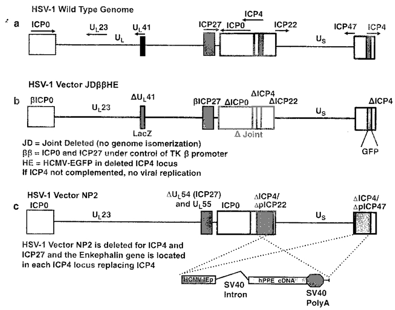
Fourth, the engineering of nonreplicating HSV results in a vector with a large transgene capacity that is incapable of reactivation in vivo and can provide a platform from which expression of relatively large molecules can be achieved in sensory neurons.16 During HSV replication, viral gene products are expressed in a temporally ordered cascade.18 Five IE gene products including the infected cell protein (ICP) 0, ICP4, ICP22, ICP27 and ICP47 are first expressed. With the exception of ICP47, these IE gene products trigger the expression of early (E) genes that code for proteins involved in DNA replication and late (L) genes coding for structural HSV proteins.18 As the viral thymidine kinase (tk) is required for replication in neurons, recombinants defective in tk can be propagated in culture and can replicate in skin, but not in the DRG. Highly defective vectors can be created by deletion of essential IE genes from the HSV genome. These mutants can only be propagated in cell lines permanently transfected with the missing gene, which can provide this product in trans following vector infection.19 Such defective mutants cannot replicate in any other cell type, including neurons, in vivo.19 Vectors have also been constructed by deleting the two essential IE genes, ICP4 and ICP27, and modifying the promoters of IE genes, ICP22 and ICP47, such that they demonstrate early transcriptional kinetics;20 these modifications effectively ablate the expression of the IE gene products, ICP4, ICP27, ICP22 and ICP4721 in noncomplementing cells. The large transgene capacity results from replication-defective HSV vectors produced in this manner. Furthermore, this transgene capacity can be increased substantially by removing the inverted repeat joint sequence separating the unique long (UL) and unique short (Us) components of the genome.22 This deletion stabilizes the genome by preventing UL - and Us-related recombination events that lead to UL/Us inversions in orientations that naturally occur during vector propagation in complementing cell lines. Figure 1 shows the design of HSV vectors applied to gene therapy studies in animal models and humans and vectors with improved capacity and stability.
Figure 1.
Replication defective herpes simplex virus (HSV) vector designs. (a) Configuration of wild-type (WT) HSV 1 genome. The boxes represent inverted repeats flanking the unique long (UL) and unique short (US) genome elements. The joint region separates UL, and US. The essential immediate early (IE) genes are ICP27 and ICP4. ICP0, ICP22 and ICP47 are nonessential IE genes. ICP4 and ICP0 are diploid in the genome because they are located in inverted repeat regions. The viral thymidine kinase (tk) gene and the virus host shut-off function (vhs) are located in the U1, segment. All of these genes are manipulated or deleted to create vectors. Deletion of ICP4 and ICP27 render the virus highly replication defective whereas deletion of the nonessential JE genes can compromise the virus life cycle in vivo but do not prevent virus replication in cell culture. (b) Configuration of the JDββHE. This vector is deleted for ICP4 and replaced by the reporter gene GFP. The vector is deleted for the joint region removing ICP4 and ICP0. The vhs gene is deleted and replaced by the reporter gene expression cassette LacZ. The remaining copy of ICP0 and the IE gene ICP27 have their IE gene promoters replaced by the early (E) tk gene promoter limiting their expression to early gene kinetics and are not expressed unless the essential IE gene ICP4 is complemented in engineered Vero cells. This type of vector can be used to express genes in embryonic stem cells. (c) Configuration of the pain vector NP2 employed to treat chronic pain. This vector is deleted for ICF27, ICP4, ICP47 and ICP22. The therapeutic gene preproenkephalin is present in two copies replacing both ICP4 loci.
Therapeutic target identification in primary afferents
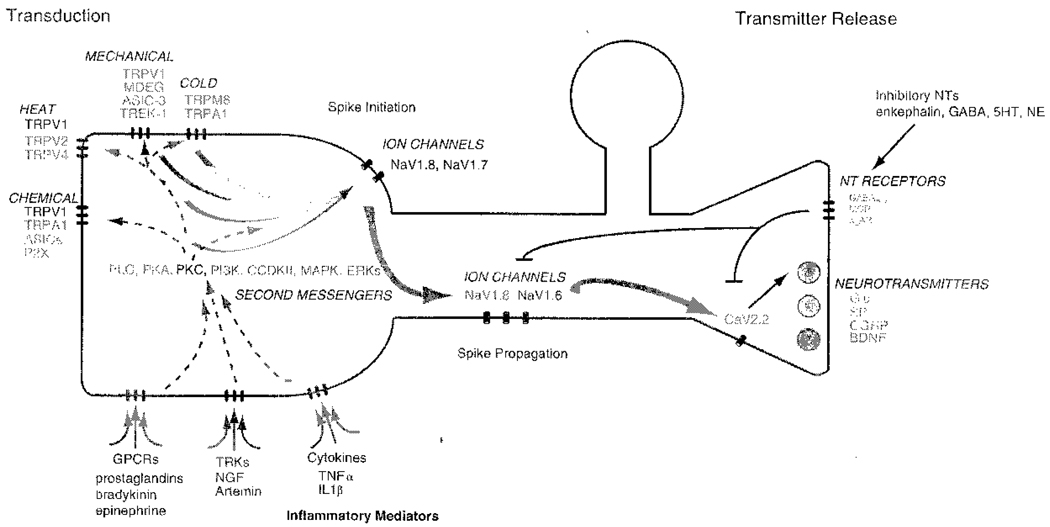
Potential peripheral nervous system targets for the treatment of chronic pain can be evaluated within the framework of the distinct events that are necessary for a nociceptor to convey information to the CNS about noxious stimuli impinging on peripheral tissues (summarized in Figure 2). First, 'energy' from the stimulus (mechanical, thermal or chemical) must be converted into an electrical signal. This process, referred to as signal transduction, results in a generator potential or depolarization of the peripheral terminal. Second, the generator potential must initiate an action potential or spike, the rapid 'all or nothing' change in membrane potential that constitutes the basic unit of electrical activity in the nervous system. This process is sometimes referred to as transformation. Third, the action potential must be successfully propagated from the peripheral terminal to the central terminal. And fourth, the propagated action potential invading the central terminal must drive a sufficient increase in intracellular calcium ions to enable release of enough transmitter to initiate the whole process once again in the second-order neuron. Additional processes important for the modulation of sensory input to the spinal cord include: (1) integration of activity within the DRG (that is, impact of action potential invasion on transcription and translation, cross-excitation and ectopic action potential generation); (2) axonal transport and trophic signaling; (3) neuron–glial interaction and neuroimmune interaction and (4) second messenger cascades underlying the actions of pronociceptive signaling molecules; only the last of these processes are illustrated in Figure 2. Distinct sets of proteins underlie each of these processes, and are, therefore potential targets of a wide variety of therapeutic interventions.3
Figure 2.
Summary of nociceptor pathways and targets for chronic pain intervention. Four events arc essential for the transmission of noxious or painful stimuli from the periphery to the central nervous system along primary afferent neurons. These include (1) transduction, (2) spike initiation, (3) spike propagation and (4) transmitter release from the central terminals. Several of the specific proteins thought to underlie these processes and play particularly important roles in nociceptive afferents are indicated. Proteins underlying these four events may be modulated by distinct classes of inflammatory mediators acting through specific receptors on primary afferent neurons. These include the G-protein coupled receptors (GPCRs), receptor tyrosine kinases (TRKs) and cytokines which act through several receptor types. Inflammatory mediators initiate a variety of second messenger cascades, several of which ultimately converge on the epsilon isoform of protein kinase C (PKC), which are responsible for the modification of receptor and/or channel properties underlying changes in afferent excitability. In addition to the pronociceptive processes initiated by inflammatory mediators, there are also antinociceptive processes, commonly initiated at the central terminals of nociceptive afferents by inhibitory neurotransmitters (NTs), acting through an additional set of GPCR and ionotropic receptors. Inhibitory NTs can both block transmitter release from the primary afferent, secondary to the inhibition of Ca2+ influx via voltage-gated Ca2+ channels such as the N-type channel CaV2.2, as well as inhibit the modulatory effects of pronociceptive inflammatory mediators. Several of the proteins involved in these processes function at points of convergence of many different processes. Abbreviations: TRPV1, 2 and 4, transient receptor potential vanilloid type 1, 2 and 4; TRPA1, transient receptor potential ankeryin rich type 1; ASICs, acid sensing ion channels; P2X, ionotropic purinergic receptor type 2; MDEG, mouse homolog of degenerin family members isolated in C, elegans; TRPM8, transient receptor potential melastatin type 8. Spike Initiation: NaV1.7, 1.8, voltage-gated sodium channel alpha subunits 7 and 8; NaV1.6, voltage-gated sodium channel alpha subunit 6; GABA, γ-aminobuteric acid; 5HT, 5-hydroxy triptophan or serotin; NE, norepinephrin; GAGAA,B, A (ionotropic) and B (metabotropic) type GABA receptors; MOR, μ opioid receptor; α2AR, α2 adrenergic receptor; Glu, glutamate; SP, substance P; CGRP, calcitonin gene-related peptide; BDNF, brain-derived neurotrophic factor; NGF, nerve growth factor; TNIFα; tumor necrosis factor-α; IL1β, interleukin 1β; PLC, phospholypase C; PKA, cyclic AMP dependent protein kinase, P13K, phosphatidylinositol 3-kinase; CCDKII, calcium and calmodulin dependent protein kinase Il; MAPK, mitogen activated protein kinase; ERK, extracellular signal related kinase.
The question, of course, is which of the multitude of proteins underlying this array of process are likely to have the greatest efficacy and specificity in the treatment of chronic pain? We suggest that the proteins or processes that are both specific to PAN signaling and that also serve as 'points of convergence' for many distinct processes are the most likely to provide specificity and selectivity. With respect to selective patterns of expressions, despite the fact that there is considerable redundancy throughout the nervous system, as noted above, there are a number of proteins that are preferentially, if not exclusively, present in PANS and are critical to nociceptive signaling. These include the voltage-gated sodium channel NaV1.8 and the transient receptor potential vanilloid type 1 (TRPVI) receptor.3. The importance of a 'point of convergence' is illustrated through consideration of the initial processes underlying nociceptive signaling within the context of the fact that the majority of PAN are referred to as polymodal nociceptors because they are responsive to thermal, mechanical and chemical stimuli. In such a neuron, selective blockade of the proteins underlying mechanotransduction will still leave a neuron hyperresponsive to thermal and chemical. stimuli In contrast, all three modalities converge on the machinery underlying spike initiation. Thus, the selective blockade of proteins responsible for spike initiation in nociceptive afferents would eliminate all forms of stimulus-evoked activities in these neurons.
HSV-based approaches
Initial strategies for the treatment of pain incorporating HSV were based on the notion that nociceptive signaling in the dorsal horn constituted a major point of convergence as nociceptive dorsal horn neurons receive input from an array of PANs. That the spinal administration of a number of analgesic compounds provides better pain relief with fewer side effects23 served as clinical evidence in support of this initial approach. Consequently, our group and others have used HSV-based vectors to overexpress inhibitory neurotransmitters in primary afferents to attenuate nociceptive neurotransmission at the first synapse between the PAN and the second order neuron in the spinal cord. The first of these transmitters to be expressed was enkephalin,24 which was subsequently demonstrated to reduce C-fiber-evoked activity in the dorsal horn,23 the second phase of formalin evoked nociceptive activity,26 thermal and mechanical hypersensitivity associated with chronic inflammation,8 as well as mechanical allodynia associated with the spinal nerve ligation model of chronic neuropathic pain.27 Others have since shown that primary erythermalgia (PE)-expressing HSV vectors produce analgesia in visceral pain models of cystitis and pancreatitis28.29 and extended studies to primates 30 Subsequently the antinociceptive efficacy of HSV-mediated expression of endomorphin-2,31 glutamic acid decarboxylase (which results in an increase in GABA synthesis and release from primary afferent neurons),32,33 and antiinflammatory cytokines such as interleukin (IL)-4, IL-10 or IL-13 (used to block central neuroimmune activation),34–37 have each been shown to have efficacy in several different animal models of acute and chronic pain. Along the lines of these latter experiments, we were also able to demonstrate analgesic efficacy with HSV-mediated expression of the soluble form of the tumor necrosis factor-α (TNF-α) receptor.38
Strategies designed to alter the underlying biology and function specifically in PAN are only now being investigated and represent a vast area of exploration. Channel proteins are perhaps the most logical targets to modify with HSV gene therapy, because, as noted above, several of these are located at points of convergence where their function is essential for normal nociceptive processing. Proof of concept is again provided by clinical experience where a diverse array of compounds currently used to treat pain, such a tricyclic antidepressants, local anesthetics and antiepileptics are thought to have analgesic efficacy because of the ability to block ion channels.39 As stressed earlier, HSV gene transfer has the distinct advantage over the available pharmacological approaches because it enables the manipulation of ion channels in afferents responsible for the pain, thereby avoiding side effects associated with blockade of channels that may be widely distributed throughout the body. Perhaps the first channel targeted in such a manner was the sodium channel subunit NaV1.7, a channel that has unique features enabling it to play a significant role in spike initiation.11 That this channel plays a critical role in nociceptor activity is highlighted by the recent discovery of individuals possessing both gain-of-function and loss-of-function point mutations in this subunit. Strikingly, two distinct pain syndromes—PE and paroxysmal extreme pain disorder—reveal the specific impact of gain-of-function mutations.40 Furthermore, in contrast to the impact of the gain–of-function mutations, loss–of-function mutations that result in nonfunctional channels, are associated with a complete insensitivity to pain.41 Not surprisingly, knockdown of NaVI.7 via the HSV-mediated expression of an antisense oligodeoxynucleotide was shown to block inflammatory hyperalgesia.42 Pilot data collected by our group have demonstrated that HSV vectors expressing zinc finger protein transcription repressors targeted against the NaV1.8 sodium channel can reduce transcription of this protein in vitro and reduce nociceptive behaviors in a rat model of neuropathic pain.43
As the only sensation associated with TRPVI activation is pain, this receptor has received considerable attention from pain researchers. Cloned in 1997, data from an array of studies suggest that TRPV1 may play a particularly important role in pain associated with tissue injury.11 It is activated by exogenous compounds such as capsaicin and resiniferotoxin that produce both pain and hypersensitivity when applied at relatively low concentrations as well as endogenous compounds associated with tissue injury ranging from protons to lipids.44 Recent evidence suggests that TRPV1 is present on the central terminals of nociceptive afferents where it also facilitates transmission of noxious mechanical stimuli.45 Importantly, data from null-mutant mice and TRPV1-selective antagonists suggest this channel plays a particularly important role in inflammatory hypersensitivity serving as a common target for signaling cascades initiated from several diverse molecules including bradykinin, neurotrophic factors and cytokines such as TNFα.11 Small molecule blockers of TRPV1 have subsequently been shown to have analgesic efficacy in array of chronic pain models.46 Unfortunately, however, systemic administration of these compounds produces hyperthermia, which has been the cause for termination of early clinical trials46 and again highlights an advantage of an HSV-based approach.
We have employed two distinct HSV based strategies designed to disrupt TRPVI signaling. The first of these involved a dominant-negative approach to target protein kinase C-ε (PKCε), an isoenzyme in PANs that appears to serve as the penultimate target downstream from an array of pronociceptive mediators including bradykinin, endothelins and proteases.47 Although there is evidence that activation of PKCε is involved in the regulation of other channels such as NaV1.8 thought to play a critical role on chronic pain states,47 the link between this PKC isoform and TRPV1 has been most firmly established PKCε phosphorylates TRPV1 at residues S502 and S80048–50 resulting in a receptor sensitization which is thought to play a critical role in thermal hypersensitivity observed in the presence of inflammation,3 bladder cystitis51 and diabetic neuropathy45 We employed an HSV vector expressing a dominant-negative form of PKCε (vector vHDNP) that is unable to phosphorylate TRPV1 but can compete for binding sites with native PKCε.52 Infection of DRG neurons with vHDNP in vitro reduced capsaicin (CAPS)-evoked current amplitudes, enhanced TRPVI desensitization rates and attenuated the ability of phorbol ester to sensitize TRPV1. Importantly, although targeted infection of DRG neurons innervating the rat hindpaw with vHDNP resulted in a small but significant increase in the paw withdrawal latency to noxious thermal stimuli, CAPS-induced thermal hyperalgesia was virtually eliminated in these rats.52
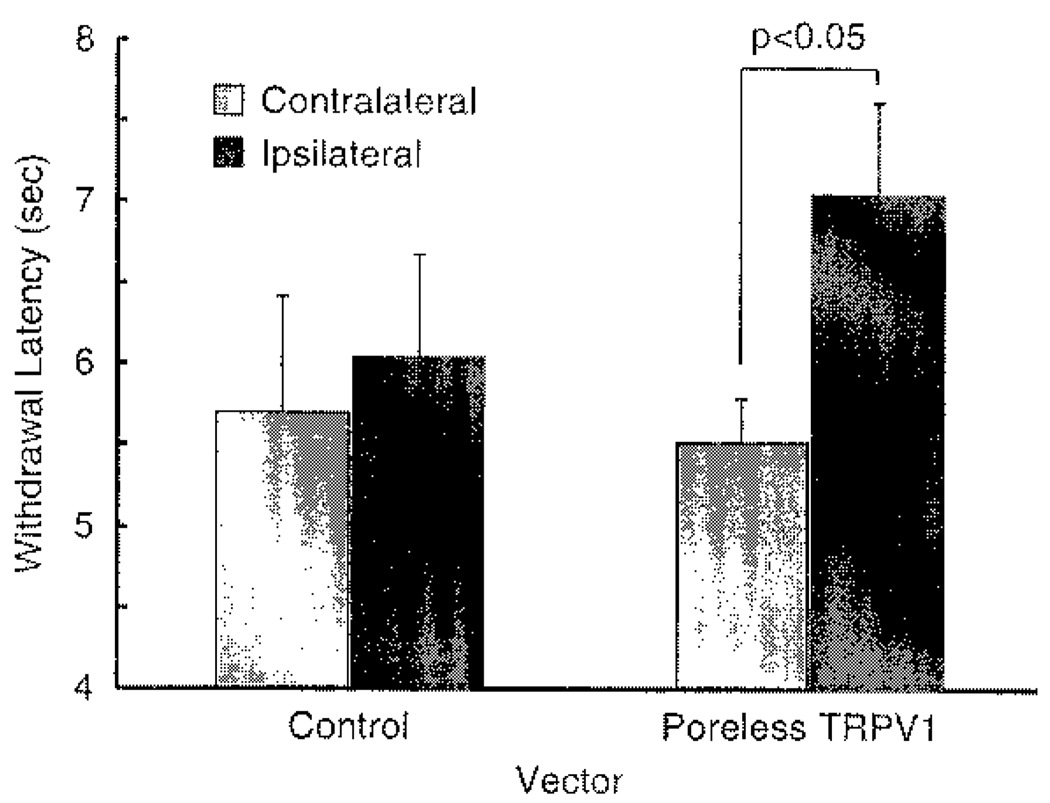
The second approach was more specific for TRPV1 and involved a dominant-negative inhibitor of TRPV referred to as poreless TRPV1.53 In vitro studies using mixed infections with an HSV vector expressing wild-type TRPV1 (vector vTTHR) and an HSV vector expressing the poreless TRPV1 (vector vHP) demonstrated reduced responsiveness to CAPS with a subsequent reduction in calcium influx as measured by cobalt assay.53 This suggested that the poreless TRPV1 subunit coassembled with wild-type receptor subunits creating a defective channel. This was further demonstrated in vivo where mice injected with vTH in their hindpaw demonstrated a significant decrease in thermal sensitivity (Figure 3). Experiments are ongoing to determine the efficacy of this construct in models of chronic inflammatory and neuropathic pain.
Figure 3.
Inhibition of chronic pain signaling using the defective transient receptor potential vanilloid type 1 (TRPV1) channel 'poreless'. Mice injected with a herpes simplex virus (HSV) vector expressing a poreless form of TRPV1 demonstrate decreased sensitivity to thermal pain. Male C57B1/6 mice were injected subcutaneously into the right rear hindpaw with 1 × 10⁷ plaque-forming units of either a control vector (DAPO, N = 4) or a vector expressing a poreless TRPV1 (vHP, N= 4). One week later, thermal response was assessed by placing each mouse into a plexiglass enclosure on a glass surface maintained at 30 °C. After a 15 min accommodation period, a light beam was focused onto the midplantar area of each hindpaw and the amount of time it took each animal to move its paw from the heat source was measured. The average of three trials per hindpaw for each animal was used to determine each animal's thermal response. Values represent mean ± s.e.m.
Novel approaches
Although previous HSV-based approaches to treat chronic pain highlight the therapeutic potential of the HSV-based platform, they highlight two potential limitations: (1) the majority of approaches rely on generating constitutive alterations in afferent function, precluding dynamic control of the antinociceptive machinery and (2) all approaches rely on the assumption that the 'right' afferents are infected, with little consideration of the fact that it might be desirable, if not necessary to infect specific populations of afferents to provide maximum pain relief. We suggest that it is possible to address both of these limitations through targeted manipulations of the HSV construct, which can then be used to deliver 'druggable targets' to the primary afferent. There are two general strategies that can be used to target specific subpopulations of primary afferents. One of these is to use PAN-specific promoters, such as that for NaV1.8 to drive transgene expression in NaV1.8-expressing neurons. The second is to use cell-specific surface receptors to which viral binding/entry can be directed in combination with the modification of viral glycoproteins to recognize novel receptors while eliminating the binding of these viral ligands to native receptors. Novel targeting ligands have either been inserted into the amino terminus of HSV glycoprotein C (gC)54 or gD55 to achieve targeting of HSV to cell-specific receptors. A 'druggable target' could involve the delivery of a ligand-gated ion channel not normally present in sensory neurons. A similar approach is now being used throughout neurobiology with the expression of the ivermectin channel, an invertebrate-specific glutamate-gated chloride channel, which can be used to selectively inactivate specific neurons. Depending on the receptor selectivity of such a channel, it should be possible to selectively activate the channel, and therefore block activity in channel-expressed afferents, via the systemic administration of an otherwise inert compound.
HSV as a means of identification of novel therapeutics
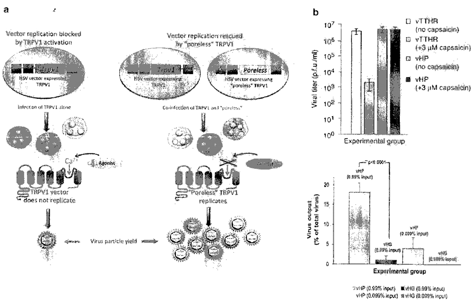
Apart from using known modulators of channel function, HSV vectors offer a new approach to identify previously unknown gene products that have a role in ion channel function (activation, sensitization, desensitization) and perhaps channel assembly, trafficking and turnover. Our method to discover modulators of ion channel activity is based on the selective survival of cells that have been coinfected with two HSV vectors, one that expresses an ion channel of interest and another that expresses a modulatory gene product. Replication-defective HSV vectors can express a functional calcium channel such as TRPV1 in vitro.53 The channel can be expressed in cells that complement the defective viral functions in trans resulting in virus replication. However treatment of infected cells with a channel agonist, in this case capsaicin, activates the TRPV1 channel resulting in rapid calcium ion influx and cell death due to osmotic shock. As a consequence virus growth is greatly reduced. Channel antagonists (such as ruthenium red) prevent ion channel activation, allowing virus replication to proceed. The ability of the channel vectors to form plaques in the presence of an antagonist raised the possibility that virus replication could be used as a sensitive readout for negative regulators of channel function. Identification of novel modulatory genes can be achieved by coinfecting cells with an HSV vector expressing the channel of interest and a set of unknown HSV vectors containing transgenes representing a library of sensory nerve-expressed genes. As these vectors cannot replicate in noncomplementing cells such as neurons, the modulatory gene products can ultimately be directly studied for their effects on endogenously expressed ion channels after vector-mediated gene delivery.
As proof of principle for this approach we modeled the potential for selecting vectors expressing gene-encoded inhibitors of TRPVI channel activity at the posttranslational level by mixed infections of a vector expressing TRPVI (vector vTT) and vector vHP. Coinfection of vTT with vHP at ratios of either 100:1 or 1000:1 demonstrated that vHP was readily detected with a minimum of potential false positives. This vHP output could be enriched with a second round of capsaicin selection, allowing sequential selection and enrichment strategies to be used (Figure 4). Experiments are in progress to test this method using additional model antagonist genes that inhibit ion channel function. The method described here suggests that vectors expressing highly diverse cDNA or siRNA libraries can be used in HSV-based selection, where up to one million library members can be screened in a single tissue culture plate for genes that inhibit agonist-induced channel activation. Alternatively, the library can be designed to consist of antisense mRNAs to identify new genes that are essential for ion channel activation.
Figure 4.
Selection method to discover genes that negatively modulate transient receptor potential vanilloid type I (TRPVI). (a) Schematic description of the genetic selection for a TRPVI dominant negative subunit. Specific herpes simplex vines type 1 (HSV-1) genetic elements within the viral genome were engineered to express either wild-type TRPVI or a nonfunctional 'poreless' mutant of TRPV1, which is impermeable to Ca2+. HSV-1 with only wild-type TRPVI was unable to propagate in the presence of agonists. Coinfection with a vector encoding the functionally inactive poreless TRPVI (vHP) and a vector encoding wild-type TRPVI rescued viral replication in the presence of TRPVI agonist as a result of the formation of functionally inactive heteromeric channels. The poreless TRPV1 vector is found at a high titer in the selected culture, (b) Selection for vHP in coinfection experiments. Top panel shows the loss of virus yield from Vero cells infected with TRPV1 vector following capsaicin treatment. Virus replication is reduced by calcium influx and cell death due to osmotic shock. Bottom panel shows virus output after two rounds of selection with 3 mM capsaicin for coinfections of either vHP (percentage of total virus input was 0.99 or 0.099%)+vTTHR or vHC (percentage of total virus input was 0.99 or 0.099%)+vTTHR. Error bars, ± s.d. Adapted from Nat Methods 2007; 4: 692–693.
Summary and conclusions
In summary, chronic pain continues to be a major health issue with few, if any, effective therapeutic interventions devoid of serious side effects. The primary afferent remains a viable target for the development of novel therapeutic interventions for two main reasons: (1) there is compelling evidence to suggest that changes in primary afferent structure and function are critical for both the initiation and maintenance of many chronic pain states and (2) blocking pain at the primary afferent should provide effective pain release in the absence of CNS side effects. Furthermore, it is becoming increasingly clear that specific subpopulations of afferents are responsible for specific 'types' of pain. Although HSV-mediated disruption of nociceptive signaling has been successfully employed in a number of preclinical models of chronic pain, these previous strategies have several important limitations including the need for repeated inoculation, the absence of temporal control over the actions of the therapeutic molecules delivered, and the absence of control over which afferents are targeted. There are clear strategies to address these limitations in the next generation of HSV-based interventions. Importantly, this vector may also provide the framework within which to identify the next generation of therapeutic targets.
Acknowledgements
We acknowledge the substantial contributions of many collaborators in the work reviewed including David Fink, Marina Mata, Shuanglin Hao, Munmun Chattopadhyay, Zhigang Zhou and Xiangmin Peng (University of Michigan), Bill Goins, Paola Grandi, Justus Cohen, George Huang, Ali Ozuer, Mingdi Zhang, Rebecca Sullenberger, and Michael Cascio (University of Pittsburgh), Rahul Srinivasin (Cal Tech), April Craft (University of Toronto) and Darren Wolfe, James Wechuck and David Krisky (Diamyd Inc., Pittsburgh, PA). Work described in this review from our laboratories was supported by the following grants: JRG, (NIH:DK07402604), MG (NIH:NS044992-02) and JCG (NIH: 2PO1NS040923:RO1119298:U54AR050733;PO1DK044935: RO1 NS059003: PO1 CA06924).
References
- 1.Niv D, Devor M. Chronic pain as a disease in its own right. Pain Pract. 2004;4:179–181. doi: 10.1111/j.1533-2500.2004.04301.x. [DOI] [PubMed] [Google Scholar]
- 2.Dworkin RH, Gnann JW, Jr, Oaklander AL, Raja SN, Schmader KB, Whitley RJ. Diagnosis and assessment of pain associated with herpes zoster and postherpetic neuralgia. J Pain. 2008;9:S37–S44. doi: 10.1016/j.jpain.2007.10.008. [DOI] [PubMed] [Google Scholar]
- 3.Gold MS, Caterina MJ. Molecular biology of nociceptor transduction. In: Basbaum AI, Bushnell MC, editors. Handbook of the Senses. vol. 5. San Diego: Academic Press; 2008. pp. 43–74. [Google Scholar]
- 4.Hansson P, Lacerenza M, Marchettini P. Aspects of clinical and experimental neuropathic pain: the clinical perspective. In: Hansson P, Fields HL, Hill RG, Marchettini P, editors. Neuropathic Pain: Pathophysioloy and Treatment. vol. 21. Seattle: IASP Press; 2001. pp. 1–18. [Google Scholar]
- 5.Barrett AM, Lucern MA, Le T, Robinson RI, Dworkin RH, Chappell AS. Epidemiology, public health burden, and treatment of diabetic peripheral neuropathic pain: a review. Pain Med. 2007;8 Suppl 2:S50–S62. doi: 10.1111/j.1526-4637.2006.00179.x. [DOI] [PubMed] [Google Scholar]
- 6.Turk DC. Clinical effectiveness and cost-effectiveness of treatments for patients with chronic pain. Clin J Pain. 2002;18:355–365. doi: 10.1097/00002508-200211000-00003. [DOI] [PubMed] [Google Scholar]
- 7.Caterina MJ, Gold MS, Meyer RA. Molecular biology of nociceptors. In: Hunt S, Koltzenburg M, Hunt SP, Rothwell NJ, editors. The Neurobiology of Pain. Oxford: Oxford University Press; 2005. pp. 1–33. [Google Scholar]
- 8.Braz J, Beaufour C, Coutaux A, Epstein AL, Cesselin F, Hamon M, et al. Therapeutic efficacy in experimental polyarthritis of viral-driven enkephalin overproduction in sensory neurons. J Neurosci. 2001;21:7881–7888. doi: 10.1523/JNEUROSCI.21-20-07881.2001. [DOI] [PMC free article] [PubMed] [Google Scholar]
- 9.Schmelz M, Schmid R, Handwerker HO, Torebjork HE. Encoding of burning pain from capsaicin-treated human skin in two categories of unmyelinated nerve fibres. Brain. 2000;123(Part 3):560–571. doi: 10.1093/brain/123.3.560. [DOI] [PubMed] [Google Scholar]
- 10.Xing H, Chen M, Ling J, Tan W, Gu JG. TRPM8 mechanism of cold allodynia after chronic nerve injury. J Neurosci. 2007;27:13680–13690. doi: 10.1523/JNEUROSCI.2203-07.2007. [DOI] [PMC free article] [PubMed] [Google Scholar]
- 11.Gold MS, Chessell I, Devor M, Dray A, Gereau RW, Kane S, et al. Peripheral nervous system targets: rapporteur report. In: Campbell JN, Basbaum AI, Dray A, Dubner R, Dworkin RH, Sang CN, editors. Emerging Strategies for the Treatment of Neuropathic Pain. Seattle: IASP Press; 2006. pp. 3–36. [Google Scholar]
- 12.Dubner R. The neurobiology of persistent pain and its clinical implications. Suppl Clin Neurophysiol. 2004;57:3–7. doi: 10.1016/s1567-424x(09)70337-x. [DOI] [PubMed] [Google Scholar]
- 13.Gracely RH, Lynch SA, Bennett GJ. Painful neuropathy: altered central processing maintained dynamically by peripheral input. Pain. 1992;51:175–194. doi: 10.1016/0304-3959(92)90259-E. [DOI] [PubMed] [Google Scholar]
- 14.Hill JM, Ball MJ, Neumann DM, Azcuy AM, Bhattacharjee PS, Bouhanik S, et al. The high prevalence of herpes simplex virus type 1 DNA in human trigeminal ganglia is not a function of age or gender. J Virol. 2008;82:8230–8234. doi: 10.1128/JVI.00686-08. [DOI] [PMC free article] [PubMed] [Google Scholar]
- 15.Jacobs A, Breakefield XO, Fraefel C. HSV 1-based vectors for gene therapy of neurological diseases and brain tumors: part I. HSV-1 structure, replication and pathogenesis. Neoplasia. 1999;1:387–401. doi: 10.1038/sj.neo.7900055. [DOI] [PMC free article] [PubMed] [Google Scholar]
- 16.Goins WF, Wolfe D, Krisky DM, Bai Q, Burton EA, Fink DJ, et al. Delivery using herpes simplex virus: an overview. Methods Mot Biol. 2004;246:257–299. doi: 10.1385/1-59259-650-9:257. [DOI] [PubMed] [Google Scholar]
- 17.Chattopadhyay M, Goss J, Wolfe D, Goins WC, Huang S, Glorioso JC, et al. Protective effect of herpes simplex virus-mediated neurotrophin gene transfer in cisplatin neuropathy. Brain. 2004;127:929–939. doi: 10.1093/brain/awh103. [DOI] [PubMed] [Google Scholar]
- 18.Burton EA, Wechuck JB, Wendell SK, Goins WF, Fink DJ, Glorioso JC. Multiple applications for replication-defective herpes simplex virus vectors. Stern Cells. 2001;19:358–377. doi: 10.1634/stemcells.19-5-358. [DOI] [PubMed] [Google Scholar]
- 19.DeLuca NA, McCarthy AM, Schaffer PA. Isolation and characterization of deletion mutants of herpes simplex virus type 1 in the gene encoding immediate-early regulatory protein ICP4. J Virol. 1985;56:558–570. doi: 10.1128/jvi.56.2.558-570.1985. [DOI] [PMC free article] [PubMed] [Google Scholar]
- 20.Krisky DM, Wolfe D, Coins WF, Marconi PC, Ramakrishnan R, Mata M, et al. Deletion of multiple immediate-early genes from herpes simplex virus reduces cytotoxicity and permits long-term gene expression in neurons. Gene Therapy. 1998;5:1593–1603. doi: 10.1038/sj.gt.3300766. [DOI] [PubMed] [Google Scholar]
- 21.Wolfe D, Goins WF, Yamada M, Moriuchi S, Krisky DM, Oligino TJ, et al. Engineering herpes simplex virus vectors for CNS applications. Exp Neurol. 1999;159:34–46. doi: 10.1006/exnr.1999.7158. [DOI] [PubMed] [Google Scholar]
- 22.Craft AM, Krisky DM, Wechuck JB, Lobenhofer EK, Jiang Y, Wolfe DP, et al. HSV mediated expression of Pax3 and MyoD in embryoid bodies results in lineage-related alterations in gene expression profiles. Stem Cells. 2008;26:3119–3129. doi: 10.1634/stemcells.2008-0417. [DOI] [PubMed] [Google Scholar]
- 23.Kaplan KM, Brose WG. Intrathecal methods. Neurosurg Cfin N Am. 2004;15:289–296. doi: 10.1016/j.nec.2004.02.011. [DOI] [PubMed] [Google Scholar]
- 24.Antunes Bras JM, Epstein AL, Bourgoin S, Hamon M, Cesselin F, Pohl M. Herpes simplex virus 1-mediated transfer of preprocnkephalin A in rat dorsal root ganglia. J Neurochem. 1998;70:1299–1303. doi: 10.1046/j.1471-4159.1998.70031299.x. [DOI] [PubMed] [Google Scholar]
- 25.Wilson SP, Yeomans DC, Bender MA, Lu Y, Goins WF, Glorioso JC. Antihyperalgesic effects of infection with a preproenkephalin-encoding herpes virus. Proc Natl Aced Sci USA. 1999;96:3211–3216. doi: 10.1073/pnas.96.6.3211. [DOI] [PMC free article] [PubMed] [Google Scholar]
- 26.Goss JR, Mata M, Goins WF, Wu HH, Glorioso JC, Fink DJ. Antinociceptive effect of a genomic herpes simplex virus-based vector expressing human proenkephalin in rat dorsal root ganglion. Gene Therapy. 2001;8:551–556. doi: 10.1038/sj.gt.3301430. [DOI] [PubMed] [Google Scholar]
- 27.Hao S, Mata M, Goins W, Glorioso JC, Fink DJ. Transgene-mediated enkephalin release enhances the effect of morphine and evades tolerance to produce a sustained antiallodynic effect in neuropathic pain. Pain. 2003;102:135–142. doi: 10.1016/s0304-3959(02)00346-9. [DOI] [PubMed] [Google Scholar]
- 28.Chuang YC, Yang LC, Chiang PH, Kang HY, Ma WL, Wu PC, et al. Gene gun particle encoding preproenkephalin cDNA produces analgesia against capsaicin-induced bladder pain in rats. Urology. 2005;65:804–810. doi: 10.1016/j.urology.2004.10.070. [DOI] [PubMed] [Google Scholar]
- 29.Lu Y, McNearney TA, Lin W, Wilson SP, Yeomans DC, Westlund KN. Treatment of inflamed pancreas with enkephalin encoding HSV-1 recombinant vector reduces inflammatory damage and behavioral sequelae. Mot Ther. 2007;15:1812–1819. doi: 10.1038/sj.mt.6300228. [DOI] [PMC free article] [PubMed] [Google Scholar]
- 30.Yeomans DC, Lu Y, Laurito CE, Peters MC, Vota-Vellis G, Wilson SP, et al. Recombinant herpes vector-mediated analgesia in a primate model of hyperalgesia. Mot Ther. 2006;13:589–597. doi: 10.1016/j.ymthe.2005.08.023. [DOI] [PubMed] [Google Scholar]
- 31.Wolfe D, Hao S, Hu J, Srinivasan R, Goss J, Mata M, et al. Engineering an endomorphin-2 gene for use in neuropathic pain therapy. Pain. 2007;133:29–38. doi: 10.1016/j.pain.2007.02.003. [DOI] [PubMed] [Google Scholar]
- 32.Hao S, Mata M, Wolfe D, Huang S, Glorioso JC, Fink DJ. Gene transfer of glutamic acid decarboxylase reduces neuropathic pain. Ann Neurol. 2005;57:914–918. doi: 10.1002/ana.20483. [DOI] [PMC free article] [PubMed] [Google Scholar]
- 33.Liu J, Wolfe D, Hao S, Huang S, Glorioso JC, Mata M, et al. Peripherally delivered glutamic acid decarboxylase gene therapy for spinal cord injury pain. Mol Ther. 2004;10:57–66. doi: 10.1016/j.ymthe.2004.04.017. [DOI] [PubMed] [Google Scholar]
- 34.Poole S, Cunha FQ, Selkirk S, Lorenzetti BB, Ferreira SH. Cytokine-mediated inflammatory hyperalgesia limited by interleukin-10. Br J Pharmacol. 1995;115:684–688. doi: 10.1111/j.1476-5381.1995.tb14987.x. [DOI] [PMC free article] [PubMed] [Google Scholar]
- 35.Cunha FQ, Poole S, Lorenzetti BB, Veiga FH, Ferreira SH. Cytokine-mediated inflammatory hyperalgesia limited by interleukin-4. Br J Pharmacol. 1999;126:45–50. doi: 10.1038/sj.bjp.0702266. [DOI] [PMC free article] [PubMed] [Google Scholar]
- 36.Lorenzetti BB, Poole S, Veiga FH, Cunha FQ, Ferreira SH. Cytokine-mediated inflammatory hyperalgesia limited by interleukin-13. Eur Cytokine Netw. 2001;12:260–267. [PubMed] [Google Scholar]
- 37.Hao S, Mata M, Glorioso JC, Fink DJ. Gene transfer to interfere with TNFalpha signaling in neuropathic pain. Gene Therapy. 2007;14:1010–1016. doi: 10.1038/sj.gt.3302950. [DOI] [PubMed] [Google Scholar]
- 38.Goss JR, Fetterfolf C, Gains W, Wolfe D, Huang S, Krisky D, et al. Additive effects of HSV vectors expressing procnkephalin, interleukin-4, and tumor necrosis factor alpha soluble receptor to treat bone cancer pain. Mol Pain. 2004;9 S1:214. [Google Scholar]
- 39.Amir R, Argoff CE, Bennett GJ, Cummins TR, Durieux ME, Gerner P, et al. The role of sodium channels in chronic inflammatory and neuropathic pain. J Pain. 2006;7:S1–S29. doi: 10.1016/j.jpain.2006.01.444. [DOI] [PubMed] [Google Scholar]
- 40.Waxman SG. Channel, neuronal and clinical function in sodium channelopathies: from genotype to phenotype. Nat Neurosci. 2007;10:405–409. doi: 10.1038/nn1857. [DOI] [PubMed] [Google Scholar]
- 41.Cox JJ, Reimann F, Nicholas AK, Thornton G, Roberts E, Springell K, et al. An SCN9A channelopathy causes congenital inability to experience pain. Nature. 2006;444:894–898. doi: 10.1038/nature05413. [DOI] [PMC free article] [PubMed] [Google Scholar]
- 42.Yeomans DC, Levinson SR, Peters MC, Koszowski AG, Tzabazis AZ, Gilly WF, et al. Decrease in inflammatory hyperalgesia by herpes vector-mediated knockdown of Na vi.7 sodium channels in primary afferents. Hum Gene Ther. 2005;16:271–277. doi: 10.1089/hum.2005.16.271. [DOI] [PubMed] [Google Scholar]
- 43.Goss JR, Zhang S, Qiao J, Galilei J, Krisky DM, Huang S. Engineered Zinc Finger Protein Transcription Factors as a Potential Therapy for Neuropathic Pain. 12th World Congress on Pain, Glasgow, Scotland. 2008 [Google Scholar]
- 44.Venkatachalam K, Montell C. TRP channels. Annu Rev Biochem. 2007;76:387–417. doi: 10.1146/annurev.biochem.75.103004.142819. [DOI] [PMC free article] [PubMed] [Google Scholar]
- 45.Honore P, Wismer CT, Mikusa J, Zhu CZ, Zhong C, Gauvin DM, et al. A-425619 [1-isoquinolin-5-yl-3-(4-trifluoromethyl-benzyl)-ureal], a novel transient receptor potential type V1 receptor antagonist, relieves pathophysiological pain associated with inflammation and tissue injury in rats. J Pharmacol Exp Ther. 2005;314:410–421. doi: 10.1124/jpet.105.083915. [DOI] [PubMed] [Google Scholar]
- 46.Wong GY, Gavva NR. Therapeutic potential of vanilloid receptor TRIPVl agonists and antagonists as analgesics: recent advances and setbacks. Brain Res Rev. 2008 doi: 10.1016/j.brainresrev.2008.12.006. e-pub ahead of print 25 December 2008. [DOI] [PubMed] [Google Scholar]
- 47.Hucho T, Levine JD. Signaling pathways in sensitization: toward a nociceptor cell biology. Neuron. 2007;55:365–376. doi: 10.1016/j.neuron.2007.07.008. [DOI] [PubMed] [Google Scholar]
- 48.Numazaki M, Tominaga T, Toyooka H, Tominaga M. Direct phosphorylation of capsaicin receptor VRl by protein kinase Cepsilon and identification of two target serine residues. J Biol Chem. 2002;277:13375–13378. doi: 10.1074/jbc.C200104200. [DOI] [PubMed] [Google Scholar]
- 49.Bhave G, Hu HJ, Glauner KS, Zhu W, Wang H, Brasier DJ, et al. Protein kinase C phosphorylation sensitizes but does not activate the capsaicin receptor transient receptor potential vanilloid 1 (TRPV1) Proc Natl Acad Sci USA. 2003;100:12480–12485. doi: 10.1073/pnas.2032100100. [DOI] [PMC free article] [PubMed] [Google Scholar]
- 50.Mandadi S, Tominaga T, Numazaki M, Murayama N, Saito N, Armati PJ, et al. Increased sensitivity of desensitized TRPV1 by PMA occurs through PKCepsilon-mediated phosphorylation at S800. Pain. 2006;123:106–116. doi: 10.1016/j.pain.2006.02.016. [DOI] [PubMed] [Google Scholar]
- 51.Sculptoreanu A, de Groat WC, Buffington CA, Birder LA. Protein kinase C contributes to abnormal capsaicin responses in DRG neurons from cats with feline interstitial cystitis. Neurosci Lett. 2005;381:42–46. doi: 10.1016/j.neulet.2005.01.080. [DOI] [PMC free article] [PubMed] [Google Scholar]
- 52.Srinivasan R, Wolfe D, Goss J, Watkins S, de Groat WC, Sculptoreanu A, et al. Protein kinase C epsilon contributes to basal and sensitizing responses of TRPV1 to capsaicin in rat dorsal root ganglion neurons. Eur I Neurosci. 2008;28:1241–1254. doi: 10.1111/j.1460-9568.2008.06438.x. [DOI] [PMC free article] [PubMed] [Google Scholar]
- 53.Srinivasan R, Huang S, Chaudhry S, Sculptoreanu A, Krisky D, Cascio M, et al. An HSV vector system for selection of ligand-gated ion channel modulators. Nat Methods. 2007;4:733–739. doi: 10.1038/nmeth1077. [DOI] [PMC free article] [PubMed] [Google Scholar]
- 54.Grandi P, Wang S, Schuback D, Krasnykh V, Spear M, Curicl DT, et al. HSV-1 virions engineered for specific binding to cell surface receptors. Mof Ther. 2004;9:419–427. doi: 10.1016/j.ymthe.2003.12.010. [DOI] [PubMed] [Google Scholar]
- 55.Zhou G, Roirman B. Characterization of a recombinant herpes simplex virus 1 designed to enter cells via the IL1.3Ralpha2 receptor of malignant glioma cells. I Virol. 2005;79:5272–5277. doi: 10.1128/JVI.79.9.5272-5277.2005. [DOI] [PMC free article] [PubMed] [Google Scholar]


