Abstract
A number of risk indices have been formulated in an attempt to predict risk of a major hemorrhage in an individual on warfarin therapy. No single index to date is able to reliably predict this risk in an individual patient. Although most warfarin related hemorrhages are gastrointestinal or intracranial in origin this case represents a particularly rare entity of a major hemorrhage presenting as an encysted empyema. To the best of our knowledge this has never before been described.
Key Words: Empyema, hematology, lung, thoracotomy
Introduction
Recurrent spontaneous pneumomediastinum (SPM) is an exceptional event and little is known about it. We report an exceptional case of patient who presented with early recurrence of SPM following spasm coughing. In both episodes, SPM resolved with medical treatment.
Case report
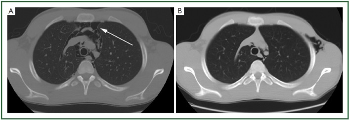
In December 2010, a 16 years-old man was admitted to a local hospital for complaints of chest pain, dyspnea, and neck pain presenting after spasm coughing. His medical history was unremarkable. Chest X-Ray and CT scan diagnosed the presence of pneumomediastinum without signs of pneumothorax and/or pleural effusion (Figure 1A). Thus, the patient was transferred to our hospital for diagnosis and treatment. Bronchosopic was attempted, but no endotracheal lesions were found. Gastrografin swallow excluded any esophageal perforation. Thus, the diagnosis of SPM was made. Medical treatment was done (reassurance, bed rest, oxygen, analgesic, and antibiotic therapy) with gradual improvement of clinical condition. Chest CT scan performed after 9 days, showed the complete disappearance of pneumomediastinum with no evidence of bullae (Figure 1B).
Figure 1.
Massive emphysema of the superior mediastinum, around the trachea and vessels (A).Complete disappearence of pneumomediastinum (B).
Following four days, the patient presented again at our unit for complaint of persistent chest pain and dyspnea which presented again after coughing. Radiological evaluation diagnosed the presence of pneumomediastinum (Figure 2A). All laboratory and diagnostic tests were within normal. Yet, the patient underwent bronchial hyperreactivity test which resulted as negative Thus, he was managed as before and discharged after 7 days. CT scan diagnosed the complete resolution of pneumomediastinum without sequel (Figure 2B).
Figure 2.
Presence of emphysema within superior mediastinum, around the trachea (A), and its complete reassorbation 7 days after (B).
Discussion
SPM was firstly described by Louis Hamman in 1939 (1). It is defined as the presence of interstitial air in the mediastinum without any apparent precipitating factor (2). Alveolar rupture leads to the accumulation of air in the interstitium that circulates centripetally through the venous sheats to the hilum and mediastinum (3). This occurs because the pressure in the mediastinum is lower than that of lung periphery. The most common symptoms are chest pain, dyspnea, and neck pain as observed in our case. If chest X-ray is negative, chest CT scan should be performed because up to 30% of patients with SPM have normal chest –X-ray as reported by Kaneki (4); yet, chest CT is useful to exclude undiagnosed chest pathologies. SPM is a benign condition that generally resolves with medical treatment (5,6). In our experience, we have observed five cases of SPM, of which one occurred just following extubation in a patient undergoing elective breast reduction for gynecomastasia (7). Our experience confirms the previously reported benign nature of this condition.
Recurrent SPM is an exceptional event, and little is known about it; in English literature only three cases of recurrent SPM are reported in the series of Albonik et al. (8), Gerazouniset al. (9), and Maciaet al. (1), respectively. It seems reasonable to think that recurrences of SPM are facilitated by the persistence of predisposing factor (e.g., asthma, interstitial lung disease, pneumonia, bullous lung, and radiation therapy for lung cancer) or when a casual situation happens again.
In the present case, at the time of the first episode of pneumomediastinum differential diagnosis includes pulmonary disease, tracheal bronchial rupture, and Boerhaave’s syndrome. However, radiological findings show the presence of pneumomediastinum without any signs of other diseases. Additional diagnostic tests including contrast-enhanced swallow study and bronchoscopy illustrate no-significant abnormalities. Thus, the diagnosis of SPM is made according to the criteria of Maciaet al. (2) as following: (i) the presence of a clinical picture consistent with pneumomediastinum; (ii) the absence of a clearly defined triggering cause; (iii) the presence of interstitial air in the mediastinum; (iv) and the patient older than 13 years of age. The medical treatment is made with resolution of pneumomediastinum 9 days later.
When the patient returns again to our unit for the presence of pneumomediastinum, we focus regarding the possible cause of the early recurrence of SPM.
The presence of predisposing factor as asthma, interstitial lung disease, pneumonia, bullous lung, and radiation therapy for lung cancer are ruled-out by clinical history and radiological findings. Yet, bronchial hyperreactivity tests result as negative. Thus, in our case the recurrent episodes of coughing may cause SPM as confirmed by review of patient’s history, but remains unclear how spasm coughing may cause the alveolar rupture in absence of respiratory disease and of use of illicit drug, often reported as predisposing factor of pneumomediastinum.
Finally, our case shows that the recurrence of SPM is a possible event also in patient without predisposing factors. An open question is the follow-up of such patients. Certainly, we should tell the patient with an episode of SPM to avoid certain practices as scuba diving especially if above-mentioned predisposing factors are present.
Acknowledgements
Disclosure: The authors declare no conflict of interest.
References
- 1.Hamman L.Spontaneous mediastinal emphysema. Bull Johns Hopkins Hospital 1939;64:1-21 [Google Scholar]
- 2.Macia I, Moya J, Ramos R, et al. Eur J CardiothoracSurg 2007;31:1110-4 [DOI] [PubMed] [Google Scholar]
- 3.Al-Mufarrej F, Badar J, Gharagozloo F, et al. Spontaneous pneumomediastinum: diagnostic and therapeutic interventions. J CardiothoracSurg 2008 3;3:59. [DOI] [PMC free article] [PubMed]
- 4.Kaneki T, Kubo K, Kawashima A, et al. Spontaneous pneumomediastinum in 33 patients: yield of chest computed tomography for the diagnosis of the mild type. Respiration 2000;67:408-11 [DOI] [PubMed] [Google Scholar]
- 5.Turban JW. Spontaneous pneumomediastinum from running sprints. Case Report Med 2010;2010. pii: 927467. [DOI] [PMC free article] [PubMed]
- 6.Gunluoglu MZ, Cansever L, Demir A, et al. Diagnosis and treatment of spontaneous pneumomediastinum. Thorac Cardiovasc Surg 2009;57:229-31 [DOI] [PubMed] [Google Scholar]
- 7.Fiorelli A, Brongo S, D’Andrea F, et al. Negative-pressure pulmonary edema presented with concomitant spontaneous pneumomediastinum: Moore meets Macklin. Interact Cardiovasc Thorac Surg 2011;12:633-5 [DOI] [PubMed] [Google Scholar]
- 8.Abolnik I, Lossos IS, Breuer R. Spontaneous pneumomediastinum: a report of 25 cases. Chest 1991;100:93-5 [DOI] [PubMed] [Google Scholar]
- 9.Gerazounis M, Athanassiadi K, Kalantzi N, et al. Spontaneous pneumomediastinum: a rare benign entity. J Thorac Cardiovasc Surg 2003;126:774-6 [DOI] [PubMed] [Google Scholar]


