Abstract
In mammals, DNA methylation is restricted to cytosines of CpG dinucleotides, which are frequently found in short genomic regions including gene promoters. Methylation within CpG-rich regions around promoters tends to repress gene expression; thus, the CpG islands of housekeeping genes are normally unmethylated. We previously described a testis-specific single-exon gene containing a CpG-rich sequence that is methylated and thus repressed in somatic cells, whereas its expression in spermatogenic cells requires that it be hypomethylated. However, the relationship among the specific expression of spermatogenic genes, their methylation dynamics, and their CpG frequencies are poorly understood. Here, we analyzed the methylation patterns of the sphort genomic region around the transcription start site in spermatogenic cell-specific single-exon genes of various CpG contents. By using UniGene and Ensembl database analyses of the mouse genome and reverse transcription-PCR, we identified 39 single-exon genes that are exclusively expressed in spermatogeniccells. Regardless of their specific expression characteristics, genes containing higher (7 to 14 CpGs in 200 bp; mean = 12) and lower (2 to 6 CpGs in 200 bp; mean = 3.1) number ofCpG were hypo- and hyper-methylated, respectively, in all cell types examined, including spermatogeniccells. We found that genes with intermediate number of CpG (2 to 11 CpGs in 200 bp; mean = 6.9) are methylated in somatic cells, but not in male germ cells. These results suggest that DNA methylation dynamics of spermatogenic cell-specific single-exon genes are associated with CpG content, and the methylation status are stably maintained throughout male germ cell development.
Introduction
In mammals, DNA methylation occurs at cytosine residues in CpG dinucleotides and is heritable during cell division. CpG cytosine methylation is thus an epigenetic marker that is essential for normal embryonic development [1]. This epigenetic modification of DNA is catalyzed by three DNA methyltransferases (Dnmts); Dnmt3a and Dnmt3b initiate de novo methylation and thereby establish new methylation patterns [2], and Dnmt1 is primarily involved in maintaining the methylation pattern [3]. In addition, another member of the Dnmt3 family, Dnmt3L, lacks the Dnmt catalytic domain and is specifically expressed during gametogenesis in the stages during which genomic imprints are established [4], [5].
Several studies have highlighted the importance of DNA methylation in male germ cells. Although mice with conventional null mutations of Dnmt3a die at approximately 3 weeks of age [2], conditional mutations of Dnmt3a targeted to the germ line result in a defect in spermatogenesis [6]. In the conditionally mutated germ cells, lack of methylation at imprinted genes and some repetitive sequences are observed [6], [7]. Male mice lacking Dnmt3L are infertile because they lack mature germ cells [4], [5] and display abnormal meiotic chromosome segregation [8], [9]. In these Dnmt3L-mutant mice, several repetitive and non-repetitive sequences, including imprint genes, in male germ cells are incompletely methylated [6]–[11]. These findings suggest that appropriate DNA methylation patterns are important for male germ-cell development.
The crucial role of DNA methylation in male germ cells is also supported by the presence of distinct methylation patterns. Some reports have shown that certain genome-wide DNA methylation patterns are unique to spermatozoa [11]–[13]. This unique state of DNA methylation might arise immediately after the genome-wide reprogramming event that occurs specifically in the primordial germ cells (PGCs) of the developing embryo between 10.5 and 12.5 days post-coitus (dpc) [14], when the DNA methylation patterns in imprinted and germ cell-specific genes are erased and reestablished [7], [15], [16]. Alternatively, de novo methylation and demethylation may occur in distinct CpG regions during spermatogenesis, as suggested by the observation that the methylation status of a few CpGs changes in spermatogenic cells [17].
Spermatogenesis is a highly programmed developmental process in which self-renewal and mitotic cell division in spermatogonia, meiotic cell division in spermatocytes, and spermiogenesis in spermatids act sequentially to produce spermatozoa. The temporal and spatial expressions of the specific genes that manage the organization of this complex process are strictly regulated.
Testis-specific expression of these genes is usually regulated by relatively short sequences in the gene promoter [18]–[20]. In addition to this type of regulatory mechanism involving the binding of transcription factors to cis-elements, we previously suggested that another type of mechanism, involving DNA methylation-induced gene suppression, might play a crucial role in gene expression during spermatogenesis. We found that the CpG-rich region in the spermatid-specific single-exon gene, Tact1/Actl7b, is methylated in somatic cells but not in spermatocytes and spermatids. Furthermore, transfection experiments with in vitro-methylated constructs indicated that methylation of Tact1/Actl7b represses its promoter activity in somatic cells [21]. These results revealed that germ cell-specific demethylation on the short genomic sequences near the promoter may be regulated for efficient transcription of the intronless gene. So far, several testis-sepcific single-exon genes have been found [22]–[25], but only limited analyses of their methylation status genes have been reported [26], [27].
In this study, we identified all of the single-exon genes that are expressed exclusively inspermatogenic cells and examined their methylation profiles around the promoter to explore the methylation dynamics of testis-specific genes during spermatogenesis. We found that these genes had a stable methylation status in the male germ line and that they could be sorted into three categories based on their methylation profiles: constitutively hypomethylated, constitutively hypermethylated, and germ line-specific hypomethylated genes. Furthermore, we found an association between their distinct methylation profiles and their CpG content. Genes with higher and lower number of CpG were constitutively hypo- and hypermethylated, respectively, and genes with intermediate number of CpG were hypomethylated in germ line cells and hypermethylated in somatic cells. These results suggest that the methylation status of spermatogenic cell-specific genes is regulated by their CpG content and that the hypomethylated status of the genes with intermediate CpG content might regulate their germ cell-specific expression.
Results
Identification of Spermatogenic Cell-specific Intronless Genes
To identify spermatogenic cell-specific intronless genes in the mouse genome, we first collected a dataset of testis-specific genes from the UniGene database (Mus musculus, UniGene build #159). We examined the structures of 246 testis-specific genes using GenBank and Ensembl database searches and found that 62 of them had open reading frames uninterrupted by introns, identifying them as candidate functional intronless genes (see Table S1). Of these genes, two (1700084P21Rik and Tcp10a) were not assembled at genome annotation status, and the genomic structures of nine (1700012O15Rik, 1700042G15Rik, 1700066D14Rik, AA066038, 4930571K23Rik, 4933414I15Rik, 1700110M21Rik, Cypt12, and an unnamed gene) were not conclusively determined. The remaining 51 genes were taken as single-exon genes; 19 of these had been previously identified.
To confirm the expression of these intronless genes in the testis, we designed PCR primer sets and performed semiquantitative reverse transcription (RT)-PCR using total RNA prepared from various organs (Figure 1, Figure S1). In addition to testes from wild-type mice, we also used testes from W/Wv mutant mice, which almost entirely lack germ cells as adults. Intense PCR signals were observed for all 51 genes in the testis, consistent with previous reports and expressed sequence-tag data for these genes in the UniGene database. Twelve of the genes (1700009J07Rik, Csl, Prm3, LOC622019, 1700011K15Rik, 1700054O13Rik, 1700019M22Rik, 1700013N18Rik, Spaca4, 1700024P04Rik, 1700010M22Rik, and Hils1) were also expressed in other tissues. The remaining 39 genes were expressed only in wild-type testis and were used as spermatogenic cell-specific intronless genes, although one of them, Ftmt, has been reported to be expressed in some other tissues [28].
Figure 1. Spermatogenic cell-specific intronless genes are expressed in testis but not in any other tissues.
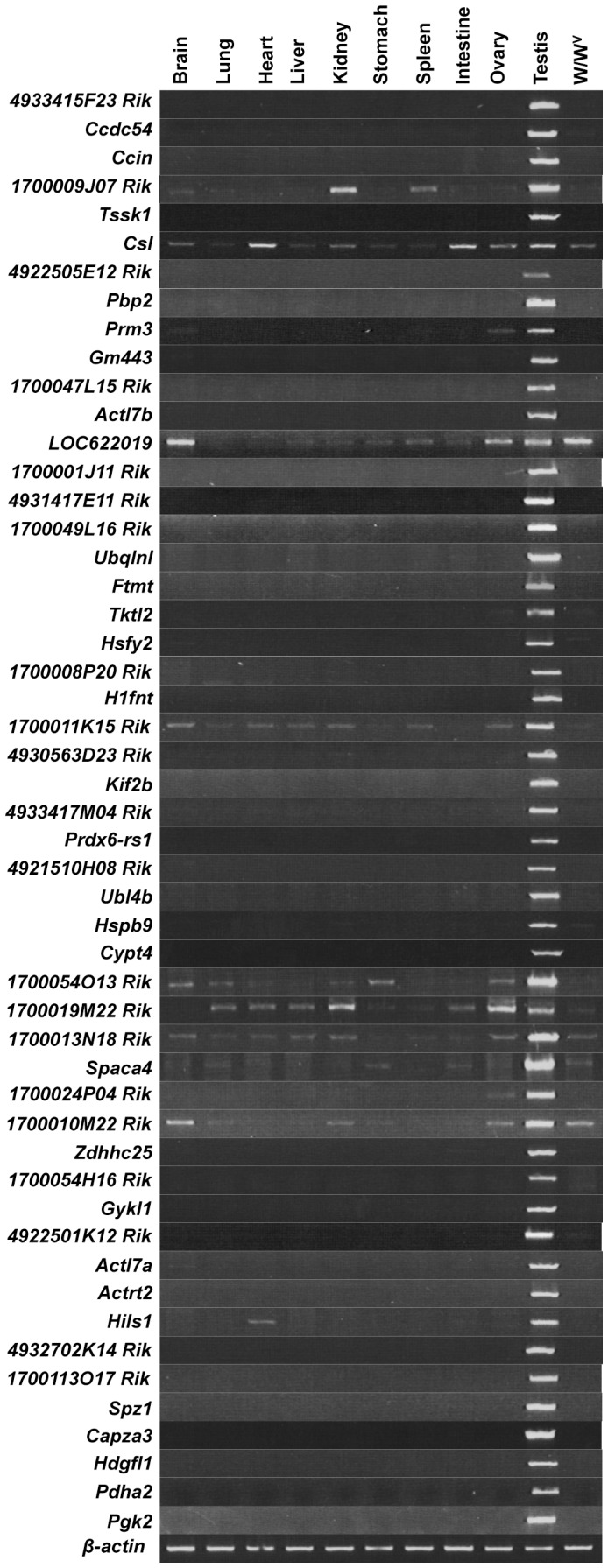
Semiquantitative RT-PCR for the 51 intronless genes identified in Table S1 was carried out using total RNA isolated from adult mouse tissues. Thirty-nine genes were exclusively detected in testis and chosen for further study. The intensity of the β-actin band was used as a quantitative standard. The absence of genomic DNA in RNA samples is shown in Figure S1.
Characterization of Spermatogenic Cell-specific Intronless Genes
The intronless spermatogenic cell-specific genes identified in this study are shown in Table 1. A large number of intronless genes arise from retroposition of parental gene transcripts [29]. Since intronless genes are expected to be non-chromosomally-linked paralogs of their multiple exon-containing parental genes, we performed an Ensembl database analysis and homology search using BLASTP to find putative parental genes for the identified intronless genes. Paralogs were identified for 32 of the 39 genes (82%), suggesting that most spermatogenic cell-specific intronless genes are retrogenes generated from parental genes. However, we could not exclude the possibility that 10 of the genes (Tssk1, Pbp2, Actl7a, Actl7b, Ubqlnl, Ftmt, Gykl1, Actrt2, 4932702k14Rik, and 1700113O17Rik) might have arisen through segmental gene duplication from the other intronless paralogs. We were unable to find paralogs for the remaining seven intronless spermatogenic cell-specific genes, which might have diverged so much from their paralogs as to be unrecognizable by our methods.
Table 1. Identified spermatogenic cell-specific intronless genes.
| Gene symbol | Chr | Ensemble ID | Paralogue | Orthologue | |||
| Gene symbol | Exon | Chr | Rat | Human | |||
| 4933415F23Rik | 1 | ENSMUSG00000073730 | Ppp1r14b (74%) | 4 | 19 | RGD1309049 (78%) | |
| Ccdc54 | 16 | ENSMUSG00000050685 | Ccdc54 (82%) | CCDC54 (64%) | |||
| Ccin | 4 | ENSMUSG00000070999 | Klhl11 b (27%) | 2 | 11 | Ccin (99%) | CCIN (93%) |
| Tssk1 | 16 | ENSMUSG00000041566 | Tssk5 b (38%) | 8 | 15 | Tssk1 (96%) | TSSK1B (84%) |
| 4922505E12Rik | 1 | ENSMUSG00000043429 | LOC289334 (80%) | C1orf65 (55%) | |||
| Pbp2 | 6 | ENSMUSG00000047104 | Pepb1 (79%) | 4 | 5 | Pbp2 (88%) | |
| Gm443 | 5 | ENSMUSG00000038044 | Cct8 (29%) | 15 | 16 | MGC125233 (81%) | CCT8L1 (61%) |
| 1700047L15Rik | 12 | ||||||
| Actl7b | 4 | ENSMUSG00000070980 | Actin family (<39%) | Actl7b (85%) | ACTL7B (83%) | ||
| 1700001J11Rik | 9 | ENSMUSG00000066639 | Rnf19a b (84%) | 10 | 15 | ||
| 4931417E11Rik | 6 | ENSMUSG00000056197 | 1200003C05Rik (71%) | 7 | 12 | RGD1562515 (91%) | |
| 1700049L16Rik | 10 | ENSMUSG00000043859 | Hn1l (88%) | 5 | 17 | RGD1559903 (81%) | |
| Ubqlnl | 7 | ENSMUSG00000051437 | Ubqln1 b (36%) | 11 | 13 | RGD1307202 (82%) | UBQLNL (45%) |
| Ftmt | 18 | ENSMUSG00000024510 | Fth1 (59%) | 4 | 19 | Ftmt (95%) | FTMT (67%) |
| Tktl2 | 8 | ENSMUSG00000025519 | Tkt (67%) | 14 | 14 | Tktl2 (94%) | TKTL2 (80%) |
| Hsfy2 | 1 | ENSMUSG00000045336 | Hsf5 (13%) | 6 | 11 | Hsfy1 b (95%) | |
| 1700008P20Rik | 7 | ENSMUSG00000055826 | Tesc (52%) | 8 | 5 | RGD1311676 (83%) | |
| H1fnt | 15 | ENSMUSG00000048077 | H1fnt (76%) | H1FNT (42%) | |||
| 4930563D23Rik | 16 | ENSMUSG00000051728 | LOC498063 (86%) | ||||
| Kif2b | 11 | ENSMUSG00000046755 | Kif2a (55%) | 20 | 13 | Kif2b (94%) | KIF2B (78%) |
| 4933417M04Rik | 1 | ENSMUSG00000073479 | OTTMUSG00000005491 (37%) | 2 | 11 | RGD1561646 (30%) | FAM71A (47%) |
| Prdx6-rs1 | 2 | ENSMUSG00000050114 | Prdx6 (86%) | 5 | 1 | Pseudo | |
| 4921510H08Rik | 10 | ENSMUSG00000047025 | LOC500825 (91%) | C12orf12 (62%) | |||
| Ubl4b | 3 | ENSMUSG00000055891 | Ubl4 (34%) | 4 | X | LOC684560 b (96%) | UBL4B (62%) |
| Hspb9 | 11 | ENSMUSG00000017832 | Hspb8 b (27%) | 3 | 5 | Hspb9 (76%) | HSPB9 (54%) |
| Cypt4 | 9 | ENSMUSG00000047995 | Cypt3 (80%) | 2 | X | ||
| Zdhhc25 | 15 | ENSMUSG00000054117 | Zdhhc7 b (41%) | 8 | 8 | LOC300323 b (83%) | |
| 1700054H16Rik | 11 | Pom121 b (28%) | 13 | 5 | DKFZp564 b (42%) | ||
| Gykl1 | 18 | ENSMUSG00000053624 | Gyk (82%) | 21 | X | Gk-rs1 (93%) | |
| 4922501K12Rik | 14 | ENSMUSG00000078127 | Gm93 (17%) | 4 | 8 | 4922501K12Rik (67%) | C10orf73 (47%) |
| Actl7a | 4 | ENSMUSG00000070979 | Actin family (<37%) | Actl7a (96%) | ACTL7A (85%) | ||
| Actrt2 | 4 | ENSMUSG00000051276 | Actin family (<49%) | Actrt2 (95%) | ACTRT2 (80%) | ||
| 4932702K14Rik | 17 | ENSMUSG00000046173 | Pabpc1 (80%) | 15 | 15 | Pabpc3 (94%) | |
| 1700113O17Rik | 2 | ENSMUSG00000062651 | H2A histone family (<39%) | ||||
| Spz1 | 13 | ENSMUSG00000046957 | Spz1 (76%) | SPZ1 (48%) | |||
| Capza3 | 6 | ENSMUSG00000041791 | Capza2 (35%) | 10 | 6 | Capza3 (97%) | CAPZA3 (91%) |
| Hdgfl1 | 13 | ENSMUSG00000045835 | Hdgf (33%) | 6 | 3 | Pwwp1 (68%) | HDGFL1 (36%) |
| Pdha2 | 3 | ENSMUSG00000047674 | Pdha1 (75%) | 11 | X | Pdha2 (92%) | PDHA2 (74%) |
| Pgk2 | 17 | ENSMUSG00000031233 | Pgk1 (83%) | 11 | X | Pgk2 (90%) | PGK2 (85%) |
Putative paralogs and orthologs were basically identified based on Ensemble database search.
BLASTP was carried out to identify paralogs and orthologs in the case that paralogs and orthologs were not registered in Ensemble database.
The most identical gene was represented when several candidates were registered in the database. Paralogs in a gene family were represented by their family name. Percentages indicate identities of amino acid sequence for each intronless gene.
Analysis of the chromosomal locations of the 32 retroposed gene pairs revealed an apparently random insertion of the retrogenes into almost all chromosomes except chromosomes 12 and 19 and the sex chromosomes. In contrast, a disproportionately large number of putative parental genes were found on the X chromosome. On the basis of the assumption that the number of parental genes on calculated that the mouse X chromosome has generated a 4-fold excess of spermatogenic cell-specific retrogenes, consistent with a previous report [30].
We also examined orthologs of the intronless spermatogenic cell-specific genes in human and rat. Of the 39 mouse genes, we found that 23 (59%) had orthologs in both the human and rat genomes (Table 1), suggesting that these genes originated before the divergence of the primate and rodent lineages. Of these orthologous genes, two (Pdha2 and Pgk2) had high levels of deduced amino acid identity with both their paralogs and their orthologs, implying that they have functions similar to those of their paralogs. For six genes (Ccin, Tssk1, Actl7a, Actl7b, Actrt2, and Capza3) the levels of identity were markedly higher for their orthologs than their paralogs, suggesting that they had acquired divergent functions. Four of the genes (1700047L15Rik, 1700001J11Rik, Cypt4, and 1700113O17Rik) had no orthologs in rat or human, suggesting that they are mouse-specific. However, it is unlikely that these four genes were generated after the divergence of mouse from rat, since their levels of deduced amino acid identity with their paralogs are low.
Timing of Intronless Gene Expression during Spermatogenesis
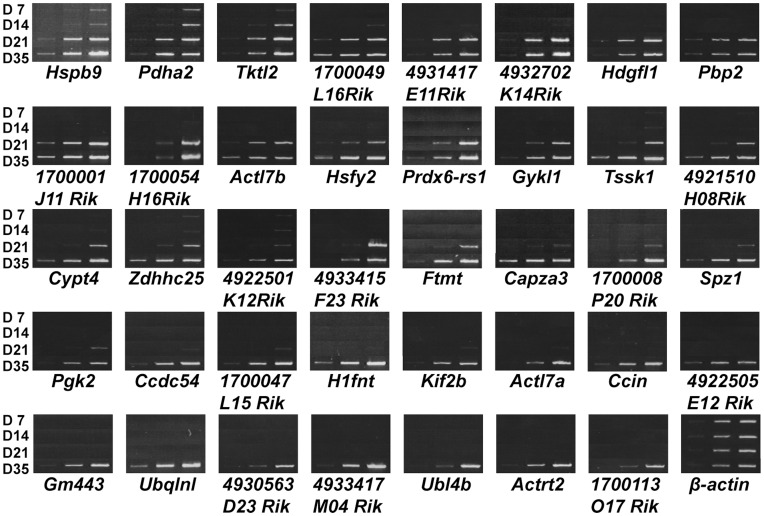
To investigate the expression profiles of the spermatogenic cell-specific intronless genes during spermatogenesis, we performed RT-PCR analysis using total RNA isolated from testes of 7-, 14-, 21-, and 35-day-old mice (D7, D14, D21, and D35 mice, respectively). The first spermatogenic cycle initiates a few days after birth and proceeds during postnatal development; consequently, the juvenile mouse testis at each of these days contains a different subset of spermatogenic cell types. Type A and B spermatogonia are present on D7. On D14, meiotic prophase is initiated, and the germ cells become pachytene spermatocytes. Haploid spermatids appear in increasing numbers on D21, and all stages of spermatogenic cells, including spermatozoa, appear on D35 [31].
As shown in Figure 2 (see also Figure S2), expression of all the genes was upregulated during the course of testis development. Signals for Hspb9 and Pdha2 were detected in D7 and D14 testes and were higher in D21 and D35 testes. Expression of Tktl2 was observed at D14 and was higher at D21 and D35. For most of the genes, strong signals of varying intensities were detected on D21, indicating expression at the late spermatocyte stage. The remaining genes were not detected until D35, indicating expression exclusive to the round spermatid stage. The observed expression profiles were consistent with previous reports [32]–[39] and conclusively indicated that spermatogenic cell-specific intronless genes have a strong bias for expression during the late meiotic spermatocyte to haploid spermatid stages.
Figure 2. Expression levels of 39 spermatogenic cell-specific intronless genes are increased in later stages of spermatogenesis.
Semiquantitative RT-PCR was carried out using total RNA isolated from D7, D14, D21, and D35 testes. The number of PCR cycles increases from left to right in each panel, as described in Table S2. The β-actin band represents equal expression levels for each stage. The absence of genomic DNA in RNA samples is shown in Figure S2.
Methylation Profile of Intronless Genes during Spermatogenesis
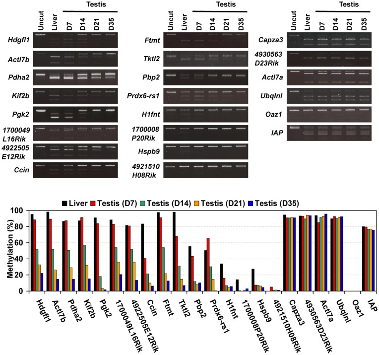
We previously demonstrated that DNA methylation of the CpG-rich region in the Tact1/Actl7b gene represses its expression in somatic cells and that demethylation is necessary for its expression in spermatogenic cells [21]. In another study, gradual loss of methylation was shown to occur in the Pgk2 gene during male germ cell development [27]. Moreover, several studies showed that the expression of the testis-specific genes were usually regulated by short genomic region including the promoter [18]–[20]. These observations prompted us to investigate whether demethylation is a general requirement for expression of spermatogenic cell-specific intronless genes. To address this issue, we carried out combined bisulfite restriction analysis (COBRA) of 20 selected intronless genes using genomic DNA from D7, D14, D21, and D35 mouse testis and from adult mouse liver. Nineteen of these genes contained restriction enzyme sites within 100 bp of the transcription start sites (Figure S3). The 20th gene, Actl7b, had a restriction site for COBRA at position +340; its methylation status is known to be coordinately regulated with two CpG sites within 100 bp of the transcription start site [21].
The twenty genes were sorted into three groups based on their observed methylation patterns in D7, D14, D21, and D35 testis and adult liver (Figure 3). The first group comprised four genes (H1fnt, 1700008P20Rik, Hspb9, and 4921510H08Rik) that were hypomethylated in all samples. Their low methylation level essentially resembled that of Oaz1, which has a COBRA restriction site within a CpG island, although H1fnt, 1700008P20Rik, and Hspb9 were moderately methylated in liver. In contrast, the four genes of the second group (Capza3, 4930563D23Rik, Actl7a, and Ubqlnl) were hypermethylated in all tissues examined, similar to endogenous IAP retroviruses, which are known to be highly methylated in both somatic tissues and testis [40]. The methylation statuses of the eight genes in the first and second groups were essentially stable in spermatogenic cells and somatic cells.
Figure 3. Methylation profiles of spermatogenic cell-specific intronless genes are classified three groups in juvenile mouse testis.
COBRA was carried out to examine the methylation status of the selected genes, the CpG island in the Oaz1 gene, and constitutively methylated endogenous IAP retroviruses in genomic DNA isolated from D7, D14, D21, and D35 testes and adult liver. Sodium bisulfite-treated DNA was amplified with specific primers (details in Table S3) and digested with HpyCH4 IV (recognition site: ACGT), Taq I (TCGA) or Acc II (CGCG).
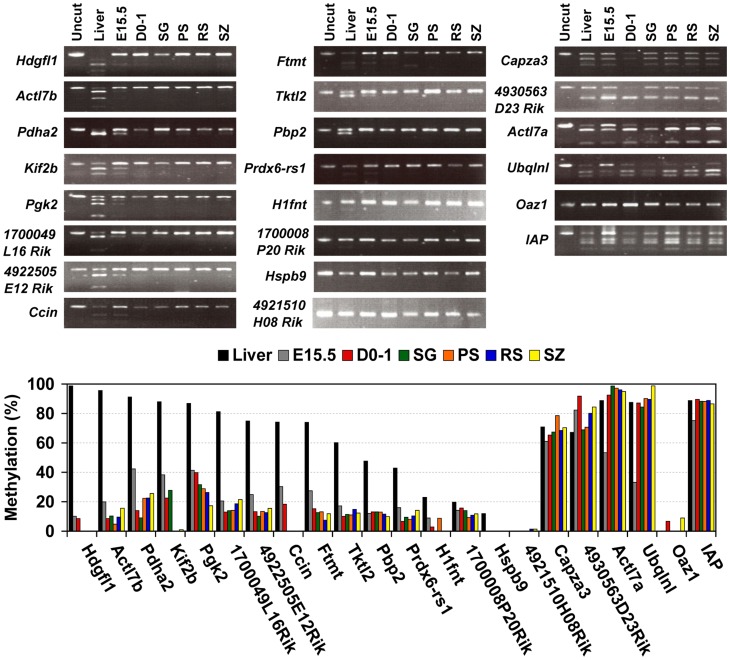
The remaining 12 genes (Hdgfl1, Actl7b, Pdha2, Kif2b, Pgk2, 1700049L16Rik, 4922505E12Rik, Ccin, Ftmt, Tktl2, Pbp2, and Prdx6-rs1), which formed the third group, were methylated at high levels in liver and D7 testis. The methylation levels of these genes gradually decreased in accordance with testis maturation, during which time the fraction of germ cells in the testis cell population increases [31] and spermatogenesis proceeds. To determine whether methylation levels of these genes were constitutively low in germ cells or if they decreased during spermatogenesis, we examined the methylation status of these genes in spermatogenic cells at each stage, including spermatogonia, pachytene spermatocytes, round spermatids, and spermatozoa. We found that they were highly methylated in liver but hypomethylated or nonmethylated in spermatogenic cells (Figure 4). Furthermore, their methylation levels were also low in pre-spermatogonia, including embryonic and neonatal gonocytes, suggesting that they were hypomethylated in a germ cell-specific manner. In contrast, methylation levels of the genes in the first and second groups were similar in isolated germ cells and in testis, although Actl7a and Ubqlnl methylation levels might have decreased transiently in embryonic germ cells.
Figure 4. Methylation profiles of spermatogenic cell-specific intronless genes are classified three groups in male germ cells.
COBRA was carried out to examine the methylation status of the selected genes, the CpG island in the Oaz1 gene, and constitutively methylated IAP in genomic DNA isolated from 15.5 dpc embryonic (E15.5) and neonatal (D0–1) gonocytes, spermatogonia (SG), pachytene spermatocytes (PS), round spermatids (RS), epididymal spermatozoa (SZ), and adult liver. The sodium bisulfite reaction and restriction enzyme digestion were carried out as described in Figure 3. The COBRA results are also represented by bar graphs.
Distinct Methylation Profiles Correlate with CpG Content
The presence of three distinct types of methylation profiles among the spermatogenic cell-specific intronless genes implies that the genes in these groups interact differently with the regulatory machinery. Methylation level has been shown to be related to the number of CpG dinucleotides within distinct genome regions [41]. To gain insight into the variety of methylation profiles, we determined the number of CpG dinucleotides in a 200-bp segment containing the COBRA restriction sites. As shown in Figure 5, the frequency of CpG dinucleotides in the genes differed among the three groups. Genes belonging to the constitutively hypermethylated group had low CpG content; in 200 bp, they had 2 to 6 CpGs (mean = 3.1). Genes of the constitutively hypomethylated group had high CpG content; in 200 bp, there were 7 to 14 CpGs (mean = 12). These results suggest that genomic regions with lower CpG number are stably methylated in any tissue, whereas those with higher CpG number tend to maintain a hypomethylated state. The genes that were demethylated in male germ cells but methylated in somatic cells had intermediate CpG number (2–11 CpGs in 200 bp; mean = 6.9). The range of CpG content in this intermediate group included genes of low CpG frequency (<6 in 200 bp) but no genes of very high frequency (>12 in 200 bp).
Figure 5. Methylation levels of the three categorized genes depend on their CpG contents.
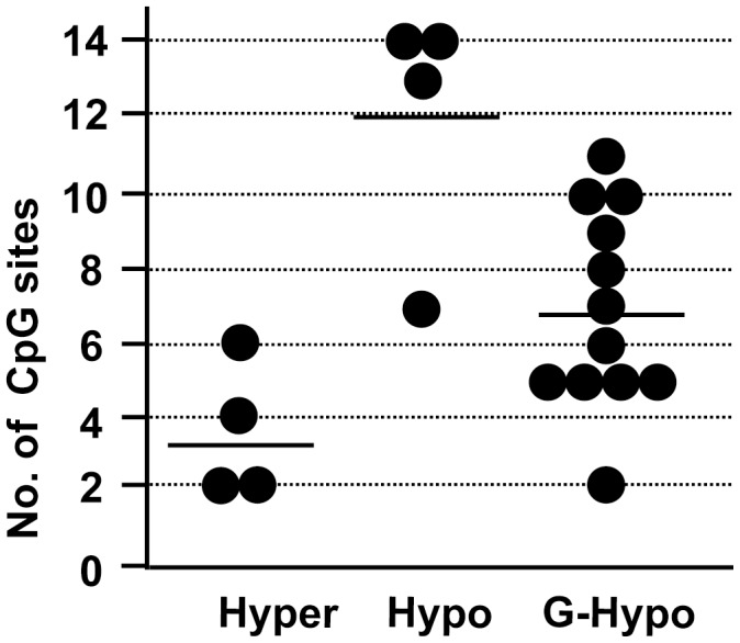
The number of CpG sites within a 200-bp segment containing the COBRA restriction sites in the 20 intronless genes described in Figures 3 and 4 was determined, and the results plotted as a histogram. Each dot indicates a gene in each group classified according to the methylation profiles shown in Figures 3 and 4. Hyper, constitutive hypermethylated; Hypo, constitutive hypomethylated; G-Hypo, germ cell-specific hypomethylated.
Discussion
Spermatogenic Cell-specific Intronless Genes
In our analysis of the mouse UniGene database (UniGene build #159), we identified 246 testis-specific genes. To select the primary hits from the database searches, we restricted the number of expressed sequence-tag entries to at least 10. Using RT-PCR, we determined that 39 of these intronless genes were expressed in spermatogenic cells. Of these genes, 36 (92%) were expressed in mice older than 21 days, suggesting that most testis-specific genes selected in this analysis were expressed during the late-meiotic and post-meiotic stages of spermatogenesis.
Previous UniGene searches yielded 24 and 28 uncharacterized genes specifically expressed in spermatocytes and round spermatids, respectively [42], [43]. Our list included 14 (58.3%) of the spermatocyte-specific genes and 8 (28.6%) of the spermatid-specific genes. Of the remaining 28 previously identified genes, 26 were not included in our list because they had either been eliminated from the most recent version of the database (9 genes), had entries in various tissues other than testis (10 genes), or had a low number of entries (9 genes). Although the remaining two genes (Mm. 45611 and 437189) satisfied our criteria, our analysis did not select them, for unknown reasons.
One-fourth (62/246) of all the testis-specific genes we identified consisted of a single exon. In contrast, only 5.6% (2/36) of spermatogonially expressed germ cell-specific genes are intronless [44]. Therefore, a lack of introns may be characteristic of genes expressed during later stages of spermatogenesis. In addition, a large percentage of testis-specific intronless genes are probably retroposons (32/39, 82%), indicating that retrogenes tend to be expressed at later stages of spermatogenesis.
Retrogenes generally give rise to pseudogenes; as inserted cDNA is reverse-transcribed from mRNA, it lacks a genuine promoter sequence and, therefore, cannot be expressed in any tissue. Thus, our results suggest that the late stage of spermatogenesis provides an appropriate environment for retroposon transcription and facilitates gene expression from promoter-like sequences. Furthermore, these genes might have acquired their high level of transcriptional activity in pachytene spermatocytes or round spermatids by accumulating favorable mutations in their promoters during evolution. Otherwise, this stage of germ cells might possess an accessible environment for retroposition. It is possible that the retrogenes were simply inserted into chromatin regions that are transcription-prone.
Regulation of Methylation Status
We classified the identified spermatogenic cell-specific intronless genes into three distinct groups on the basis of their DNA methylation profiles, which were associated with their CpG content. The genes with lower CpG number were constitutively hypermethylated in somatic and germ cells. In contrast, genes with higher CpG number were hypomethylated in both somatic and germ cells. These two groups accounted for 40% of the spermatogenic cell-specific intronless genes.
We also demonstrated that the remaining intronless genes those with intermediate CpG number were hypomethylated in spermatogenic cells and hypermethylated in somatic cells. These observations agree with the reported promoter methylome analysis in which methylated DNA from human primary fibroblasts and sperm was immunoprecipitated and analyzed using microarrays [41]. Other studies have shown that the haploid-specific genes with lower CpG number Prm1, Prm2, and Tnp2, which have two exons are hypermethylated throughout spermatogenesis [45], [46], and another haploid-specific, two-exon gene with intermediate CpG number, Tnp1, is hypomethylated during spermatogenesis [46]. Taken together, these analyses suggest that a correlation between methylation status and CpG content is a common feature of the mammalian genome. We also found that the intronless genes with intermediate CpG number were hypomethylated in gonocytes in neonatal males and in male embryos at 15.5 dpc, indicating that these genes are hypomethylated throughout the male germ line.
Dynamic changes in DNA methylation occur during PGC development in the genital ridge and in early embryogenesis. The differentially methylated regions of some imprinted genes and some repetitive sequences are demethylated in PGC in the post-migrating stage but not in somatic cells [15], [16]. These observations suggest that genome-wide demethylation occurs in germ cells at these stages, during which testis-specific hypomethylated intronless genes may also be demethylated. The hypomethylated status of the genes and PGC expression of the Mvh gene [47] are maintained during spermatogenesis until spermatozoa form, although the differential methylation regions of paternally imprinted genes are remethylated after 14.5 dpc [7].
Dnmt3L, an activator of Dnmt3a and Dnmt3b [48]–[52], is indispensable for methylation of imprinted genes and repetitive sequences in gonocytes [7], indicating that Dnmt3L/Dnmt3a and Dnmt3L/Dnmt3b complexes can target these genome regions but not testis-specific hypomethylated intronless genes. It was also reported that methylation of histone H3 Lysine 4 blocks the association of Dnmt3L to the chromatin [53], suggesting that testis-specific hypomethylated intronless genes were marked with the histone modification preceded to the de novo methylation. In addition, genome-wide remethylation occurs during early embryogenesis. Although intronless genes with higher CpG number, are hypomethylated in all cell types, testis-specific hypomethylated intronless genes are hypermethylated in somatic cells, indicating that genome sequences of the genes might be a determinant of de novo methylation during development, as mentioned previously [41].
Relationship between DNA Methylation and Gene Expression
DNA methylation is thought to affect transcriptional silencing by two mechanisms. First, methylated CpGs can directly inhibit the association of some transcriptional factors with their regulatory sequences [54]; second, proteins that recognize methyl-CpG can elicit the repressive potential of methylated DNA [55], [56]. In the latter mechanism, methyl-CpG-binding proteins bound to methylated CpGs are thought to recruit transcriptional co-repressors to modify the surrounding chromatin, creating a gene-silencing environment [57]–[62]. In the present study, however, we showed that intronless genes with lower CpG number are expressed in round spermatids, in which they have hypermethylated CpGs. It appears that a high density of methylated CpGs is required for methylation-dependent gene silencing. Alternatively, the positions of the methylated CpGs might not be appropriate for gene silencing.
Some promoters of testis-specific genes can activate transcription in cultured somatic cells [21], [63], [64]. We previously demonstrated that methylation of the testis-specific Tact1/Actl7b gene with intermediate number of CpG dramatically reduced its expression relative to an unmethylated control in a transient transfection assay [21]. More than 40 cancer/testis antigens have been identified; this class of tumor antigen is encoded by genes that are normally expressed in testis germ cells but not in normal somatic tissues [65], and promoter demethylation appears to be a key element in their expression. The cancer/testis antigen genes are methylated in normal somatic tissues and are activated by demethylation during spermatogenesis. The global hypomethylation that occurs during carcinogenesis is associated with their expression [66], [67]. These observations suggest that DNA methylation of testis-secific hypomethylated genes in somatic cells play a role in gene silencing.
However, certain mechanisms other than DNA methylation might be involved in repression of testis-specific hypomethylated intronless genes in male germ cells, despite their hypomethylated states. Since a crucial epigenetic mark of transcriptional silencing is the methylation of histone H3 Lysine 9 (H3K9) by the histone lysine methyltransferase G9a [68], [69], this methylation might be involved in the repression the genes. The genome-wide pattern of dimethylated H3K9 is established in pre-leptotene spermatocytes by G9a and is erased in pachytene spermatocytes by the mono/dimethylated H3K9-specific demethylase JHDM2A [70]. In addition, in one study, H3K9 methylation was correlated with Tnp1 and Prm1 gene silencing in the round spermatids of mutant mice with a disrupted JHDM2A gene [71]. On the other hand, in another study, dimethylated H3K9 was not enriched in the proximity of the Prm1 gene promoter in spermatogenic cells at any stage [72]. These inconsistent data show that the exact nature of the link between histone modifications and silencing of testis-specific hypomethylated genes in male germ cells remains to be elucidated.
Materials and Methods
Tissue Preparation and Germ Cell Isolation
All tissues and cells were isolated from Slc: ICR mice (Japan SLC, Inc.). TRIzol reagent (Invitrogen) was used to isolated total RNA from adult mouse tissues, including brain, lung, heart, liver, kidney, stomach, spleen, ovary, wild-type testis, and W/Wv testis, and from testes of D7, D14, D21, and D35 mice.
Gonocytes and spermatogonia were isolated by FACS. Testes from 15.5 dpc embryos and from neonates at day 0–1 and 7 were dissociated with 0.1% trypsin (Nacalai Tesque) at 32°C for 10 min. The trypsin reaction was stopped by the addition of DMEM containing 10% FBS and passed through nylon mesh. The dissociated testis cells were incubated with 10% FBS/DMEM at 32°C for 15 min and then washed three times with ice-cold phosphate-buffered saline containing 0.1% bovine serum albumin and 0.01% sodium azide.
The isolated testis cells were incubated with anti-EpCAM antibody (Santa Cruz Biotechnology) for 60 min and then with Alexa Fluor 488-conjugated goat anti-rat IgG (Molecular Probes) and used for FACS analysis (FACS Aria, BD Biosciences). The purity of the germ cells was determined by immumostaining using anti-GCNA1 antibody and Alexa Fluor 594-conjugated goat anti-rat IgM (Molecular Probes). Approximately 80% of the cells exhibited signals for both EpCAM and GCNA1.
Pachytene spermatocytes and round spermatids were also isolated using FACS. The tunica albuginea was removed from adult testis, and the seminiferous tubules were dissociated using sequential enzymatic digestion by collagenase (100 U/ml, Worthington) at 32°C for 15 min and then by 0.1% trypsin at 32°C for 15 min. Five minutes into the trypsin treatment, DNase I (2 µg/ml; Sigma) was added. The trypsin reaction was stopped by the addition of trypsin inhibitor (0.1%; Nacalai Tesque), and the resulting suspension was passed through nylon mesh. The cells were then incubated with PBS containing 1% FBS, 1 µg/ml Hoechst 33342, and 2 µg/ml propidium iodide (Invitrogen) at 32°C for 20 min. FACS Aria was performed with a 407-nm violet laser. The pachytene spermatocytes and round spermatids were successfully separated by their DNA content and collected [73]. The purity of the cell preparations was evaluated by immunostaining of SCP3 for pachytene spermatocytes (75%) and by examination of cell morphology for round spermatids (90%). Epididymal spermatozoa were collected using a standard protocol.
All animal experiments were approved by the Animal Care and Use Committee of the Research Institute for Microbial Diseases, Osaka University.
Reverse Transcription (RT)-PCR
One microgram of total RNA was treated with DNase I at 37°C for 20 min, and the cDNA was reverse-transcribed using SuperScript III (Invitrogen) at 50°C for 60 min. Ex Taq Polymerase (TaKaRa) was then used for PCR with the primers and conditions shown in Table S2.
Combined Bisulfite Restriction Analysis (COBRA)
One microgram of genomic DNA was treated with bisulfite using an EZ DNA methylation Kit (Zymo Research), and 50 ng of the bisulfite-treated DNA was used for PCR with the primers and conditions shown in Table S3. The PCR products were ethanol-precipitated and digested with HpyCH4 IV (NEB) and AccII (TaKaRa) at 37°C and with TaqI (TaKaRa) at 60°C for 1 h. The digested PCR products were separated by 1.6%-agarose gel electrophoresis. Signal intensities were measured using an Image Reader FLA-7000 (Fujifilm).
Supporting Information
The absence of genomic DNA in 51 RNA samples. RT-PCRs were performed using RNA samples with or without reverse transcriptase reaction.
(TIF)
Schematic representation of CpG sites of 20 selected spermatogenic cell-specific intronless genes. CpG sites within 200 bp of the transcription start site are shown with vertical lines. A dotted arrow indicates the transcription start sites. Restriction enzyme sites are indicated by capital letters. A, AccII; H, HpyCH4 IV; T, TaqI.
(TIF)
Testis-specific genes in the mouse.
(DOC)
Primer sequence and parameters for RT-PCR.
(DOC)
Primer sequence and parameters for bisulfite PCR.
(DOC)
Acknowledgments
We thank Drs. G. C. Enders and S. Chuma for their kind gifts of anti-GCNA1 antibody and anti-SCP3 antibody, respectively. We also thank K. Nakamura and Y. Kabumoto for FACS analysis.
Funding Statement
Grant-in-Aid for Science Research from the Ministry of Education, Culture, Sports, Science and Technology Japan. The funders had no role in study design, data collection and analysis, decision to publish, or preparation of the manuscript.
References
- 1. Bird A (2002) DNA methylation patterns and epigenetic memory. Genes Dev 16: 6–21. [DOI] [PubMed] [Google Scholar]
- 2. Okano M, Bell DW, Haber DA, Li E (1999) DNA methyltransferases Dnmt3a and Dnmt3b are essential for de novo methylation and mammalian development. Cell 99: 247–257. [DOI] [PubMed] [Google Scholar]
- 3. Robert MF, Morin S, Beaulieu N, Gauthier F, Chute IC, et al. (2003) DNMT1 is required to maintain CpG methylation and aberrant gene silencing in human cancer cells. Nat Genet 33: 61–65. [DOI] [PubMed] [Google Scholar]
- 4. Bourc’his D, Xu GL, Lin CS, Bollman B, Bestor TH (2001) Dnmt3L and the establishment of maternal genomic imprints. Science 294: 2536–2539. [DOI] [PubMed] [Google Scholar]
- 5. Hata K, Okano M, Lei H, Li E (2002) Dnmt3L cooperates with the Dnmt3 family of de novo DNA methyltransferases to establish maternal imprints in mice. Development 129: 1983–1993. [DOI] [PubMed] [Google Scholar]
- 6. Kaneda M, Okano M, Hata K, Sado T, Tsujimoto N, et al. (2004) Essential role for de novo DNA methyltransferase Dnmt3a in paternal and maternal imprinting. Nature 429: 900–903. [DOI] [PubMed] [Google Scholar]
- 7. Kato Y, Kaneda M, Hata K, Kumaki K, Hisano M, et al. (2007) Role of the Dnmt3 family in de novo methylation of imprinted and repetitive sequences during male germ cell development in the mouse. Hum Mol Genet 16: 2272–2280. [DOI] [PubMed] [Google Scholar]
- 8. Bourc’his D, Bestor TH (2004) Meiotic catastrophe and retrotransposon reactivation in male germ cells lacking Dnmt3L. Nature 431: 96–99. [DOI] [PubMed] [Google Scholar]
- 9. Webster KE, O’Bryan MK, Fletcher S, Crewther PE, Aapola U, et al. (2005) Meiotic and epigenetic defects in Dnmt3L-knockout mouse spermatogenesis. Proc Natl Acad Sci USA 102: 4068–4073. [DOI] [PMC free article] [PubMed] [Google Scholar]
- 10. Hata K, Kusumi M, Yokomine T, Li E, Sasaki H (2006) Meiotic and epigenetic aberrations in Dnmt3L-deficient male germ cells. Mol Reprod Dev 73: 116–122. [DOI] [PubMed] [Google Scholar]
- 11. Oakes CC, La Salle S, Smiraglia DJ, Robaire B, Trasler JM (2007) A unique configuration of genome-wide DNA methylation patterns in the testis. Proc Natl Acad Sci USA 104: 228–233. [DOI] [PMC free article] [PubMed] [Google Scholar]
- 12. Shiota K, Kogo Y, Ohgane J, Imamura T, Urano A, et al. (2002) Epigenetic marks by DNA methylation specific to stem, germ and somatic cells in mice. Genes Cells 7: 961–969. [DOI] [PubMed] [Google Scholar]
- 13. Eckhardt F, Lewin J, Cortese R, Rakyan VK, Attwood J, et al. (2006) DNA methylation profiling of human chromosomes 6, 20 and 22. Nat Genet 38: 1378–1385. [DOI] [PMC free article] [PubMed] [Google Scholar]
- 14. Reik W, Dean W, Walter J (2001) Epigenetic reprogramming in mammalian development. Science 293: 1089–1093. [DOI] [PubMed] [Google Scholar]
- 15. Hajkova P, Erhardt S, Lane N, Haaf T, El-Maarri O, et al. (2002) Epigenetic reprogramming in mouse primordial germ cells. Mech Dev 117: 15–23. [DOI] [PubMed] [Google Scholar]
- 16. Maatouk DK, Kellam LD, Mann MRW, Lei H, Li E, et al. (2006) DNA methylation is a primary mechanism for silencing postmigratory primordial germ cell genes in both germ cell and somatic cell lineages. Development 133: 3411–3418. [DOI] [PubMed] [Google Scholar]
- 17. Oakes CC, La Salle S, Smiraglia DJ, Robaire B, Trasler JM (2007) Developmental acquisition of genome-wide DNA methylation occurs prior to meiosis in male germ cells. Dev Biol 307: 368–379. [DOI] [PubMed] [Google Scholar]
- 18. Ike A, Ohta H, Onishi M, Iguchi M, Nishimune Y, et al. (2004) Transient expression analysis of the mouse ornithine decarboxylase antizyme haploid-specific promoter using in vivo electroporation. FEBS Lett 559: 159–164. [DOI] [PubMed] [Google Scholar]
- 19. Somboonthum P, Ohta H, Yamada S, Onishi M, Ike A, et al. (2005) cAMP-responsive element in TATA-less core promoter is essential for haploid-specific gene expression in mouse testis. Nucleic Acids Res 33: 3401–3411. [DOI] [PMC free article] [PubMed] [Google Scholar]
- 20. Tang H, Kung A, Goldberg E (2008) Regulation of murine lactate dehydrogenase C (Ldhc) gene expression. Biol Reprod 78: 455–461. [DOI] [PMC free article] [PubMed] [Google Scholar]
- 21. Hisano M, Ohta H, Nishimune Y, Nozaki M (2003) Methylation of CpG dinucleotides in the open reading frame of a testicular germ cell-specific intronless gene, Tact1/Actl7b, represses its expression in somatic cells. Nucleic Acids Res 31: 4797–4804. [DOI] [PMC free article] [PubMed] [Google Scholar]
- 22. Kleene KC, Mulligan E, Steiger D, Donohue K, Mastrangelo MA (1998) The mouse gene encoding the testis-specific isoform of Poly(A) binding protein (Pabp2) is an expressed retroposon: intimations that gene expression in spermatogenic cells facilitates the creation of new genes. J Mol Evol 47: 275–281. [DOI] [PubMed] [Google Scholar]
- 23. Yoshimura Y, Tanaka H, Nozaki M, Yomogida K, Shimamura K, et al. (1999) Genomic analysis of male germ cell-specific actin capping protein α. Gene 237: 193–199. [DOI] [PubMed] [Google Scholar]
- 24. Hisano M, Yamada S, Tanaka H, Nishimune Y, Nozaki M (2003) Genomic structure and promoter activity of the testis haploid germ cell-specific intronless genes, Tact1 and Tact2 . Mol Reprod Dev 65: 148–156. [DOI] [PubMed] [Google Scholar]
- 25. Onishi M, Yasunaga T, Tanaka H, Nishimune Y, Nozaki M (2004) Gene structure and evolution of testicular haploid germ cell-specific genes, Oxct2a and Oxct2b . Genomics 83: 647–657. [DOI] [PubMed] [Google Scholar]
- 26. Iannello RC, Gould JA, Yang JC, Giudice A, Medcalf R, et al. (2000) Methylation-dependent silencing of the testis-specific Pdha-2 basal promoter occurs through selective targeting of an activating transcription factor/cAMP-responsive element-binding site. J Biol Chem 275: 19603–19608. [DOI] [PubMed] [Google Scholar]
- 27. Geyer CB, Kiefer CM, Yang TP, McCarrey JR (2004) Ontogeny of a demethylation domain and its relationship to activation of tissue-specific transcription. Biol Reprod 71: 837–844. [DOI] [PubMed] [Google Scholar]
- 28. Santambrogio P, Biasiotto G, Sanvito F, Olivieri S, Arosio P, et al. (2007) Mitochondrial ferritin expression in adult mouse tissues. J Histochem Cytochem 55: 1129–1137. [DOI] [PMC free article] [PubMed] [Google Scholar]
- 29. Brosius J (1999) RNAs from all categories generate retrosequences that may be exapted as novel genes or regulatory elements. Gene 238: 115–134. [DOI] [PubMed] [Google Scholar]
- 30. Emerson JJ, Kaessmann H, Betran E, Long M (2004) Extensive gene traffic on the mammalian X chromosome. Science 303: 537–540. [DOI] [PubMed] [Google Scholar]
- 31. Bellve AR, Cavicchia JC, Millette CF, O’Brien DA, Bhatnagar YM, et al. (1977) Spermatogenic cells of the prepuberal mouse. Isolation and morphological characterization. J Cell Biol 74: 68–85. [DOI] [PMC free article] [PubMed] [Google Scholar]
- 32. Hickox DM, Gibbs G, Morrison JR, Sebire K, Edgar K, et al. (2002) Identification of a novel testis-specific member of the phosphatidylethanolamine binding protein family, pebp-2. Biol Reprod 67: 917–927. [DOI] [PubMed] [Google Scholar]
- 33. Yang F, Skaletsky H, Wang PJ (2007) Ubl4b, an X-derived retrogene, is specifically expressed in post-meiotic germ cells in mammals. Gene Expr Patterns 7: 131–136. [DOI] [PMC free article] [PubMed] [Google Scholar]
- 34. Iannello RC, Dahl HH (1992) Transcriptional expression of a testis-specific variant of the mouse pyruvate dehydrogenase E1α subunit. Biol. Reprod 47: 48–58. [DOI] [PubMed] [Google Scholar]
- 35. Tanaka H, Iguchi N, de Carvalho CE, Tadokoro Y, Yomogida K, et al. (2003) Novel actin-like proteins T-ACTIN 1 and T-ACTIN 2 are differentially expressed in the cytoplasm and nucleus of mouse haploid germ cells. Biol Reprod 69: 475–482. [DOI] [PubMed] [Google Scholar]
- 36. Thomas KH, Wilkie TM, Tomashefsky P, Bellve AR, Simon MI (1989) Differential gene expression during mouse spermatogenesis. Biol Reprod 41: 729–739. [DOI] [PubMed] [Google Scholar]
- 37. Martianov I, Brancorsini S, Catena R, Gansmuller A, Kotaja N, et al. (2005) Polar nuclear localization of H1T2, a histone H1 variant, required for spermatid elongation and DNA condensation during spermiogenesis. Proc Natl Acad Sci USA 102: 2808–2813. [DOI] [PMC free article] [PubMed] [Google Scholar]
- 38. Tanaka H, Iguchi N, Isotani A, Kitamura K, Toyama Y, et al. (2005) HANP1/H1T2, a novel histone H1-like protein involved in nuclear formation and sperm fertility. Mol Cell Biol 25: 7107–7119. [DOI] [PMC free article] [PubMed] [Google Scholar]
- 39. Lecuyer C, Dacheux JL, Hermand E, Mazeman E, Rousseaux J, et al. (2000) Actin-binding properties and colocalization with actin during spermiogenesis of mammalian sperm calicin. Biol Reprod 63: 1801–1810. [DOI] [PubMed] [Google Scholar]
- 40. Walsh CP, Chaillet JR, Bestor TH (1998) Transcription of IAP endogenous retroviruses is constrained by cytosine methylation. Nat Genet 20: 116–117. [DOI] [PubMed] [Google Scholar]
- 41. Weber M, Hellmann I, Stadler MB, Ramos L, Paabo S, et al. (2007) Distribution, silencing potential and evolutionary impact of promoter DNA methylation in the human genome. Nat Genet 39: 457–466. [DOI] [PubMed] [Google Scholar]
- 42. Hong S, Choi I, Woo JM, Oh J, Kim T, et al. (2005) Identification and integrative analysis of 28 novel genes specifically expressed and developmentally regulated in murine spermatogenic cells. J Biol Chem 280: 7685–7693. [DOI] [PubMed] [Google Scholar]
- 43. Choi E, Lee J, Oh J, Park I, Han C, et al. (2007) Integrative characterization of germ cell-specific genes from mouse spermatocyte UniGene library. BMC Genomics 8: 256. [DOI] [PMC free article] [PubMed] [Google Scholar]
- 44. Wang PJ, Pan J (2007) The role of spermatogonially expressed germ cell-specific genes in mammalian meiosis. Chromosome Res 15: 623–632. [DOI] [PubMed] [Google Scholar]
- 45. Choi YC, Aizawa A, Hecht NB (1997) Genomic analysis of the mouse protamine 1, protamine 2, and transition protein 2 gene cluster reveals hypermethylation in expressing cells. Mamm Genome 8: 317–323. [DOI] [PubMed] [Google Scholar]
- 46. Trasler JM, Hake LE, Johnson PA, Alcivar AA, Millette CF, et al. (1990) DNA methylation and demethylation events during meiotic prophase in the mouse testis. Mol Cell Biol 10: 1828–1834. [DOI] [PMC free article] [PubMed] [Google Scholar]
- 47. Imamura M, Miura K, Iwabushi K, Ichisaka T, Nakagawa M, et al. (2006) Transcriptional repression and DNA hypermethylation of a small set of ES cell marker genes in male germline stem cells. BMC Dev Biol 6: 34. [DOI] [PMC free article] [PubMed] [Google Scholar]
- 48. Chedin F, Lieber MR, Hsieh CL (2002) The DNA methyltransferase-like protein DNMT3L stimulated de novo methylation by Dnmt3a. Proc Natl Acad Sci USA 99: 16916–16921. [DOI] [PMC free article] [PubMed] [Google Scholar]
- 49. Chen ZX, Mann JR, Hsieh CL, Riggs AD, Chedin F (2005) Physical and functional interactions between the human DNMT3L protein and members of the de novo methyltransferase family. J Cell Biochem 95: 902–917. [DOI] [PubMed] [Google Scholar]
- 50. Kareta MS, Botello ZM, Ennis JJ, Chou C, Chedin F (2006) Reconstitution and mechanism of the stimulation of de novo methylation by human DNMT3L. J Biol Chem 281: 25893–25902. [DOI] [PubMed] [Google Scholar]
- 51. Suetake I, Shinozaki F, Miyagawa J, Takeshima H, Tajima S (2004) DNMT3L stimulates the DNA methylation activity of Dnmt3a and Dnmt3b through a direct interaction. J Biol Chem 279: 27816–27823. [DOI] [PubMed] [Google Scholar]
- 52. Suetake I, Morimoto Y, Fuchikami T, Abe K, Tajima S (2006) Stimulation effect of dnmt3L on the DNA methylation activity of Dnmt3a2. J Biochem 140: 553–559. [DOI] [PubMed] [Google Scholar]
- 53. Ooi SK, Qiu C, Bernstein E, Li K, Erdjument-Bromage H, et al. (2007) DNMT3L connects unmethylated lysine 4 of histone H3 to de novo methylation of DNA. Nature 448: 714–717. [DOI] [PMC free article] [PubMed] [Google Scholar]
- 54. Watt F, Molloy PL (1988) Cytosine methylation prevents binding to DNA of a HeLa cell transcription factor required for optimal expression of the adenovirus major late promoter. Genes Dev 2: 1136–1143. [DOI] [PubMed] [Google Scholar]
- 55. Boyes J, Bird A (1991) DNA methylation inhibits transcription indirectly via a methyl-CpG binding protein. Cell 64: 1123–1134. [DOI] [PubMed] [Google Scholar]
- 56. Hendrich B, Bird A (1998) Identification and characterization of a family of mammalian methyl-CpG binding proteins. Mol Cell Biol 18: 6538–6547. [DOI] [PMC free article] [PubMed] [Google Scholar]
- 57. Jones PL, Veenstraa GJ, Wade PA, Vermaak D, Kass SU, et al. (1998) Methylated DNA and MeCP2 recruit histone deacetylase to repress transcription. Nat Genet 19: 187–191. [DOI] [PubMed] [Google Scholar]
- 58. Nan X, Ng HH, Johnson CA, Laherty CD, Turner BM, et al. (1998) Transcriptional repression by the methyl-CpG-binding protein MeCP2 involves a histone deacetylase complex. Nature 393: 386–389. [DOI] [PubMed] [Google Scholar]
- 59. Ng HH, Zhang Y, Hendrich B, Johnson CA, Turner BM, et al. (1999) MBD2 is a transcriptional repressor belonging to the MeCP1 histone deacetylase complex. Nat Genet 23: 58–61. [DOI] [PubMed] [Google Scholar]
- 60. Sarraf SA, Stancheva I (2004) Methyl-CpG binding protein MBD1 couples histone H3 methylation at lysine 9 by SETDB1 to DNA replication and chromatin assembly. Mol Cell 15: 595–605. [DOI] [PubMed] [Google Scholar]
- 61. Wade PA, Gegonne A, Jones PL, Ballestar E, Aubry F, et al. (1999) Mi-2 complex couples DNA methylation to chromatin remodeling and histone deacetylation. Nat Genet 23: 62–66. [DOI] [PubMed] [Google Scholar]
- 62. Zhang Y, Ng HH, Erdjument-Bromage H, Tempst P, Bird A, et al. (1999) Analysis of the NuRD subunits reveals a histone deacetylase core complex and a connection with DNA methylation. Genes Dev 13: 1924–1935. [DOI] [PMC free article] [PubMed] [Google Scholar]
- 63. Singal R, vanWert J, Bashambu M, Wolfe SA, Wilkerson DC, et al. (2000) Testis-specific histone H1t gene is hypermethylated in nongerminal cells in the mouse. Biol Reprod 63: 1237–1244. [DOI] [PubMed] [Google Scholar]
- 64. Iannello RC, Young J, Sumarsono S, Tymms MJ, Dahl HH, et al. (1997) Regulation of Pdha-2 expression is mediated by proximal promoter sequences and CpG methylation. Mol Cell Biol 17: 612–619. [DOI] [PMC free article] [PubMed] [Google Scholar]
- 65. Simpson AJ, Caballero OL, Jungbluth A, Chen YT, Old LJ (2005) Cancer/testis antigens, gametogenesis and cancer. Nat Rev Cancer 5: 615–625. [DOI] [PubMed] [Google Scholar]
- 66. Weber J, Salgaller M, Sammid D, Johnson B, Herlyn M, et al. (1994) Expression of the MAGE-1 tumor antigen is up-regulated by the demethylating agent 5-aza-2′-deoxycytidine. Cancer Res 54: 1766–1771. [PubMed] [Google Scholar]
- 67. De Smet C, De Backer O, Faraoni I, Lurquin C, Brasseur F, et al. (1996) The activation of human gene MAGE-1 in tumor cells is correlated with genome-wide demethylation. Proc Natl Acad Sci USA 93: 7149–7153. [DOI] [PMC free article] [PubMed] [Google Scholar]
- 68. Roopra A, Qazi R, Schoenike B, Daley TJ, Morrison JF (2004) Localized domains of G9a-mediated histone methylation are required for silencing of neuronal genes. Mol Cell 14: 727–738. [DOI] [PubMed] [Google Scholar]
- 69. Nishio H, Walsh MJ (2004) CCAAT displacement protein/cut homolog recruits G9a histone lysine methyltransferase to repress transcription. Proc Natl Acad Sci USA 101: 11257–11262. [DOI] [PMC free article] [PubMed] [Google Scholar]
- 70. Tachibana M, Nozaki M, Takeda N, Shinkai Y (2007) Functional dynamics of H3K9 methylation during meiotic prophase progression. Embo J 26: 3346–3359. [DOI] [PMC free article] [PubMed] [Google Scholar]
- 71. Okada Y, Scott G, Ray MK, Mishina Y, Zhang Y (2007) Histone demethylase JHDM2A is critical for Tnp1 and Prm1 transcription and spermatogenesis. Nature 450: 119–123. [DOI] [PubMed] [Google Scholar]
- 72. Martins RP, Krawetz SA (2007) Decondensing the protamine domain for transcription. Proc Natl Acad Sci USA 104: 8340–8345. [DOI] [PMC free article] [PubMed] [Google Scholar]
- 73. Bastos H, Lassalle B, Chicheportiche A, Riou L, Testart J, et al. (2005) Flow cytometric characterization of viable meiotic and postmeiotic cells by Hoechst 33342 in mouse spermatogenesis. Cytometry A 65: 40–49. [DOI] [PubMed] [Google Scholar]
Associated Data
This section collects any data citations, data availability statements, or supplementary materials included in this article.
Supplementary Materials
The absence of genomic DNA in 51 RNA samples. RT-PCRs were performed using RNA samples with or without reverse transcriptase reaction.
(TIF)
Schematic representation of CpG sites of 20 selected spermatogenic cell-specific intronless genes. CpG sites within 200 bp of the transcription start site are shown with vertical lines. A dotted arrow indicates the transcription start sites. Restriction enzyme sites are indicated by capital letters. A, AccII; H, HpyCH4 IV; T, TaqI.
(TIF)
Testis-specific genes in the mouse.
(DOC)
Primer sequence and parameters for RT-PCR.
(DOC)
Primer sequence and parameters for bisulfite PCR.
(DOC)


