Abstract
Background and aims
The aim of the present study was to compare the effect of application of an image processing mode of a colorizer on the efficacy of the detection of interproximal carious lesions viewed in direct digital radiography.
Materials and methods
A total of 102 proximal surfaces of extracted human premolars on direct digital images were evaluated by three observers with and without the application of pseudocolor filter. The teeth were sectioned and viewed microscopically to determine the gold standard. The kappa value agreement ratios were calculated.
Results
Sensitivity and specificity values for normal digital and colorized images were 66.7%, 60%, 80.5%, and 50%, respectively. However, there were no statistically significant differences between the two types of images (P = 0.12).
Conclusion
In this study application of pseudocolor filter on digital radiographic images failed to result in significantly improved caries detection.
Keywords: Dental caries, digital radiography, pseudocolor
Introduction
Dental caries, a chronic infectious disease, is very common and affects 95% of the population; it is still a major cause of tooth loss.1 The growing sophistication in available interventions for prevention and non-surgical treatment of dental caries is matched by a similar increase in the available methods for diagnosis of carious lesions.2 Diagnosis of posterior approximal carious lesions by means of bite-wing radiographs is an approved clinical method.3
Although radiographic examination by means of conventional dental films is still a useful diagnostic tool, radiography has many limitations, including the need for ionizing radiation, physical limitations based on anatomic considerations, and the high degree of inter- and intra-examiner variability.4
Direct digital sensors for intraoral radiography are very sensitive and their use may lead to significant reduction of exposure time. Most in vitro studies have shown similar results with direct digital radiography and conventional radiographic films for the detection of approximal caries.3
Shrout et al5 in 1996 showed that digital manipulation of captured images enhances caries detection accuracy; however, one of the three observers who could achieve this capability successfully was a maxillofacial radiologist.
In 1999 a study by Eickholz6 on application of different digital filters on bite-wing radiographs of extracted teeth reported the inability of FRICOM software filters to improve approximal caries diagnosis. Hack7 showed that contrast enhancement of digital images could improve approximal caries diagnosis. Gakenheimer8 reported the LOGICON software capability to improve caries diagnosis up to 20%.
Koob et al3 and Hack9 could not improve approximal caries diagnosis in their study by application of noise reduction and gray scale reversal, respectively.
However, information about the use of "pseduocolor" tool to improve direct digital radiographic detection of proximal caries has not been reported yet.10
Materials and methods
We used teeth extracted during routine clinical treatment for our evaluation. The sample consisted of 51 unrestored teeth with non-cavitated interproximal surfaces based on visual inspection. The teeth had been stored in saline with thymol (1%) added to prevent bacterial growth. Tooth surfaces ranged from sound to discolored after cleaning, with white/brown discoloration.
Every three teeth were mounted in dental stone blocks, simulating the clinical situation where teeth would be in proximal contact. Each block had a code and a leaded marker, determining the mesial aspect of the teeth
Image acquisition
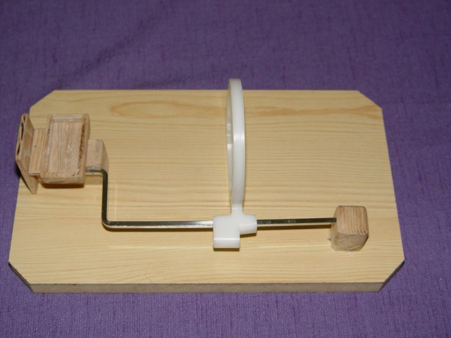
In order to standardize projection geometry, an optical bench was constructed, consisting of a positioning ring (Rinn Corporation, USA) for the x-ray tube in combination with the corresponding film holder mounted on a wooden platform; a wooden box was placed near it for the placement of dental casts (Figure 1). By placing each dental cast in the corresponding box, projection geometry was obtained, in which the central ray passed orthogonally through the interproximal contact. The resultant focus-to-object distance was 15 cm. Digital images of the teeth were acquired by using a dental x-ray unit (ELITYS Trophy, TRX 708, CROISSY BEAUBOURG, France) for all exposures operating at 70 kVp, 8 mA, and 0.0.1 sec with 2.0 mm aluminum equivalent filtration.
Figure 1.
The optical bench which was constructed and used as a film holder.
Direct digital images were obtained with a direct digital sensor (RVG UI6, Trophy, Valle, France).
Viewing sessions
Three observers (a maxillofacial radiologist, an operative dentistry specialist and a dentist) were recruited for this study. They were asked to score the presence or absence of caries in the proximal surfaces of the teeth.
The observers were instructed to assess only proximal surfaces coronal to the cemento-enamel junction. They also were told that any decalcification should be considered, regardless of its size, or its management strategy. Observers viewed images in 2 viewing sessions for the two imaging modes, with a 2-week interval.
Histological examination
Subsequent to imaging, the teeth were sectioned mesiodistally into two sections, using a ground section device (DEMCO Non-stop E6-230, USA). Tooth sections were examined under a stereomicroscope at ×20 magnification by two observers. The results were registered in a 2-point scale, in which 0 equals absence of caries and 1 equals presence of caries.
Statistical analysis
Observers’ assessments using each of the imaging modes were compared with the baseline data to determine the diagnostic performance, using “kappa” correlation coefficient. Statistical significance was defined at α=0.05.
Results
According to the microscopic assessment, 30 (29.4%) of a total of 102 evaluated interproximal surfaces were intact, whereas 72 (70.6%) exhibited carious lesions.
Inter-observer agreement according to correlation coefficient was computed too (Tables 1 and 2), which showed that inter-observer agreement between observers 2 and 3 was better than the inter-observer agreement of observers 1 and 2. Kappa correlation coefficient of observers demonstrated good observer agreement.
Table 1. Inter-observer agreement percentage and correlation coefficient of caries detection in gray scale mode of digital imaging.
| Observers | Agreement percent | Kappa correlation coefficient |
| 1&2 | 67.6 | 0.33 |
| 1&3 | 85.2 | 0.70 |
| 2&3 | 74.5 | 0.40 |
Table 2. Inter-observer agreement percentage and correlation coefficient of caries detection in colored mode of digital imaging.
| Observers | Agreement percent | Kappa correlation coefficient |
| 1&2 | 68.6% | 0.27 |
| 1&3 | 60.7% | 0.11 |
| 2&3 | 62.7% | 0.38 |
There was a weak correlation between each of the imaging mode and gold standard, as shown in Table 3.
Table 3. Sensitivity, specificity, positive (PPV) and negative predictive values (NPV), agreement percentage and correlation coefficient with histology of each of the imaging modes.
| Imaging mode | Agreement percent with histology | NPV | PPV | Specificity | Sensitivity | Kappa correlation coefficient with histology |
| Gray scale | 64.7% | 42.8% | 80% | 60% | 66.7% | 0.24 |
| Colored | 71.5% | 51.7% | 72.2% | 50% | 80.5% | 0.30 |
Sensitivities, specificities, and positive and negative predictive values were computed for each of the imaging modes and for each of the observers (Table 4).
Table 4. Sensitivity, specificity, positive (PPV) and negative predictive values (NPV), agreement percent and correlation coefficient with histology of each of the imaging modes for each of the observers.
| Parameters / Observers | Agreement percentage with histology | NPV | PPV | Specificity | Sensitivity | Kappa correlation coefficient with histology |
| Observer 1 | 62.7% | 40.9% | 79.3% | 60% | 63.9% | 0.20 |
| Observer 2 | 67.6% | 46.3% | 81.9% | 63.3% | 69.5% | 0.54 |
| observer 3 | 61.7% | 40% | 78.9% | 60% | 62.5% | 0.19 |
Comparison of the correct answers as shown in Table 5 were in favor of no statistically significant differences between the two imaging modes (P = 0.12)
Table 5. Distribution of correct answers according to imaging mode.
| Colored /Gray scale | Negative | Positive | Total |
| Positive | 23 | 48 | 71 |
| Negative | 15 | 16 | 31 |
| Total | 38 | 64 | 102 |
Discussion
Radiography is still the diagnostic standard in the detection of inaccessible approximal caries, and presently conventional dental films are frequently replaced by digital imaging systems.11
In addition to many advantages of digital imaging, post-processing of the image is a point of interest, which means alteration of captured images with different software filters in order to improve the quality of the image or to analyze its contents.12 The rationale for the study was to provide evidence for the clinicians that post-processing filter of psudocolor has a diagnostic performance comparable to their well-known traditional gray scale views, which has not been approved yet.11 , 12
Extensive carious lesions are rarely misdiagnosed on a radiograph; therefore, in this study we used extracted teeth whose approximal surfaces were either intact or had discoloration without cavitations on visual inspection.13
The number of the teeth in our study was 52 and the number of the observers was 3, selected based on the report of Hintz et al.14
However, caries diagnosis is a contrast-dependent task and we were unable to find significant differences between gray-scale and colored images in this aspect, which might be explained by unfamiliarity of the clinician’s eyes with colored images, their conception, analysis and interpretation.15 , 16 , 17 Therefore, as shown in the results, inter-observer agreement in gray-scale images was better than this value in colored images.
Conclusion
In conclusion, as shown previously by Koob,3 Shorut5 and Haak7 post-processing by digital images failed to result in significant improvements in the accuracy of caries detection. It is possible that alterations in pseudocolor filter in order to differentiate more densities from each other and operators' familiarity with colored images will make this tool efficient and easy to use.
References
- 1.Cawson RA, Odell EM. Cawson’s Essential of Oral Pathology and Oral Medicine. 7th ed. New York: Churchill Livingston; 2002. 36 [Google Scholar]
- 2.Bader JD, Shugars DA, Bonito AJ. Systematic review of selected dental caries diagnosicand management methods. J Dent Educ . 2001;65:960–86. [PubMed] [Google Scholar]
- 3.Koob A, Sanden E, Hassfeld S, Staehle HJ, Eickholz P. Effect of digital filtering on the measurement of the depth of proximal caries under different exposure conditions. Am J Dent . 2004;17:388–93. [PubMed] [Google Scholar]
- 4.Stooky GK, Jackson RD, Zandona AG, Analoui M. Dental caries diagnosis. Dent Clin North Am . 1999;43:665–77. [PubMed] [Google Scholar]
- 5.Shrout MK, Russell CM, Potter BJ, Powell BJ, Hildebolt CF. Digital enhancement of radiographs: Can it improve caries diagnosis? J Am Dent Assoc . 1996;127:469–73. doi: 10.14219/jada.archive.1996.0238. [DOI] [PubMed] [Google Scholar]
- 6.Eickhilz P, Kolb I, Lenhard M, Hassfeld S, Staehle H. Digital radiography of interproximal caries: effect of different filters. Caries Res . 1999;33:234–41. doi: 10.1159/000016522. [DOI] [PubMed] [Google Scholar]
- 7.Haak R, Wicht MJ, Noack MJ. Conventional, digital and contrast enhanced bite wing radiographsin the descisionto restore approximal carious lesions. Caries Res . 2001;35:193–9. doi: 10.1159/000047455. [DOI] [PubMed] [Google Scholar]
- 8.Gakenheimer DC. The efficacy of computerized caries detector in intra oral digital radiography. J Am Dent Assoc 2. 2004;17:388–93. doi: 10.14219/jada.archive.2002.0303. [DOI] [PubMed] [Google Scholar]
- 9.Haak R, Wicht MJ. Grey-scale reversed radiographic display in the detection of approximal caries. J Dent . 2005;33:65–71. doi: 10.1016/j.jdent.2004.08.003. [DOI] [PubMed] [Google Scholar]
- 10.Langland OE, Langlaus RP, Preece JW. Principles of Dental Imaging. 2nd ed. Philadelphia: Lippincott Williams & Wilkins; 2002. 283 [Google Scholar]
- 11.Seneadza V, Koob A, Kaltschmitt J, Staehle HJ, Duwenhoegger J, Eickholz P. Digital enhancement of radiographs for assessment of interproximal dental caries. Dentomaxillofac Radio . 2008;37:142–8. doi: 10.1259/dmfr/51572889. [DOI] [PubMed] [Google Scholar]
- 12.White SC, Pharoah MJ. Oral Radiology: Principles and Interpretations. 5th ed. London: Mosby; 2004. 335 [Google Scholar]
- 13.Hellén-Halme K, Petersson A, Warfvinge G, Nilsson M. Effect of ambient light and monitor brightness and contrast settings on the detection of approximal caries in digital radiographs: an in vitro study. Dentomaxillofacl Radiol . 2008;37:380–4. doi: 10.1259/dmfr/26038913. [DOI] [PubMed] [Google Scholar]
- 14.Hintze H, Frydenberg M, Wenzel A. Influence of number of surfaces and observers on statistical power in a multiobserver ROC radiographic caries detection study. Caries Res . 2003;37:200–5. doi: 10.1159/000070445. [DOI] [PubMed] [Google Scholar]
- 15.Nair MK, Nair UP. An in-vitro evaluation of Kodak Insight and Ektaspeed Plus film with a CMOS detector for natural proximal caries: ROC analysis. Caries Res. 2001;35:354–9. doi: 10.1159/000047474. [DOI] [PubMed] [Google Scholar]
- 16.Khan EA, Tyndall DA, Ludlow JB, Caplan D. Proximal caries detection: Sirona Sidexis versus Kodak Ektaspeed Plus. Gen Dent . 2005;53:43–8. [PubMed] [Google Scholar]
- 17.Erten H, Akarslan ZZ, Topuz O. The efficiency of three different films and rodiovisography in detecting approximal carious lesions. Quintessence Int . 2005;36:65–70. [PubMed] [Google Scholar]


