Abstract
Background and aims
One of the major problems of all-ceramic restorations is their probable fracture under occlusal force. The aim of the present in vitro study was to compare the effect of two marginal designs (chamfer and deep chamfer) on the fracture resistance of all-ceramic restorations, CERCON.
Materials and methods
This in vitro study was carried out with single-blind experimental technique. One stainless steel die with 50’ chamfer finish line design (0.8 mm deep) was prepared using a milling machine. Ten epoxy resin dies were prepared. The same die was retrieved and 50' chamfer was converted into a deep chamfer design (1 mm). Again ten epoxy resin dies were prepared from the deep chamfer die. Zirconia cores with 0.4 mm thickness and 35 µm cement space were fabricated on the epoxy resin dies (10 chamfer and 10 deep chamfer samples). The zirconia cores were cemented on the epoxy resin dies and underwent a fracture test with a universal testing machine and the samples were investigated from the point of view of the origin of the failure.
Results
The mean values of fracture resistance for deep chamfer and chamfer samples were 1426.10±182.60 and 991.75±112.00 N, respectively. Student’s t-test revealed statistically significant differences between the groups.
Conclusion
The results indicated a relationship between the marginal design of zirconia cores and their fracture re-sistance. A deep chamfer margin improved the biomechanical performance of posterior single zirconia crown restorations, which might be attributed to greater thickness and rounded internal angles in deep chamfer margins.
Keywords: CAD/CAM, dental restoration, fracture strength, zirconium oxide
Introduction
One of the major problems of all-ceramic restorations is their probable fracture under occlusal and lateral forces.1 The majority of restorations contain metal which brings about toxic, chemical and allergic affects. The difference between their color and that of the natural tooth is another problem. Most people prefer tooth-colored crowns. All-ceramic crowns have esthetic and biocompatibility.2 In recent years such restorations have been used in posterior restorations. However, some crown fractures due to the relatively low mechanical resistance of ceramic crowns have been reported, which might be attributed to the magnitude of biting forces applied on premolars and molars and to the inherent brittleness of ceramics.3,4Ceramic materials are particularly susceptible to tensile stresses, and mechanical resistance is also strongly influenced by the presence of superficial flaws and internal voids. Such defects might represent the sites of crack initiation. This phenomenon may be influenced by different factors, such as marginal design and thickness of the restoration, residual processing stress, magnitude and direction and frequency of the applied load, elastic modulus of restoration components, restoration?cement interfacial defects, and oral environmental effects.5In one research, finite element analysis (FEA) was used to study stress distribution during mastication in maxillary second premolars restored with metal-ceramic crowns and compare them to non-restored teeth. High stresses were recorded at the cervical line of restored teeth within the dentin-metal interface and within the ceramic-metal interface.6 The FEA method was used to study stress distribution in the lower first molars restored with all-ceramic crowns. The results of that study revealed concentration of stress at the cervical area.7 The aim of the present study was to evaluate the effect of marginal design of crowns on improved mechanical performance of CERCON crowns from a clinical point of view. Such a condition can be achieved preparing a deep chamfer margin in crowns instead of a chamfer and shoulder margin. Florian Beuer8 suggested that shoulder margin has a greater fracture resistance than deep chamfer and chamfer margin. Sadan et al9 proposed that both these types of finish lines are considered to be adequate for the tooth. However, Di Lorio et al10 suggested that the shoulder margin could improve the biomechanical performance of single-crown alumina restorations. De Jager et al11 discovered that for long-lasting restorations in posterior region it is advisable to make a chamfer with collar preparation. Cho L et al12 found out that the fracture strength of chamfer finish line (0.9, 1.2 mm) was greater than 1.2 mm rounded end shoulder and 1.2 shoulder finish line. Potiket et al13 suggested that a 1-mm deep shoulder finish line with a rounded internal line angle has good fracture strength for the natural teeth restored with all-ceramic crowns. Rammersberg et al14 discovered that a minimally invasive 0.5-mm axial chamfer tooth preparation has the greatest stability for posterior metal-free crowns. The aim of the present in vitro study was to compare the fracture resistance under a cyclic load applied to chamfer and deep chamfer margins of zirconia crowns.
Materials and Methods
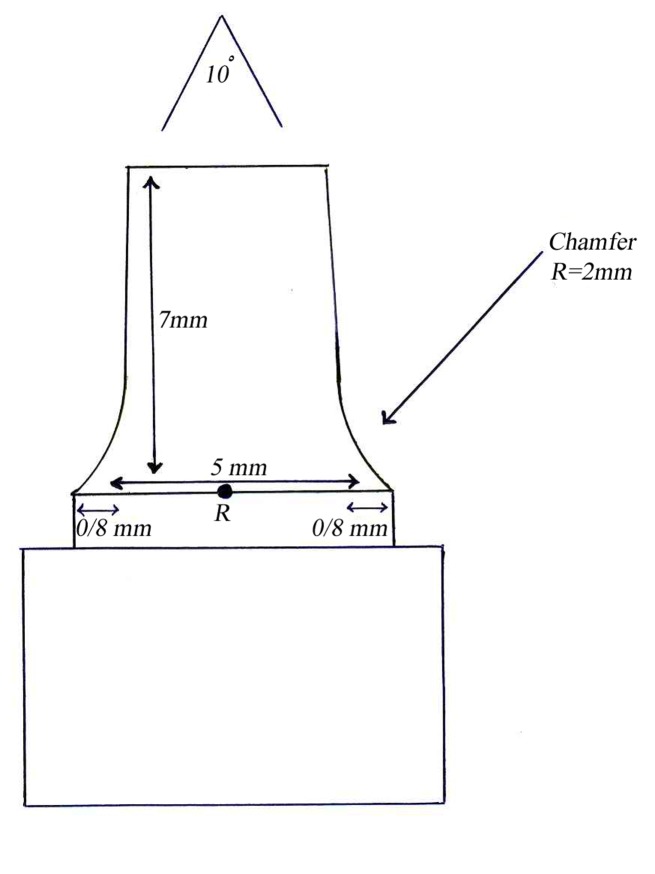
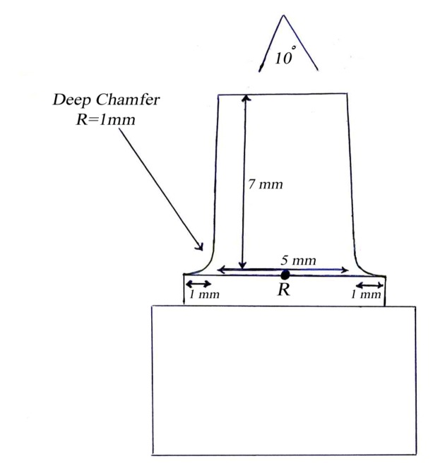
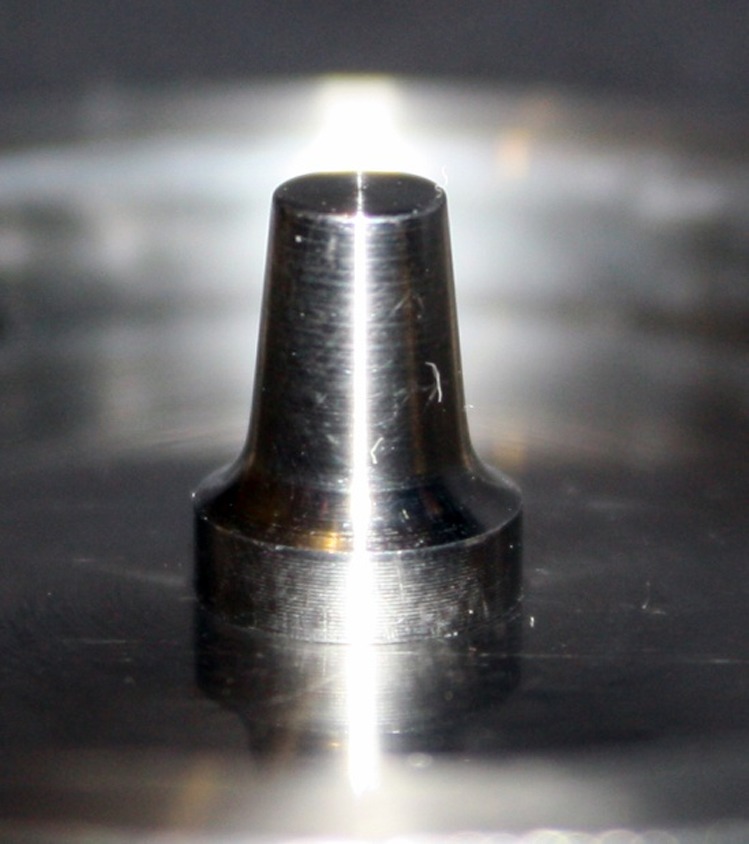
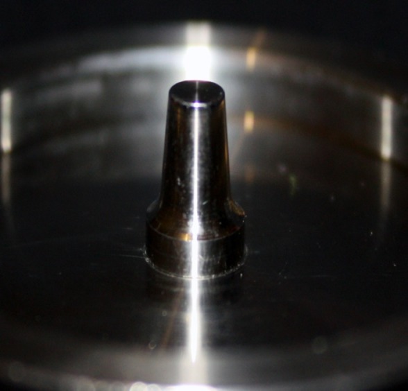
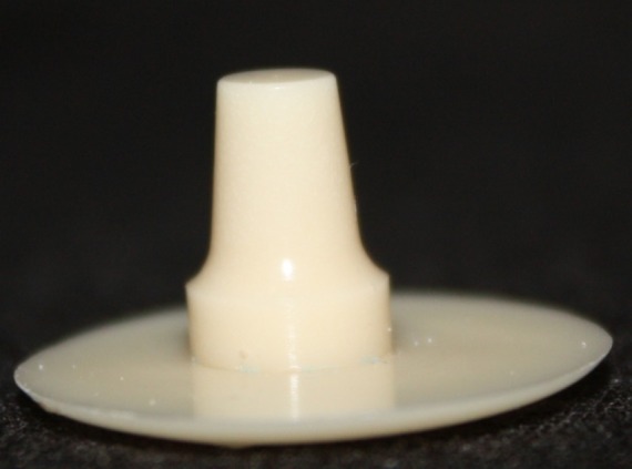
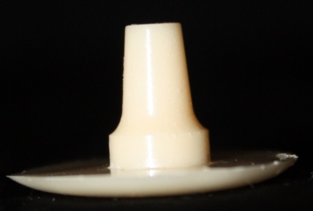
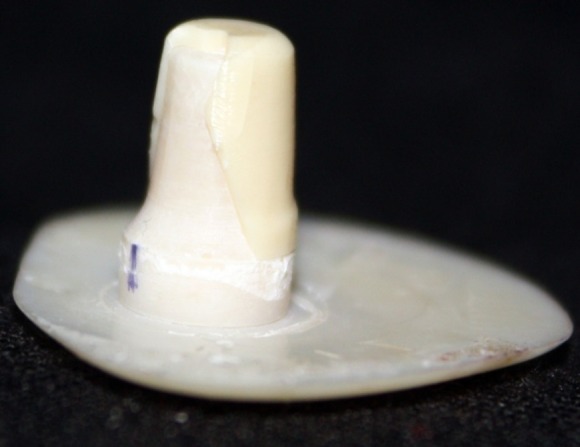
This in vitro single-blind experimental study was carried out using 1 machined standard stainless steel die with a height of 7 mm and a diameter of 5 mm.15The marginal area of the die was prepared with 50' chamfer finish line (0.8 mm deep).16,17 The axial walls were 10° convergent (Figure 1).15 Impressions were poured in Epoxy resin CW2215 (Hunstman-Germany). Afterwards, the standard die was converted into a deep chamfer with a depth of 1 mm(Figures 2a,b).16,17 Again 10 polyvinylsiloxane impressions were made and ten epoxy resin dies were created from these impressions (Figure 2c,d).8,10 Twenty copings were produced of a partially sintered ZrO2 ceramic material using CAD/CAM technology (Cercon Smart Ceramics, DeguDent, Hanau, Brain, DeguDent). The copings with 0.4 mm thicknesses8and 35 µm of cement space8weremilled out from the pre-sintered ZrO2 and the Cercon (DeguDent) heat-sintered them at 1350°C for 6 hours. Since the coping mainly determinates the overall resistance to fracture of veneered crown5,18porcelain veneering was omitted. The copings were evaluated visually; those with margin deemed visually unacceptable were rejected and another coping was made instead. Each coping was then cemented on its definitive die with GI (GC Gold Labled, Tokyo, Japan).14 Finger pressure was applied during the setting time.24 After cementation, excess luting agent was removed and the samples were stored in a saline solution at room temperature for 24 hours. Mechanical tests were carried out using a universal testing machine (GOTECH AI-700LAC, Arsona, USA). The load was applied at the center of the occlusal surface along the long axis with a crosshead speed of 0.5 mm/min until fracture occurred.19 The fracture load data were automatically recorded using Vista software. The samples were investigated from the point of view of the origin of the failure (Figure 2e,f). Data was analyzed with student's t-test at a significance level of P<0.05.
Figure 1. Diagram of chamfer (a) and deep chamfer (b) preparations.
a.
b.
a.
b.
c.
d.
e.
f.
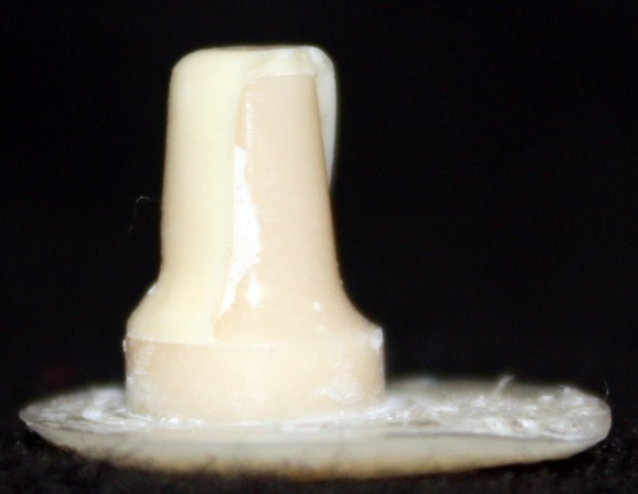
Figure 2. Standard dies of chamfer (a) and deep chamfer (b) preparations. Epoxy resin dies with chamfer (c) and deep chamfer (d) margins. Fracture area in chamfer (e) and deep chamfer (f) margins.
Results
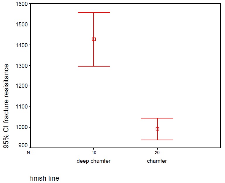
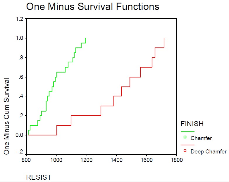
The mean ± SD of fracture resistance were 1426.10 ± 182.60 and 991.75 ± 112.00 N for the deep chamfer and chamfer margins, respectively. Not only the maximum but also the minimum fracture resistances of two groups were more than intra-oral loads. Student's t-test revealed statistically significant differences between the groups (P=0.05) (Table 1). This study was carried out with 95% confidence interval; Kaplan–Meir graph showed that deep chamfer margin tolerates more cracks till fracture than chamfer margin (Figure 3), which might be attributed to greater thickness in deep chamfer margins.
Table 1. Fracture resistance of chamfer and deep chamfer edge zirconia cores.
| Margin design | N | Mean | Std. Deviation | Std. Error | 95% Confidence Interval for Mean | Minimum | Maximum | |
| Lower Bound | Upper Bound | |||||||
| Deep chamfer | 10 | 1426.100 | 182.60671 | 57.74531 | 1295.4710 | 1556.7290 | 1100.00 | 1656.00 |
| Chamfer | 10 | 991.7500 | 112.00088 | 25.04416 | 939.3320 | 1044.1680 | 813.00 | 1196.00 |
Figure 3. Error bar and Kaplan–Meir graph for fracture resistance of deep chamfer and chamfer preparations.
finish line.
RESIST.
Discussion
One of the major problems of all-ceramic restorations is their probable fracture under occlusal and lateral forces.1The majority of restorations contain metal which brings about biologic problems and have no esthetic appearance.2 This study compared fracture resistance of chamfer and deep chamfer margins of CERCON crowns under a cyclic load. Student's t-test revealed statistically significant differences between the two groups; fracture resistance of deep chamfer margin was more than that of chamfer margin. Elastic modulus of the supported materials of the core affected the fracture resistance of the core.20 Therefore, in this study, we used epoxy resin dies that are much better brass dies.21 Another difference from clinical conditions is the unknown nature of the bond between the luting agent and die material. It is reasonable to suppose that the presence of a hybrid layer at the dentin-cement interfaces influences the biomechanical behavior of the core/supporting die system. However, both of these factors equally influenced the samples in the present study. Therefore, it is possible to make a comparison between the two groups. Fracture resistance of the two groups are more than the occlusal forces so we could use all of these marginal designs successfully in the posterior all-ceramic crowns, which are very good substitutes for PFM crowns. However, there was a statistically significant difference between the two groups, revealing that the deep chamfer has more fracture resistance than chamfer margin, which might be attributed to greater thickness in deep chamfer margins that can bear load better than chamfer margins. We used resin cements for cementation, so we had a strong unity in the margins that provided strength against fracture.22 It seems deep chamfer can bear load better, making it more fracture resistant than chamfer margin.
Conclusion
Both marginal designs had high fracture resistance that is more than biting forces so we could use both; however, because of higher fracture resistance of deep chamfer margins, this finish line is recommended to improve the biomechanical performance of posterior single zirconia restorations.
References
- 1.Anusavice KJ, Phillips RW. Phillips' Science of Dental Materials. 11th ed. St. Louis: W.B. Saunders; 2003. [Google Scholar]
- 2.Ferrance JL. Using posterior composites appropriately. J Am Dent Assoc . 1992;123:53–8. [PubMed] [Google Scholar]
- 3.Etemadi S, Smales RJ. Survival of resin-bonded porcelain veneer crowns placed with and without meta reinforcement. J Dent. 2006;34:134–45. doi: 10.1016/j.jdent.2005.05.007. [DOI] [PubMed] [Google Scholar]
- 4.Mclaren EA, White SN. Survival of Inceram crowns in a private practice. a prospective clinical trial. J Prosthet Dent. 2000;83:216–22. doi: 10.1016/s0022-3913(00)80015-3. [DOI] [PubMed] [Google Scholar]
- 5.Webber B, McDonald A, Knowles J. An in vitro study of the compressive load at fracture of procera all ceramic crowns with varying thickness of veneer porcelain. J Prosthet Dent . 2003;89:154–60. doi: 10.1067/mpr.2003.85. [DOI] [PubMed] [Google Scholar]
- 6.Aykul H, Toparli M, Dalkiz M. A calculation of stress distribution in metal-porcelain crowns by using three-dimensional finite element method. J Oral Rehabil . 2002;29:381–6. doi: 10.1046/j.1365-2842.2002.00826.x. [DOI] [PubMed] [Google Scholar]
- 7.Imanishi A, Nakamura T, Ohyama T, Nakamura T. 3-D Finite element analysis of all ceramic posterior crowns. J Oral Rehabil . 2003;30:818–22. doi: 10.1046/j.1365-2842.2003.01123.x. [DOI] [PubMed] [Google Scholar]
- 8.Florian Bever, Hans Aggstaller, Daniel Edelhoff. Effect of preparation Design on the fracture Resistance of Zirconia crown copings. Dent Mater . 2008;27:362–367. doi: 10.4012/dmj.27.362. [DOI] [PubMed] [Google Scholar]
- 9.Sadan A, Blutz MB, Lang B. Clinical consideration for densely sintered alumina and zirconia restorations. Part 1. Int J Periodontics Restorative Dent. 2005;25:213–9. [PubMed] [Google Scholar]
- 10.Di Iorio, Murmura G, Orsini G, Scarano A, Caputi S. Effect of margin design on the fracture resistance of Procera all ceram cores. an in vitro study. J Contemp Dent Pract. 2008;9:1–8. [PubMed] [Google Scholar]
- 11.De Jager, Pallav P, Feilzer AJ. The influence of design parameters on the FEA-determined stress distribution in CAD/CAM produced All ceramic dental crown. Dent Mater. 2005;21:242–51. doi: 10.1016/j.dental.2004.03.013. [DOI] [PubMed] [Google Scholar]
- 12.Cho L, Choli J, Yi YJ, Park CK. Effect of finish line variants on marginal accuracy and fracture strength of ceramic optimized polymer/fiber-reinforced composite crowns. J Prothet Dent . 2004;91:554–60. doi: 10.1016/j.prosdent.2004.03.004. [DOI] [PubMed] [Google Scholar]
- 13.Potiket N, Chiche G, Finger IM. In vitro fracture strength of teeth restored with different All ceramic crown systems. J Prosthet Dent . 2004;92:491–5. doi: 10.1016/j.prosdent.2004.09.001. [DOI] [PubMed] [Google Scholar]
- 14.Rammelsberg P, Eickemeyer G, Erdelt K, Pospiech P. Fracture resistance of posterior metal-free polymer crowns. J Prosthet Dent . 2000;84:303–8. doi: 10.1067/mpr.2000.108068. [DOI] [PubMed] [Google Scholar]
- 15.Jalalian E, Keshavarzi G. [Comparison of Heavy Chamfer and Shoulder Finish line Designs on Marginal Adaptation of All-ceramic IPS e. max press Restorations]. Journal of Dental School, Shahid Beheshti University of Medical Sciences 2005; 5:53-7. [Persian] Available from: http://www.sid.ir/fa/VEWSSID/J_pdf/54413898604.pdf [Google Scholar]
- 16.Shillingburg HT, Hobo S, Whitsett LD, Jacobi R, Brackett SE. Chicago: Quintessence. 3rd ed. Chicago: Quintessence; 1997. 139-71 [Google Scholar]
- 17.Gavelis JR, Mornecy JD, Riley ED, Sozio RB. The effect of various finish line preparation. J Prosthet Dent . 1981;45:138–45. doi: 10.1016/0022-3913(81)90330-9. [DOI] [PubMed] [Google Scholar]
- 18.Beuer F, Kerler T, Erdelt KJ, Schweiger J, Eichberger M, Gernet W. [Influence of veneering on the fracture resistance of zirconium restorations]. Dtsch Zahnarztl Z 2004;59:527-530. [German] [Google Scholar]
- 19.Jalalian E, Moghadam L. [Compare the fracture resistance of 2 all ceramic systems, IPS e. max, IPS Empress]. Journal of Dentistry, Shiraz University of Medical Sciences (JDSUMS) 2008; 9:51-7. [Persian] Available from: http://www.sid.ir/fa/VEWSSID/J_pdf/73013871806.pdf [Google Scholar]
- 20.Scherrer SS, deRijk KG. The fracture resistance of all ceramic crowns on supporting structure with different elastic moduli. Int J Prosthodont . 1993;6:462–7. [PubMed] [Google Scholar]
- 21.Ayad MF. Effect of the crown preparation margin and die type on the marginal accuracy of fiber-reinforced composite crowns. J Contemp Dent Pract . 2008;9:9–16. [PubMed] [Google Scholar]
- 22.Cho HO, Kang DW. Marginal fidelity and fracture strength of IPS-Empress 2 ceramic crowns according to different cement types. J Korean Acad Prosthodont . 2002;40:545–60. [Google Scholar]
- 23.Gibbs CH, Anusavice KJ, Young HM, Jones JS, Esquivel- Upshaw. Maximum clenching force of patients with moderate loss of posterior tooth support: a pilot study. J Prosthet Dent . 2002;88:498–502. doi: 10.1067/mpr.2002.129062. [DOI] [PubMed] [Google Scholar]
- 24.Att W, Komine F, Gerds Th, Strub JR. Marginal adaptation of three different zirconium dioxide three-unit fixed dental prostheses. J Prosthet Dent . 2009;101:239–74. doi: 10.1016/S0022-3913(09)60047-0. [DOI] [PubMed] [Google Scholar]


