

Abstract
Candida albicans is an opportunistic pathogen and is recognised and phagocytosed by macrophages. Using live-cell imaging, non-lytic expulsion/exocytosis of C. albicans from macrophages is demonstrated for the first time. Following complete expulsion, both the phagocyte and pathogen remain intact and viable. Partial engulfment of hyphal C. albicans without macrophage lysis is also demonstrated. These observations underpin the complexity of interactions between C. albicans and innate immune cells.
Keywords: Candida albicans, Macrophage, Phagocytosis, Innate immunology, Fungal infection
1. Introduction
Candida albicans inhabits the human mucosa as a commensal fungus, however superficial and systemic lethal disease can arise under circumstances of perturbed host susceptibility. Associated with its infectious capacity is a repertoire of virulence attributes, of which the dimorphic transition between budding yeast and hypha is key. This facilitates niche-specific growth, tissue penetration, dissemination and immune evasion/escape (Kumamoto and Vinces, 2005). Host defence against systemic candidiasis relies mainly on the ingestion and elimination of fungal cells by cells of the innate immune system, especially neutrophils and macrophages (Sheth et al., 2011; McKenzie et al., 2010). Our recent work carefully describes the phagocytic process during interactions of macrophages with C. albicans using sophisticated video microscopy and tracking (Lewis et al., in press). We and others have shown that engulfment of live hyphal-forming strains leads ultimately to host cell lysis and death (McKenzie et al., 2010; Lo et al., 1997). May, Casadevall and colleagues have previously observed yeast cells of Cryptococcus neoformans (Ma et al., 2006; Alvarez and Casadevall, 2006; Johnston and May, 2010) undergoing expulsion from phagocytes, rendering both host and pathogen viable. We report here non-lytic expulsion/exocytosis as an alternative outcome following phagocytosis of hyphae-producing C. albicans by the murine macrophage cell line J774.1. We show that both the fungal pathogen and the macrophage remain viable and functional (retain phagocytic ability/continued hyphal growth) after non-lytic expulsion. This is not only the first demonstration of non-lytic expulsion for a Candida species but also the first time this was observed in the context of hyphal cells.
2. Description of observations
Hyphal-mediated macrophage lysis was captured using live cell imaging (Video 1). The central macrophage internalizes C. albicans and we observed subsequent extension of hyphal filaments within the macrophage (Video 1). In a majority of cases, the membranes of the macrophage ultimately failed to restrain hyphal expansion; resulting in the eventual rupture of the phagocyte (Video 1). C. albicans escape and associated macrophage lysis are common features of C. albicans-macrophage interactions and have been studied in more detail previously (Sheth et al., 2011).
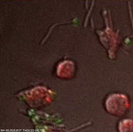
However, in a minority of cases, non-lytic expulsion/exocytosis was observed (Video 2). Although this occurred at a very low frequency (<1%, n > 50) it was observed repeatedly in various experimental conditions. The video shows that the central macrophage (stained red) engulfing a yeast cell C. albicans (stained green) followed by hyphal growth inside the macrophage. Fungal expulsion proceeded with the hypha emitted tip-first and was completed within approximately 40 min (Video 2). Following externalisation of C. albicans, both macrophage and fungus remained viable – the macrophage proceeded to undergo mitosis to yield motile daughter cells and the hypha continued to extend at normal rates (Video 2).
We also report partial engulfment of hyphal C. albicans without macrophage lysis (Video 3). In the example, a yeast cell was engulfed leaving the growing hypha exposed; this hypha was then engulfed by a second macrophage (Video 3). Extensive stretching of the host cell ensued, after which the macrophage plasma membrane retracted back from the growing hypha (Video 3). In contrast to macrophage lysis resulting from hyphal penetration of the membrane (Video 1), the host cell remained viable and active (Video 3). While a portion of the fungus remained within the macrophage, the externalised hyphal tip was later recognised and engulfed by a third macrophage as it continued to grow (Video 3).
3. Discussion
Here we report non-lytic expulsion/exocytosis of C. albicans from macrophages, the first demonstration of this process for a filamentous fungus. This process has been described previously for the facultative pathogenic yeast C. neoformans (Ma et al., 2006; Alvarez and Casadevall, 2006). Studies suggest that non-lytic expulsion, or ‘vomocytosis’ of C. neoformans occured in 7–10% of J774.1 in a 24 h period, although with greater frequency in primary cells and in vivo (Lo et al., 1997; Ma et al., 2006; Alvarez and Casadevall, 2006). Our in vitro system detected non-lytic complete expulsion of C. albicans at a frequency of <1% within a 6 h period, after which the majority of macrophages were overcome by significant hyphal growth in the culture medium. Even though non-lytic complete expulsion of C. albicans is a rare event, that is difficult to quantify, it is not an artefact and was observed repeatedly (n > 50) with control strains and glycosylation-deficient mutant strains under a range of experimental conditions. To date we have only observed non-lytic expulsion in macrophage cell lines and not in primary mouse or human macrophages but this may simply be a reflection of the limited number of interactions investigated in primary cells to date.
Increase of phagosomal pH increases the rate of C. neoformans vomocytosis from macrophages (Nicola et al., 2011). Both hyphal and yeast forms of C. albicans are know to modify the phagosomes in which they are internalised by raising pH (Fernandez-Arenas et al., 2009), therefore the occurrence and frequency of non-lytic expulsion of C. albicans may also be related to intraphagosomal pH.
The clinical relevance of this phenomenon is difficult to determine and warrants further investigation as non-lytic expulsion of hyphal C. albicans may benefit the macrophage in terms of avoiding lysis, but conversely may also benefit the fungus as it escapes the microbicidal environment of the phagosome. This study underpins the complexities of the host/fungus interaction of a species which has both commensal and infectious capacity.
4. Methods
C. albicans strain (NGY152) hitherto referred to as the “control” strain was grown in SC-Ura at 30 °C and J774.1 macrophages were cultured in supplemented DMEM medium at 37 °C with 5% CO2, as described previously (McKenzie et al., 2010). For phagocytosis assays, 1 × 10⁶ macrophages were seeded to glass based dishes and cultured overnight at 37 °C with 5% CO2. During experiments, medium was replaced with supplemented CO2-independent medium containing 1 μM Lysotracker Red (Invitrogen, Paisley, UK) (Erwig et al., 2006). C. albicans were stained with 1 mg/ml FITC cell membrane label (Sigma, UK) following manufactures instruction, in 0.05 M carbonate-bicarbonate buffer (pH 9.6) for 10 min at 20 °C in the dark, washed three times and resuspended in PBS, were added to macrophages at a 3:1 ratio. Video microscopy was conducted at 37 °C with a DeltaVision Core microscope (Applied Precision, Washington, USA) and images captured at 1 min intervals for 6 h by an EMCCD camera.
Acknowledgments
LPE is a Scottish Senior Clinical Fellow and acknowledges the support of the Chief Scientist Office (SCD/03). This work was funded by Wellcome Trust Project Grant to LPE and JQ 089930. NARG was funded by a Wellcome Trust Programme Grant (080088) and an equipment Grant (075470) (for DeltaVision), and by a FP7-2007-2013 Grant (HEALTH-F2-2010-260338-ALLFUN). We would like to thank the University of Aberdeen imaging facility, in particular Kevin MacKenzie, for helpful support and advice.
Footnotes
Supplementary data associated with this article can be found, in the online version, at doi:10.1016/j.fgb.2012.01.008.
Appendix A. Supplementary material
References
- Alvarez M., Casadevall A. Phagosome extrusion and host-cell survival after Cryptococcus neoformans phagocytosis by macrophages. Curr. Biol. 2006;16(21):2161–2165. doi: 10.1016/j.cub.2006.09.061. [DOI] [PubMed] [Google Scholar]
- Erwig L.P., McPhilips K.A., Wynes M.W., Ivetic A., Ridley A.J., Henson P.M. Differential regulation of phagosome maturation in macrophages and dendritic cells mediated by Rho GTPases and ezrin–radixin–moesin (ERM) proteins. Proc. Natl. Acad. Sci. USA. 2006;103(34):12825–12830. doi: 10.1073/pnas.0605331103. (Epub August 14 2006) [DOI] [PMC free article] [PubMed] [Google Scholar]
- Fernandez-Arenas E., Bleck C.K., Nombela C., Gil C., Griffiths G., Diez-Orejas R. Candida albicans actively modulates intracellular membrane trafficking in mouse macrophage phagosomes. Cell Microbiol. 2009;11(4):560–589. doi: 10.1111/j.1462-5822.2008.01274.x. [DOI] [PubMed] [Google Scholar]
- Johnston S.A., May R.C. The human fungal pathogen Cryptococcus neoformans escapes macrophages by a phagosome emptying mechanism that is inhibited by Arp2/3 complex-mediated actin polymerisation. PLoS Pathog. 2010;6(8) doi: 10.1371/journal.ppat.1001041. e1001041. [DOI] [PMC free article] [PubMed] [Google Scholar]
- Kumamoto C.A., Vinces M.D. Contributions of hyphae and hypha-co-regulated genes to Candida albicans virulence. Cell Microbiol. 2005;7(11):1546–1554. doi: 10.1111/j.1462-5822.2005.00616.x. [DOI] [PubMed] [Google Scholar]
- Lewis, L.E., Bain, J.M., Lowes, C., Gillespie, C., Rudkin, F.M., Gow, N.A.R., Erwig, L.-P., 2012. Stage specific assessment of Candida albicans phagocytosis by macrophages identifies cell wall composition and morphogenesis as key determinants. PLoS Pathog, in press. [DOI] [PMC free article] [PubMed]
- Lo H.J., Kohler J.R., DiDomenico B., Loebenberg D., Cacciapuoti A., Fink G.R. Nonfilamentous C. albicans mutants are avirulent. Cell. 1997;90(5):939–949. doi: 10.1016/s0092-8674(00)80358-x. [DOI] [PubMed] [Google Scholar]
- Ma H., Croudace J.E., Lammas D.A., May R.C. Expulsion of live pathogenic yeast by macrophages. Curr. Biol. 2006;16(21):2156–2160. doi: 10.1016/j.cub.2006.09.032. [DOI] [PubMed] [Google Scholar]
- McKenzie C.G., Koser U., Lewis L.E., Bain J.M., Mora-Montes H.M., Barker R.N. Contribution of Candida albicans cell wall components to recognition by and escape from murine macrophages. Infect Immun. 2010;78(4):1650–1658. doi: 10.1128/IAI.00001-10. [DOI] [PMC free article] [PubMed] [Google Scholar]
- Nicola A.M., Robertson E.J., Albuquerque P., Derengowski Lda S., Casadevall A. Nonlytic exocytosis of Cryptococcus neoformans from macrophages occurs in vivo and is influenced by phagosomal pH. mBio. 2011;2(4) doi: 10.1128/mBio.00167-11. 10.1128/mBio.00167,11. Print 2011. [DOI] [PMC free article] [PubMed] [Google Scholar]
- Sheth C.C., Hall R., Lewis L., Brown A.J., Odds F.C., Erwig L.P., Gow N.A. Glycosylation status of the C. albicans cell wall affects the efficiency of neutrophil phagocytosis and killing but not cytokine signaling. Med Mycol. 2011;49(5):513–524. doi: 10.3109/13693786.2010.551425. (Epub January 24 2011) [DOI] [PMC free article] [PubMed] [Google Scholar]
Associated Data
This section collects any data citations, data availability statements, or supplementary materials included in this article.