Abstract
Inhibition of acetylcholinesterase (AChE) and butyrylcholinesterase (BChE) is considered a promising strategy for the treatment of Alzheimer's disease (AD). This research project aims to provide a comprehensive knowledge of newly synthesized coumarin analogues with anti-AD potential. In the present work a series of 3-thiadiazolyl- and thioxo-1,2,4-triazolylcoumarins derivatives were designed, synthesized, and tested as potent inhibitors of cholinesterases. These compounds were assayed against AChE from electrophorus electricus and rabbit; and BChE from horse serum and rabbit by Ellman's method using neostigmine methylsulphate and donepezil as reference drugs. Some of the assayed compounds proved to be potent inhibitors of AChE and BChE with Ki values in the micromolar range. 4b was found to be the most active compound with Ki value 0.028 ± 0.002 μM and higher selectivity for AChE/BChE. The ability of 4b to interact with AChE was further confirmed through computational studies, in which a primary binding was proved to occur at the active gorge site, and a secondary binding was revealed at the peripheral anionic site. Structure activity relationships of prepared compounds were also discussed.
1. Introduction
Alzheimer's disease (AD), Parkinson's disease, and age-related memory disorders always remain in keen interest of researchers. AD is a progressive neurodegenerative disorder that is characterized by the appearance of neurofibrillary tangles, neuritic plagues within the brain of AD patients, [1] rapid loss of synapses, and degeneration of basal cholinergic neurons [2]. Loss of cholinergic neurons causes reduction in cortical and hippocampal levels of the neurotransmitter acetylcholine (ACh) that leads to impairment in cognitive functions as well [3–5]. On the behavioral side, confusion, irritability, anger, and inability to perform body functions properly are obvious symptoms [3, 6]. As general health care system is improving globally day by day and thus proportion of older people increases, the number of AD patients is estimated to increase considerably [7]. Thus, new drugs are required for the treatment of AD. Recently it was found that AChE could accelerate the deposition phenomenon of neuritic plagues [8, 9]. Various anti-AChE agents, that is, ensaculine, donepezil, propidium, rivastigmine, and tacrine (Figure 1) have shown slight improvement in cognitive and memory disorders [10]. However, these available nitrogen containing anti-AChE drugs have certain side effects and lesser CNS permeability. So, new drugs are required for the treatment of AD with better CNS penetration and decreased toxic effects.
Figure 1.
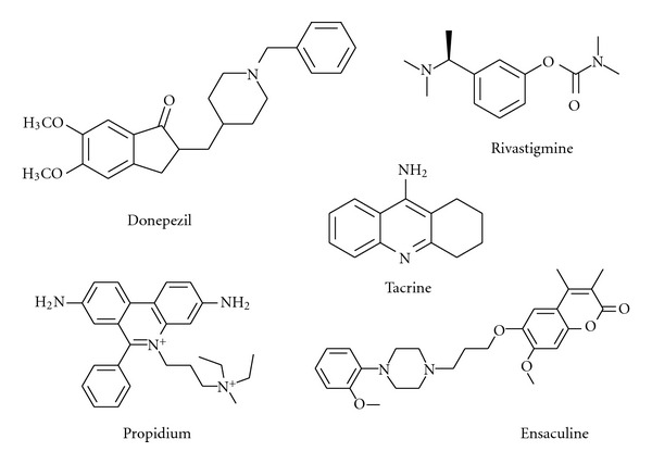
Chemical structures of some common FDA approved cholinesterase inhibitors.
Natural compounds have always served as a useful source for the study of AChE inhibitory activity. A number of phytoconstituents for instance, alkaloids (indole, isoquinoline, and steroidal), pregnane glycosides (cynantrosides), stilbenes, triterpenes, [11] ursane [12], and xanthones have shown AChE inhibitory potency. These and other such examples have urged researchers to explore nature for identification of active compounds against AD. Coumarins, an important class of natural compounds have been studied extensively due to their vast medicinal significance [11, 12]. They have been reported for their analgesic [13], antibacterial [14], anticancer [15], anticoagulant [16], anti-inflammatory [13], anthelmintic [17], antifungal [14], antihepatitis C [18], antimutagenic [19], and antituberculosis [20] activities. Coumarins have also proved as potent nonpeptidic protease inhibitors [21], heat shock protein inhibitors [22], monoamine oxidase inhibitors [23], 17β-hydroxysteroid dehydrogenase (17β-HSD) type 1inhibitors [24], and TNF-α (tumor necrosis factor-alpha) inhibitors [25]. 4-methylcoumarins having different functional groups are well known for their antioxidant and radical scavenging activities [26]. Thiazole derivatives have been studied extensively due to their versatile pharmacological significance that is, they possess antitumor [27], cardiotonic [27], anti-HIV [28], analgesic and anti-inflammatory [29] activity. They are also reported as DNA-gyrase [30] and lipoxygenase enzyme inhibitors [31]. 1, 3, 4 thiadiazole nucleus is found in a number of compounds and potent due to its anti-ulcer [32], antiallergic [33] and diuretic activities [34]. The present paper focuses on synthesis of new class of coumarins which could prove as potent therapeutic moieties against progression of AD in future.
2. Results and Discussion
2.1. Chemistry
Coumarin-3-carbohydrazide was obtained by refluxing coumarinyl ester with hydrazine hydrate according to published procedure [32]. The thiosemicarbazides (2a–g) were obtained by the stirring coumarin-3-carbohydrazide and aryl isothiocyanates (1a–g). The IR spectra of thiosemicarbazides (2a–g) revealed carbonyl absorption in the range 1644–1689 cm−1 and that of thiocarbonyl at 1235–1268 cm−1, respectively whilst the characteristic absorption bands for three secondary N-H groups appeared in the region 3143–3432 cm−1. In 1H NMR, the NH proton of amide type linkage appeared at δ 11.23–11.96 ppm and signal for two NH proton of thiourea type linkage at δ 9.99–11.14 ppm. The carbonyl and thiocarbonyl appeared at 164.7–167.0 and 176.2–181.7 ppm, respectively in 13C NMR as well as those of the aromatic carbons (Scheme 1).
Scheme 1.
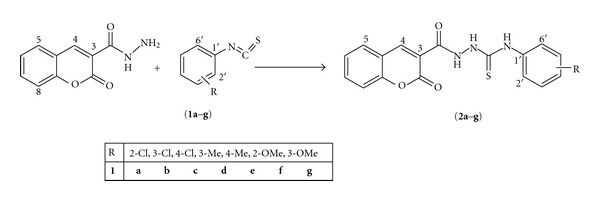
Synthesis of 3-(4-Aryl-5-thioxo-4,5-dihydro-1H-1,2,4-triazol-3-yl)-2H-chromen-2-ones.
The 4,5-disubstituted-1,2,4-triazol-3-thiones (3a–g) were synthesized by refluxing the corresponding thiosemicarbazides (2a–g) in aqueous sodium hydroxide (4N) solution (Scheme 2). The products were purified by recrystallization in aqueous ethanol. The formation of triazoles (3a–g) was indicated by the disappearance of broad peaks of NH and C=O groups of thiosemicarbazides and by appearance of (C=N) absorption in the range 1375–1413 cm−1. In 1H NMR, signal for N-H proton of triazoles appeared in the range of 11.13–13.94 ppm and the disappearance of signals for N-H protons confirmed the formation of the triazoles. In 13C NMR the disappearance of peak due to amidic (C=O) group and the appearance of (C=N) peak in the range of 156.3–159.1 ppm was detected.
Scheme 2.
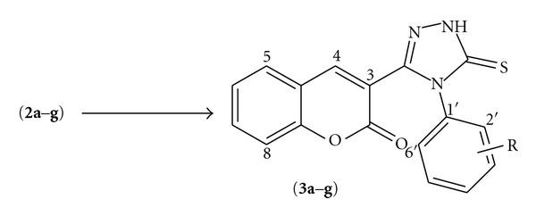
Synthesis of 3-(4-Aryl-5-thioxo-4,5-dihydro-1H-1,2,4-triazol-3-yl)-2H-chromen-2-ones.
2,5-Disubstituted-1,3,4-thiadiazoles (4a–g) were synthesized by treating the thiosemicarbazide (2a–g) with concentrated polyphosphoric acid at low temperature (Scheme 3). The disappearance of broad peaks of NH groups and amidic (C=O) group of thiosemicarbazides and appearance of (C=N) absorption in the range 1415–1471 cm−1 was noticed in IR spectra. In 1H NMR, signal for N-H proton of thiadiazoles appeared in the range of 11.14–11.86 ppm whilst in 13C NMR the disappearance of (C=O) group peak and the appearance of (C=N) peak in the range of 157.0–159.2 ppm indicated the desired conversion.
Scheme 3.

Synthesis of 3-(5-(arylamino)-1,3,4-thiadiazol-2-yl)-2H-chromen-2-ones.
2.2. In Vitro Inhibition Studies of AChE and BChE
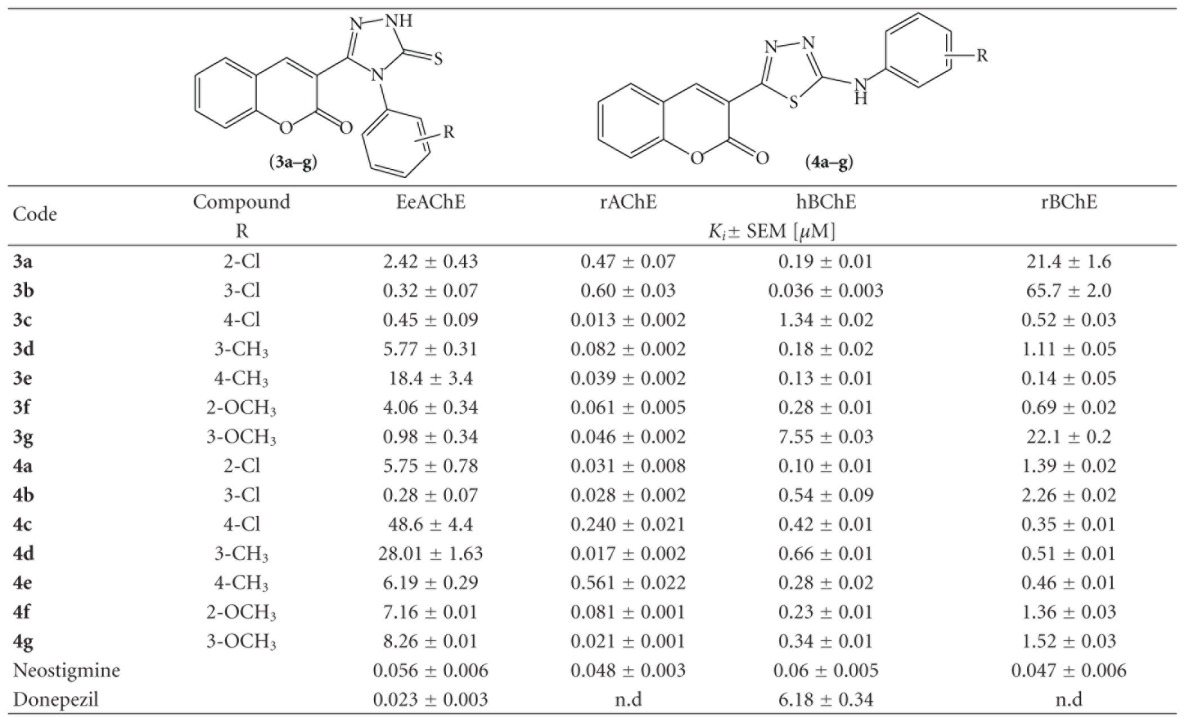
The basic structure of our compound series 3a–g is 3-(4-phenyl-5-thioxo-4,5-dihydro-1H-1,2,4-triazol-3-yl)-2H-chromen-2-one and 4a–g series is 3-(5-(phenylamino)-1,3,4-thiadiazol-2-yl)-2H-chromen-2-one. The in vitro cholinesterase activity of these new molecules was determined using spectrophotometric method with neostigmine and donepezil as reference compounds (Table 1).
Table 1.
AChE and BuChE inhibitory activities of new coumarin derivativesa.
|
aValues are expressed as the mean ± standard error of the mean of three experiments. Ki inhibitory concentration (μM) of AChE from electrophorus electricus (EeAChE) or rabbit (rAChE) and (hBChE) from horse serum or rabbit serum (rBChE).
The Ki values suggested that most of designed compounds exhibited potent and selective inhibitory activities in nanomolar range towards cholinesterases. The most potent compounds are 3c, 4b, 4d, 4c, and 3d on rabbit AChE than the reference compound, neostigmine. However, our investigated compounds were less active on EeAChE as compared to the reference drugs. It showed the selectivity of inhibition of the tested compounds on rAChE. Our compounds have different scaffold than neostigmine, therefore, they were more selective on both enzymes and showed more selectivity as compared to reference compound. Compound 3b was 2-fold more active on hBChE than the neostigmine compound, but 20-fold more active than donepezil; however, other compounds were slightly less potent on the hBChE. All compounds were less potent than the reference drugs for rBChE inhibition. Varying Ki values were observed by the attachment of different substituent to the phenyl ring of 4a–g compounds. Among the newly synthesized analogues, the most potent compound against AChE was 4b having strong electron withdrawing (–Cl) group at metaposition. However, when −Cl is shifted to para and ortho position, activity was reduced as in the case of 4a and 3d, respectively. If (–CH3) group is attached to para and meta positions activity was further decreased as observed in 4f and 4c, respectively. Reduced activity was observed when (–OCH3) group is attached to phenyl group at ortho and meta position as in compounds 4g and 4d, respectively. Substitution of various groups to the phenyl group of 3a–g compounds also effect inhibitory activity. 3b showed excellent inhibitory potency when (–Cl) is attached at meta position and slightly decreased if shifted to ortho position that is, 3a. 3g showed good activity against AChE when (–OCH3) group is attached at meta position and slightly reduced if shifted to ortho position as in case of 4e. (–CH3) group when attached at meta position showed good inhibitory activity, that is, 3f, however remarkably reduced when shifted to para position as in case of 3e. In general, all the tested compounds were also excellent inhibitors of butyrylcholinesterase. Among 3a–g compounds 3b was found to be the most potent inhibitor of BChE having (–Cl) group attached at metaposition. However, inhibitory activity was slightly reduced when –Cl is shifted to ortho and metaposition as in case of 3a and 3c. 4e showed good inhibitory activity against BChE when –OCH3 group is attached at ortho position and reduced if –OCH3 group is shifted to meta position. (–CH3) group when attached at para and metaposition showed good inhibitory activity as in compounds 3e and 3f, respectively. Among 4a–g compounds, 3d was found to be the most potent inhibitor where –Cl is attached at ortho position. However, when –Cl is shifted to meta and para position, no significant change on inhibitory potency was observed as in compounds 4b and 4a, respectively. In 4 g and 4d (–OCH3) group attached at ortho and meta position, respectively, showed good inhibitory potential against both BChE enzyme isolated from horse and rabbit serum. When –CH3 group was substituted at para and meta positions revealed excellent inhibitory potency as shown in 4f and 4c respectively.
2.3. Kinetic Characterization of AChE and BChE Inhibition
The mechanism of binding to AChE and BChE was studied by the analysis of Line-weave-Burk plots for the most potent compound 4b. The effect of different concentrations of inhibitor (from 0–100 and 0–5 nM for acetyl, and butyrylcholinesterase, resp.) on initial velocities were investigated over a range of substrate concentrations (from 200–2500 mM). It revealed that 4b was a competitive inhibitor of AChE and BChE (Figure 2). As Km value was increasing and value of Vmax remained almost same in the presence or absence of inhibitor, indication of competitive type of inhibitory mechanism against AChE and BChE.
Figure 2.
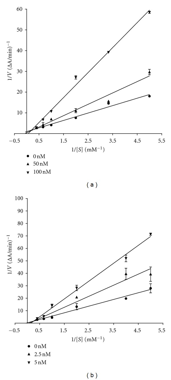
Lineweavere-Burk reciprocal plots of inhibition kinetics of (a) acetylcholinesterase and (b) butyrylcholinesterase by the compound 4b. Changes in the initial velocities of the reaction were measured at different concentrations of the inhibitors (from 0–100 and 0–5 nM for acetyl and butyrylcholinesterase, resp.) by using substrates ATCI and BTCCl.
2.4. Docking Results
In order to investigate the probable binding modes and to explain different binding affinities of the molecules, compounds were docked into binding sites of the enzymes. Newly synthesized inhibitors for AChE and BChE possessed the similar binding modes. Our docking results showed that all compounds have similar binding modes with different docking scores as shown in Table 2. Most of the compounds have been gorged into the catalytic amino acid triad [35] (Glu224, Ser225, and His 494) of AChE and (Glu225, Ser226, and His466) of BChE, respectively. The top ranked binding conformation of the most active compound of AChE and BChE is shown in Figures 3 and 4, respectively. Based on docking simulations, we can explain that strong binding affinity of 3g with BChE is due to the hydrogen bonding of coumarin ring carbonyl moiety with N-H of His-466 residue and sulfur of triazolethoiphene with side chain of one of catalytic residue Glu225, shown in Figure 4. Similarly, the most active compound 4b for AChE interacts with enzyme through hydrogen bonding interaction. Carbonyl moiety of coumarin ring makes a hydrogen bond with N-H of His494 residue as shown in Figure 3. It has been observed that AChE inhibitors have strong binding affinities with their enzymes over BChE inhibitors, which is in contrast to observed activities. This might be due to inabilities or poor performance of the docking scoring functions [36].
Table 2.
Docking scores of all synthesized compounds in AChE and BChE.
| Code | AChE | BChE |
|---|---|---|
| Chemfitness scores | ||
| 3a | 35.598 | 28.560 |
| 3b | 34.155 | 29.813 |
| 3c | 33.360 | 27.474 |
| 3d | 36.365 | 22.734 |
| 3e | 34.741 | 29.559 |
| 3f | 37.335 | 29.979 |
| 3g | 30.625 | 26.258 |
| 4a | 37.172 | 22.609 |
| 4b | 38.591 | 23.518 |
| 4c | 38.212 | 26.031 |
| 4d | 35.733 | 28.354 |
| 4e | 30.520 | 23.943 |
| 4f | 37.081 | 27.062 |
| 4g | 31.208 | 23.505 |
Figure 3.
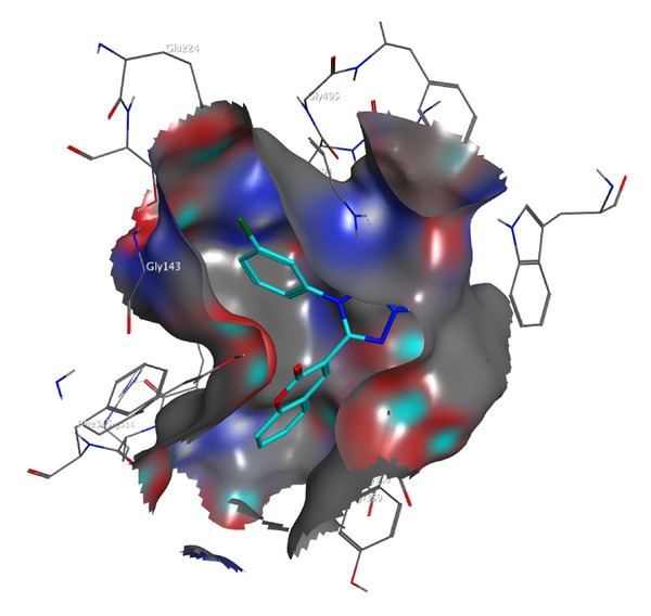
Binding mode of top ranked most active (4b) compound in the binding site of AChE enzyme.
Figure 4.
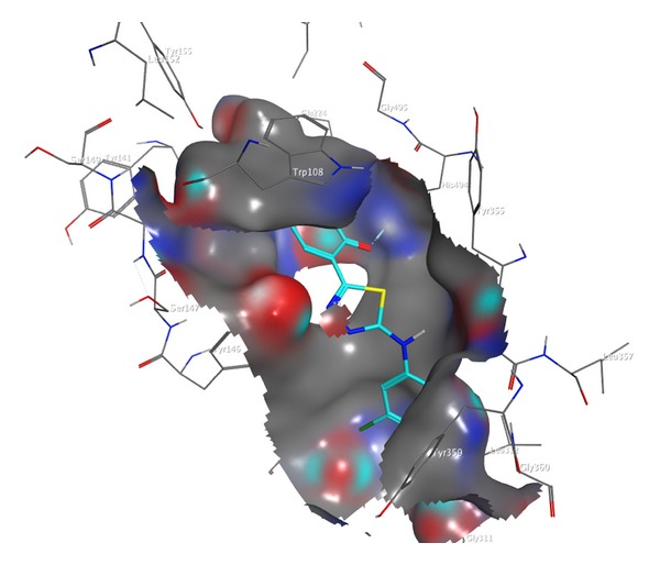
Binding mode of top ranked most active (3g) compound in the binding site of BChE enzyme.
3. Conclusion
The synthesis, biological evaluation, and molecular docking studies of fourteen novel 3-thiadiazolyl- and thioxo-1,2,4-triazolyl coumarins derivatives as potential inhibitors of cholinesterases have been reported. The cholinesterase inhibition studies revealed that all compounds were potential inhibitors of the investigated enzymes and can be used for treatment of Alzheimer's disease. Some of the investigated compounds were more potent on rabbit AChE enzyme, better than the reference drug. It was also observed that the coumarin derivatives were competitive inhibitors for the AChE and BChE enzymes. The study of their anticholinesterase activities might lead to a novel family of potent anti-AD compounds in near future.
4. Experimental
4.1. Chemistry
4.1.1. Synthesis of 1,4-Disubstituted-thiosemicarbazides (3a–g), General Procedure
A mixture of carbohydrazide (6.8 mmol) and substituted phenyl isothiocyanate (1a–g) (6.6 mmol) was stirred for 10–12 h at 50–60°C. After consumption of the starting materials, the mixture was cooled at room temperature. The methanol was evaporated on rotary evaporator leaving behind a crude product as oil that solidified on cooling and was recrystallized from a mixture of ethyl acetate and petroleum ether (4 : 1) to yield thiosemicarbazides(2a–g).
4.1.2. 4-(2-Chlorophenyl)-1-(2-oxo-2H-chromene-3-carbonyl)thiosemicarbazide (2a)
Green solid (54%): m.p. 215–217°C; Rf*: 0.54; IR (KBr, cm−1): 3296–3163 (NH), 1731 (C=O), 1673 (C=O), 1596, 1512 (C=C), 1268 (C=S); 1H NMR (300 MHz, DMSO-d6): δ 11.96 (s, 1H, NH-C=O), 10.05 (s, 2H, NH-C=S), 9.01 (s, 1H, C-H, H-4), 8.03 (d, 1H, J = 7.5 Hz, H-8), 7.71–7.69 (m, 1H, H-6), 7.37–7.09 (m, 3H, H-7, H-3′, H-5′), 6.99 (d, 1H, J = 8.1 Hz, H-5), 6.94–6.76 (m, 2H, H-4′, H-6′); 13C NMR (75 MHz, DMSO-d6): δ 181.2 (C=S), 166.9 (NH-C=O), 159.5 (C=O), 150.2, 135.3, 134.6, 132.1, 129.5, 126.6, 125.5, 124.9, 123.4, 122.4, 120.9, 118.6, 116.2 (Ar-Cs). Anal. Calcd. for C17H12ClN3O3S: C, 54.62; H, 3.24; N, 11.24, S, 8.58; Found: C, 54.54; H, 3.11; N, 11.09; S, 8.49.
4.1.3. 4-(3-Chlorophenyl)-1-(2-oxo-2H-chromene-3-carbonyl)thiosemicarbazide (2b)
Yellow solid (59%): m.p. 189–191°C; Rf*: 0.54; IR (KBr, cm−1): 3295–3163 (NH), 1724 (C=O), 1670 (C=O), 1590, 1508 (C=C), 1267 (C=S); 1H NMR (300 MHz, DMSO-d6): δ 11.35 (s, 1H, NH-C=O), 10.15 (s, 2H, NH-C=S), 9.00 (s, 1H, C-H, H-4), 7.68 (d, 1H, J = 7.8 Hz, H-8), 7.42–7.36 (m, 2H,H-6, H-7), 7.20 (d, J = 7.8 Hz, H-5), 6.99–6.89 (m, 2H, H-3′, H-5′), 6.84–6.79 (m, 2H, H-4′, H-6′); 13C NMR (75 MHz, DMSO-d6): δ 181.4 (C=S), 163.1 (NH-C=O), 159.3 (C=O), 156.9, 142.2, 133.6, 131.1, 129.0, 128.6, 125.4, 120.2, 119.8, 119.4, 118.7, 117.1, 116.1 (Ar-Cs). Anal. Calcd. for C17H12ClN3O3S: C, 54.62; H, 3.24; N, 11.24, S, 8.58; Found: C, 54.71; H, 3.17; N, 11.15; S, 8.43.
4.1.4. 4-(4-Chlorophenyl)-1-(2-oxo-2H-chromene-3-carbonyl)thiosemicarbazide (2c)
Greenish solid (56%): m.p. 177–179°C; Rf*: 0.57; IR (KBr, cm−1): 3341–3223 (NH), 1732 (C=O), 1670 (C=O), 1558, 1514, 1484 (C=C), 1268 (C=S); 1H NMR (300 MHz, DMSO-d6): δ 11.23 (s, 1H, NH-C=O), 11.11 (s, 2H, NH-C=S), 9.01 (s, 1H, C-H, H-4), 7.70 (d, 1H, J = 7.5 Hz, H-8), 7.50–7.22 (m, 5H, H-5, H-6, H-7, H-3′, H-5′), 7.00–6.94 (m, 2H, H-2′, H-6′); 13C NMR (75 MHz, DMSO-d6): δ 181.5 (C=S), 167.0 (NH-C=O), 163.2 (C=O), 149.3, 133.7, 131.3, 131.2, 129.1, 125.3, 124.3, 123.7, 123.4, 118.7, 117.0 (Ar-Cs). Anal. Calcd. for C17H12ClN3O3S: C, 54.62; H, 3.24; N, 11.24, S, 8.58; Found: C, 54.51; H, 3.32; N, 11.07; S, 8.43.
4.1.5. 1-(2-Oxo-2H-chromene-3-carbonyl)-4-m-tolylthiosemicarbazide (2d)
Yellow solid (61%): m.p. 150–152°C; Rf*: 0.50; IR (KBr, cm−1): 3300–3143 (NH), 1732 (C=O), 1674 (C=O), 1522, 1497 (C=C), 1236 (C=S); 1H NMR (300 MHz, DMSO-d6): δ 11.76 (s, 1H, NH-C=O), 9.99 (s, 2H, NH-C=S), 8.48 (s, 1H, C-H, H-4), 8.10 (d, 1H, J = 7.5 Hz, H-8), 7.41–7.36 (m, 1H, H-6), 7.25–7.20 (m, 2H, H-7, H-5′), 7.21–7.14 (m, 1H, H-2′), 7.01(d, 1H, J = 7.5 Hz, H-5), 6.89–6.81 (m, 2H, H-4′, H-6′), 2.31(s, 3H, CH3); 13C NMR (75 MHz, DMSO-d6): δ 179.2 (C=S), 164.7 (NH-C=O), 157.0 (C=O), 149.4, 138.7, 137.7, 131.7, 128.3, 126.6, 126.2, 124.5, 123.3, 122.4, 120.7, 119.7, 116.4 (Ar-Cs), 21.4 (CH3). Anal. Calcd. for C18H15N3O3S: C, 61.18; H, 4.28; N, 11.89, S, 9.07; Found: C, 61.11; H, 4.33; N, 11.75; S, 8.97.
4.1.6. 1-(2-Oxo-2H-chromene-3-carbonyl)-4-p-tolylthiosemicarbazide (2e)
White solid (59%): m.p. 179–181°C; Rf*: 0.51; IR (KBr, cm−1): 3432–3179 (NH), 1718 (C=O), 1689 (C=O), 1573, 1538, 1513 (C=C), 1259 (C=S); 1H NMR (300 MHz, DMSO-d6): δ 11.73(s, 1H, NH-C=O), 11.14 (s, 2H, NH-C=S), 9.98 (s, 1H, C-H, H-4), 7.42 (d, 1H, J = 7.5 Hz, H-8), 7.26–7.23 (m, 2H, H-7, H-6), 7.21–7.14 (m, 2H, H-2′, H-6′), 6.99 (d, 1H, J = 7.5 Hz, H-5), 6.89–6.81 (m, 2H, H-3′, H-5′), 2.30 (s, 3H, CH3); 13C NMR (75 MHz, DMSO-d6): δ 176.2 (C=S), 165.4 (NH-C=S), 157.0 (C=O), 148.2, 137.0, 134.8, 131.8, 128.9, 126.2, 123.4, 122.7, 120.7, 119.7, 116.5 (Ar-Cs), 21.1 (CH3). Anal. Calcd. for C18H15N3O3S: C, 61.18; H, 4.28; N, 11.89, S, 9.07; Found: C, 61.02; H, 4.20; N, 11.95; S, 8.92.
4.1.7. 4-(2-Methoxyphenyl)-1-(2-oxo-2H-chromene-3-carbonyl)thiosemicarbazide (2f)
White solid (57%): m.p. 198–200°C; Rf*: 0.56; IR (KBr, cm−1): 3326–3281 (NH), 1726 (C=O), 1644 (C=O), 1593, 1520 (C=C), 1235 (C=S); 1H NMR (300 MHz, DMSO-d6): δ 11.90(s, 1H, NH-C=O), 11.14 (s, 2H, NH-C=S), 9.97 (s, 1H, C-H, H-4), 7.88 (d, 1H, J = 7.8 Hz, H-8), 7.28–7.25 (m, 1H,H-6), 7.23–7.14 (m, 1H, H-7), 7.09 (d, 1H, J = 7.2 Hz, H-5), 7.00–6.96 (m, 2H, H-3′, H-6′), 6.93-6.86 (m, 2H, H-4′, H-5′), 3.35 (s, 3H, OCH3); 13C NMR (75 MHz, DMSO-d6): δ 181.7 (C=S), 166.2 (NH-C=O), 161.2 (C=O), 151.6, 139.1, 136.2, 130.4, 128.2, 126.0, 125.9, 125.7, 124.5, 120.6, 120.3, 119.9, 116.6, 111.6 (Ar-Cs), 55.7 (OCH3). Anal. Calcd. for C18H15N3O4S: C, 58.53; H, 4.09; N, 11.38, S, 8.68; Found: C, 58.67; H, 4.21; N, 11.24; S, 8.53.
4.1.8. 4-(3-Methoxyphenyl)-1-(2-oxo-2H-chromene-3-carbonyl)thiosemicarbazide (2g)
Green solid (57%): m.p. 169–172°C; Rf*: 0.56; IR (KBr, cm−1): 3324–3283 (NH), 1721 (C=O), 1652 (C=O), 1589, 1518 (C=C), 1237 (C=S); 1H NMR (300 MHz, DMSO-d6): δ 11.70 (s, 1H, NH-C=O), 10.00 (s, 2H, NH-C=S), 9.01 (s, 1H, C-H, H-4), 7.69 (d, 1H, J = 7.8 Hz, H-8), 7.40–7.37 (m, 1H, H-6), 7.30–7.17 (m, 2H, H-7, H-5), 7.00–6.91 (m, 2H, H-4′, H-5′), 6.90–6.75 (m, 2H, H-2′, H-6′), 3.76 (s, 3H, OCH3); 13C NMR (75 MHz, DMSO-d6): δ 180.7 (C=S), 163.2 (NH-C=O), 159.4 (C=O), 150.4, 140.7, 133.7, 131.3, 129.9, 129.1, 127.5, 125.7, 124.5, 120.7, 120.0, 119.6, 117.0, 111.5 (Ar-Cs), 55.5 (OCH3). Anal. Calcd. for C18H15N3O4S: C, 58.53; H, 4.09; N, 11.38, S, 8.68; Found: C, 58.45; H, 4.11; N, 11.29; S, 8.51.
4.1.9. Synthesis of 4,5-Disubstituted-1,2,4-triazol-3(4H) thiones (4a–g), General Procedure
The thiosemicarbazides (2a–g) (1.4 mmol) were refluxed (4-5 h) in aqueous sodium hydroxide solution (4N, 25 mL). The progress of reaction was monitored by TLC. After completion of the reaction, the reaction mixture was cooled to room temperature and filtered. The filtrate was neutralized with hydrochloric acid (4N) to precipitate the triazoles, which were filtered and recrystallized from aqueous ethanol.
4.1.10. 3-(4-(2-Chlorophenyl)-5-thioxo-4,5-dihydro-1H-1,2,4-triazol-3-yl)-2H-chromen-2-one (3a)
Yellow solid (63%): m.p 205–207°C; Rf*: 0.61; IR (KBr, cm−1): 3292 (NH), 1730 (C=O), 1555, 1477 (C=C), 1409 (C=N), 1253 (C=S); 1H NMR (300 MHz, DMSO-d6): δ 11.13 (s, 1H, NH), 9.01 (s, 1H, C-H, H-4), 7.63 (d, 1H, J = 7.8 Hz, H-8), 7.42–7.38 (m, 2H, H-6, H-7), 6.93 (d, 1H, J = 7.5 Hz, H-5), 7.11–7.08 (m, 2H, H-3′, H-5′), 7.03–6.99 (m, 1H, H-6′), 6.88–6.81 (m, 1H, H-4′); 13C NMR (75 MHz, DMSO-d6): δ 165.2 (C=S), 159.5 (C=O), 158.5 (C=N), 148.7, 133.6, 131.8, 131.1, 128.9, 128.6, 127.6, 126.4, 122.2, 121.4, 119.4, 119.1, 117.3, 116.9 (Ar-Cs). Anal. Calcd. for C17H10ClN3O2S: C, 57.39; H, 2.83; N, 11.81, S, 9.01; Found: C, 57.23; H, 2.71; N, 11.70; S, 8.87.
4.1.11. 3-(4-(3-Chlorophenyl)-5-thioxo-4,5-dihydro-1H-1,2,4-triazol-3-yl)-2H-chromen-2-one (3b)
Yellow solid (61%): m.p. 207–209°C; Rf*: 0.61; IR (KBr, cm−1): 3282 (NH), 1728 (C=O), 1553, 1482 (C=C), 1383 (C=N), 1267 (C=S); 1H NMR (300 MHz, DMSO-d6): δ 11.13 (s, 1H, NH), 9.00 (s, 1H, C-H, H-4), 7.68 (d, 1H, J = 7.8 Hz, H-8), 7.43–7.41 (m, 2H, H-6, H-7), 7.38–7.37 (m, 2H, H-4′, H-5′), 7.00 (d, 1H, J = 7.8 Hz,H-5), 6.97–6.95 (m, 2H, H-2′, H-6′); 13C NMR (75 MHz, DMSO-d6): δ 163.2 (C=S), 159.3 (C=O), 157.8 (C=N), 148.4, 133.7, 131.2, 130.5, 129.1, 128.6, 122.2, 120.1, 119.2, 118.6, 117.3, 117.0 (Ar-Cs). Anal. Calcd. for C17H10ClN3O2S: C, 57.39; H, 2.83; N, 11.81, S, 9.01; Found: C, 57.27; H, 2.69; N, 11.88; S, 8.92.
4.1.12. 3-(4-(4-Chlorophenyl)-5-thioxo-4,5-dihydro-1H-1,2,4-triazol-3-yl)-2H-chromen-2-one (3c)
Green solid (66%): m.p. 204–206°C; Rf*: 0.62; IR (KBr, cm−1): 3287 (NH), 1728 (C=O), 1571, 1486 (C=C), 1381 (C=N), 1269 (C=S); 1H NMR (300 MHz, DMSO-d6): δ 11.14 (s, 1H, NH), 9.01 (s, 1H, C-H, H-4), 7.69 (d, 1H, J = 7.8 Hz, H-8), 7.43–7.40 (m, 2H, H-6, H-7), 7.39–7.37 (m, 2H, H-3′, H-5′), 6.98 (d, 1H, J = 7.8 Hz, H-5), 6.96–6.94 (m, 2H, H-2′, H-6′); 13C NMR (75 MHz, DMSO-d6): δ 163.3 (C=S), 159.1 (C=O), 157.9 (C=N), 148.6, 133.7, 131.3, 130.4, 129.2, 128.7, 122.6, 120.0, 119.3, 118.7, 117.2, 117.0 (Ar-Cs). Anal. Calcd. for C17H10ClN3O2S: C, 57.39; H, 2.83; N, 11.81, S, 9.01; Found: C, 57.26; H, 2.69; N, 11.67; S, 8.92.
4.1.13. 3-(5-Thioxo-4-m-tolyl-4,5-dihydro-1H-1,2,4-triazol-3-yl)-2H-chromen-2-one (3d)
Yellow solid (59%): m.p. 217-219°C; Rf*: 0.59; IR (KBr, cm−1): 3281 (NH), 1725 (C=O), 1576, 1541 (C=C), 1413 (C=N), 1235 (C=S); 1H NMR (300 MHz, DMSO-d6): δ 11.15 (s, 1H, NH), 9.00 (s, 1H, C-H, H-4), 7.71–7.68 (m, 2H, H-8, H-6), 7.43–7.37 (m, 2H, H-7, H-5), 6.99–6.95 (m, 4H, H-2′, H-4′, H-5′, H-6′), 2.21 (s, 3H, CH3); 13C NMR (75 MHz, DMSO-d6): δ 163.3 (C=S), 160.1 (C=O), 159.1 (C=N), 149.3, 133.7, 131.3, 131.1, 129.2, 128.2, 126.1, 124.6, 122.1, 120.1, 119.3, 118.6, 117.4, 117.0 (Ar-Cs), 24.3 (CH3). Anal. Calcd. for C18H13N3O2S: C, 64.46; H, 3.91; N, 12.53, S, 9.56; Found: C, 64.51; H, 3.79; N, 12.42; S, 9.40.
4.1.14. 3-(5-Thioxo-4-p-tolyl-4,5-dihydro-1H-1,2,4-triazol-3-yl)-2H-chromen-2-one (3e)
Yellow solid (61%): m.p. 211–213°C; Rf*: 0.58; IR (KBr, cm−1): 3297 (NH), 1732 (C=O), 1557, 1513 (C=C), 1375 (C=N), 1288 (C=S); 1H NMR (300 MHz, DMSO-d6): δ 11.14 (s, 1H, NH), 9.01 (s, 1H, C-H, H-4), 7.43–7.25 (m, 4H, H-8, H-7, H-6, H-5), 7.07 (d, 2H, J = 8.1 Hz, H-2′, H-6′), 6.99 (d, 2H, J = 7.8 Hz, H-3′, H-5′), 2.23 (s, 3H, CH3); 13C NMR (75 MHz, DMSO-d6): δ 163.5 (C=S), 159.4 (C=O), 158.6 (C=N), 149.1, 137.7, 133.6, 130.9, 129.6, 128.7, 125.5, 121.0, 119.3, 118.6, 117.0, 116.0, 112.7, 116.8 (Ar-Cs), 20.8 (CH3). Anal. Calcd. for C18H13N3O2S: C, 64.46; H, 3.91; N, 12.53, S, 9.56; Found: C, 64.61; H, 3.82; N, 12.39; S, 9.41.
4.1.15. 3-(4-(2-Methoxyphenyl)-5-thioxo-4,5-dihydro-1H-1,2,4-triazol-3-yl)-2H-chromen-2-one (3f)
Yellow solid (64%): m.p. 214–216°C; Rf*: 0.51; IR (KBr, cm−1): 3275 (NH), 1730 (C=O), 1552, 1475 (C=C), 1410 (C=N), 1233 (C=S); 1H NMR (300 MHz, DMSO-d6): δ 13.94 (s, 1H, NH), 9.88 (s, 1H, C-H, H-4), 7.36–7.28 (m, 2H, H-8, H-6), 7.22–7.14 (m, 2H, H-5, H-7), 7.00–6.95 (m, 2H, H-3′, H-5′), 6.77–6.72 (m, 2H, H-4′, H-6′), 3.56 (s, 3H, OCH3); 13C NMR (75 MHz, DMSO-d6): δ 168.5 (C=S), 159.1 (C=O), 156.3 (C=N), 150.9, 139.7, 132.3, 131.4, 130.7, 126.2, 123.1, 120.5, 119.8, 118.8, 117.7, 116.0, 112.7, 113.7 (Ar-Cs), 56.0 (OCH3). Anal. Calcd. for C18H13N3O3S: C, 61.53; H, 3.73; N, 11.96, S, 9.13; Found: C, 61.43; H, 3.56; N, 11.88; S, 9.01.
4.1.16. 3-(4-(3-Methoxyphenyl)-5-thioxo-4,5-dihydro-1H-1,2,4-triazol-3-yl)-2H-chromen-2-one (3g)
Green solid (60%): m.p. 218–220°C; Rf*: 0.51; IR (KBr, cm−1): 3273 (NH), 1727 (C=O), 1551, 1477 (C=C), 1412 (C=N), 1237 (C=S); 1H NMR (300 MHz, DMSO-d6): δ 11.13 (s, 1H, NH), 9.01 (s, 1H, C-H, H-4), 7.71–7.68 (m, 2H, H-8, H-6), 7.43–7.37 (m, 2H, H-5, H-7), 7.01–6.95 (m, 2H, H-4′, H-5′), 6.70–6.36 (m, 2H, H-2′, H-6′), 3.53 (s, 3H, OCH3); 13C NMR (75 MHz, DMSO-d6): δ 163.5 (C=S), 159.1 (C=O), 156.4 (C=N), 150.8, 139.9, 133.7, 131.3, 130.4, 126.6, 123.2, 120.7, 119.7, 118.6, 117.0, 116.1, 113.5, 112.4 (Ar-Cs), 56.7 (OCH3). Anal. Calcd. for C18H13N3O3S: C, 61.53; H, 3.73; N, 11.96, S, 9.13; Found: C, 61.48; H, 3.61; N, 11.81; S, 9.16.
4.1.17. Synthesis of 2,5-disubstituted-1,3,4-thiadiazoles (4a–g), General Procedure
1,4-Disubstituted thiosemicarbazides (2a–g) (1.4 mmol), in polyphosphoric acid (0.5 mL, 2.8 mmol) were stirred overnight at 70°C. After completion of reaction, the cooled solution was poured on the crushed ice. The reaction mixture was extracted with ethyl acetate (3 × 20 mL) and combined extracts were washed with sodium bicarbonate (5%) and water until the washings were neutral. The organic layer was dried with anhydrous sodium sulphate and concentrated under reduced pressure to yield the 2,5-disubstituted-1,3,4-thiadiazoles, purified by recrystallization in ethanol.
4.1.18. 3-(5-(2-Chlorophenylamino)-1,3,4-thiadiazol-2-yl)-2H-chromen-2-one (4a)
Yellow solid (45%): m.p. 212–214°C; Rf*: 0.40; IR (KBr, cm−1): 3278 (NH), 1725 (C=O), 1563, 1529 (C=C), 1415 (C=N); 1H NMR (300 MHz, DMSO-d6): δ 11.42 (s, 1H, NH), 8.73 (s, 1H, C-H, H-4), 7.44–7.36 (m, 4H, H-6, H-8, H-3′, H-5′), 7.04–6.96 (m, 4H, H-5, H-7, H-4′, H-6′); 13C NMR (75 MHz, DMSO-d6): δ 164.7 (C=O), 159.8 (C=N-NH), 159.2 (C=N), 149.2, 133.5, 132.6, 131.9, 130.7, 129.1, 128.5, 127.8, 127.6, 120.0, 119.8, 117.8, 117.3, 116.8 (Ar-Cs). Anal. Calcd. for C17H10ClN3O2S: C, 57.39; H, 2.83; N, 11.81, S, 9.01; Found: C, 57.27; H, 2.76; N, 11.72; S, 8.87.
4.1.19. 3-(5-(3-Chlorophenylamino)-1,3,4-thiadiazol-2-yl)-2H-chromen-2-one (4b)
Yellow solid (44%): m.p. 210–212°C; Rf*: 0.40; IR (KBr, cm−1): 3271 (NH), 1728 (C=O), 1565, 1527 (C=C), 1419 (C=N); 1H NMR (300 MHz, DMSO-d6): δ 11.15 (s, 1H, NH), 9.01 (s, 1H, C-H, H-4), 7.70–7.61 (m, 4H, H-6, H-8, H-3′, H-5′), 7.50–7.36 (m, 4H, H-5, H-7, H-2′, H-6′); 13C NMR (75 MHz, DMSO-d6): δ 163.7 (C=O), 159.9 (C=N-NH), 159.1 (C=N), 148.2, 135.5, 133.6, 132.9, 130.5, 129.2, 128.6, 127.7, 126.6, 120.2, 119.1, 117.9, 117.1, 116.3 (Ar-Cs). Anal. Calcd. for C17H10ClN3O2S: C, 57.39; H, 2.83; N, 11.81, S, 9.01; Found: C, 57.23; H, 2.71; N, 11.67; S, 9.09.
4.1.20. 3-(5-(4-Chlorophenylamino)-1,3,4-thiadiazol-2-yl)-2H-chromen-2-one (4c)
Yellow solid (59%): m.p. 216–218°C; Rf*: 0.45; IR (KBr, cm−1): 3256 (NH), 1728 (C=O), 1543, 1521 (C=C), 1432 (C=N); 1H NMR (300 MHz, DMSO-d6): δ 11.15 (s, 1H, NH), 9.01 (s, 1H, C-H, H-4), 7.69 (d, 1H, J = 7.5 Hz, H-8), 7.65–7.61 (m, 1H, H-6), 7.48 (d, 2H, J = 8.4 Hz, H-3′, H-5′), 7.42–7.36 (m, 1H,H-7), 6.98 (d, 1H, J = 8.4 Hz, H-5), 6.95(d, 2H, J = 8.1 Hz, H-2′, H-6′); 13C NMR (75 MHz, DMSO-d6): δ 163.3 (C=O), 159.1 (C=N-NH), 157.3 (C=N), 148.6, 133.7, 131.3, 130.7, 129.3, 129.1, 128.3, 127.9, 120.1, 119.3, 118.6, 116.9 (Ar-Cs). Anal. Calcd. for C17H10ClN3O2S: C, 57.39; H, 2.83; N, 11.81, S, 9.01; Found: C, 57.26; H, 2.96; N, 11.74; S, 8.89.
4.1.21. 3-(5-(m-Toluidino)-1,3,4-thiadiazol-2-yl)-2H-chromen-2-one (4d)
Yellow solid (39%): m.p. 219–221°C; Rf*: 0.47; IR (KBr, cm−1): 3198 (NH), 1730 (C=O), 1600, 1561, 1501 (C=C), 1463 (C=N); 1H NMR (300 MHz, DMSO-d6): δ 11.14 (s, 1H, NH), 9.01 (s, 1H, C-H, H-4), 7.71–7.68 (m, 2H, H-8, H-6), 7.43–7.37 (m, 2H, H-7, H-5′), 6.99–6.94 (m, 4H, H-5, H-2′, H-4′, H-6′), 2.08 (s, 3H, CH3); 13C NMR (75 MHz, DMSO-d6): δ 163.2 (C=O), 159.1 (C=N-NH), 157.4 (C=N), 148.8, 133.7, 130.9, 130.2, 128.3, 127.1, 124.3, 121.9, 121.8, 120.1, 119.7, 119.5, 119.1, 118.6, 117.9, 117.3, 117.0, 116.7, 116.3, 116.0 (Ar-Cs), 31.1 (CH3). Anal. Calcd. for C18H13N3O2S: C, 64.46; H, 3.91; N, 12.53, S, 9.56; Found: C, 64.34; H, 3.98; N, 12.38; S, 9.41.
4.1.22. 3-(5-(p-Toluidino)-1,3,4-thiadiazol-2-yl)-2H-chromen-2-one (4e)
Yellow solid (56%): m.p. 191–193°C; Rf*: 0.44; IR (KBr, cm−1): 3267 (NH), 1725 (C=O), 1592, 1554 (C=C), 1471 (C=N); 1H NMR (300 MHz, DMSO-d6): δ 11.14 (s, 1H, NH), 9.70 (s, 1H, C-H, H-4), 7.72–7.64 (m, 2H, H-8, H-6), 7.45–7.24 (m, 2H, H-7, H-5), 7.17–7.13 (m, 2H, H-2′, H-6′), 7.12–7.08 (m, 2H, H-3′, H-5′), 2.27 (s, 3H, CH3); 13C NMR (75 MHz, DMSO-d6): δ 167.4 (C=O), 163.0 (C=N-NH), 157.0 (C=N), 145.8, 134.0, 131.6, 130.0, 129.0, 124.3, 121.9, 119.7, 119.5, 117.9, 117.0, 116.3 (Ar-Cs), 20.8 (CH3). Anal. Calcd. for C18H13N3O2S: C, 64.46; H, 3.91; N, 12.53, S, 9.56; Found: C, 64.32; H, 3.78; N, 12.40; S, 9.45.
4.1.23. 3-(5-(2-Methoxyphenylamino)-1,3,4-thiadiazol-2-yl)-2H-chromen-2-one (4f)
Yellow solid (49%): m.p. 206–208°C; Rf*: 0.51; IR (KBr, cm−1): 3287 (NH), 1726 (C=O), 1594, 1544 (C=C), 1466 (C=N); 1H NMR (300 MHz, DMSO-d6): δ 11.86 (s, 1H, NH), 9.96 (s, 1H, C-H, H-4), 7.69 (d, 1H, J = 7.2 Hz, H-8), 7.43–7.38 (m, 2H, H-6, H-7), 7.04–6.85 (m, 5H, H-5, H-3′, H-4′, H-5′, H-6′), 3.87 (s, 3H, OCH3); 13C NMR (75 MHz, DMSO-d6): δ 163.2 (C=O), 159.1 (C=N-NH), 157.3 (C=N), 148.6, 147.0, 133.7, 132.0, 129.5, 128.3, 128.0, 121.7, 121.0, 120.1, 118.6, 116.8, 116.4 (Ar-Cs), 56.5 (OCH3). Anal. Calcd. for C18H13N3O3S: C, 61.53; H, 3.73; N, 11.96, S, 9.13; Found: C, 61.41; H, 3.60; N, 11.83; S, 9.04.
4.1.24. 3-(5-(3-Methoxyphenylamino)-1,3,4-thiadiazol-2-yl)-2H-chromen-2-one (4g)
Yellow solid (46%): m.p. 202–204°C; Rf*: 0.51; IR (KBr, cm−1): 3284 (NH), 1729 (C=O), 1584, 1545 (C=C), 1468 (C=N); 1H NMR (300 MHz, DMSO-d6): δ 11.15 (s, 1H, NH), 9.79 (s, 1H, C-H, H-4), 7.70 (d, 1H, J = 7.2 Hz, H-8), 7.43–7.37 (m, 2H, H-6, H-7), 7.99–6.85 (m, 5H, H-5, H-2′, H-4′, H-5′, H-6′), 3.56 (s, 3H, OCH3); 13C NMR (75 MHz, DMSO-d6): δ 165.2 (C=O), 159.9 (C=N-NH), 157.7 (C=N), 147.8, 146.3, 133.6, 131.0, 129.2, 128.4, 127.7, 123.7, 121.9, 120.5, 119.6, 118.8, 116.4 (Ar-Cs), 58.6 (OCH3). Anal. Calcd. for C18H13N3O3S: C, 61.53; H, 3.73; N, 11.96, S, 9.13; Found: C, 61.46; H, 3.81; N, 11.85; S, 9.02.
*Rf solvent system (petroleum ether: ethyl acetate, 4 : 1).
4.2. Materials and Methods
4.2.1. Chemicals and Materials
Acetylcholinesterase (AChE) (EC 3.1.1.7, type VI-S from Electric Eel), butyrylcholinesterase (BChE) (EC 3.1.1.8, from horse serum). AChE and BChE were also isolated from rabbit brain and serum, respectively. Acetylthiocholine iodide (ATCI), S-butyrylthiocholine chloride (BTCCl), 5,5′-dithio-bis(2-nitrobenzoic acid) (DTNB), neostigmine methylsulfate, donepezil, dimethylsulfoxide (DMSO), and bovine serum albumin were purchased from Sigma-Aldrich (Steinheim, Germany).
4.2.2. Isolation of AChE from Rabbit Brain and BChE from Blood Serum
AChE was isolated from rabbit brain. Heads were decapitated and brains were thawed and homogenized for 30 sec in 2.5 volumes of 1 mM EDTA, 0.32 M ice cold sucrose, 10 μM tetracain (pH7.0). The homogenates were centrifuged at 105,000 ×g for 60 min (4°C). The supernatants were removed and pellets were again homogenized in same volume of 0.2% triton phosphate buffer. After centrifugation, the supernatants were again removed and pellets were rehomogenized for 10 sec in 2.5 volumes of 1% triton phosphate buffer. The final supernatants obtained after centrifugation for 60 min at 105,000 ×g were applied to an immunosorbent affinity column at a flow rate of 6.5 mL/hr approximately [37]. BChE was obtained from rabbit blood serum by applying the techniques of centrifugation. The purified enzymes were stored at –80°C.
4.2.3. Determination of AChE and BChE Inhibitory Activities in 96-Microliter Well Plates
The inhibitory activities of coumarin derivatives were measured quantitatively by Ellman's method [38] and performed with some modifications as in Ingkaninan et al. method [39] by using 96-microliter well plate. Series of newly synthesized coumarin derivatives were tested as AChE and BChE inhibitors. Initially, each compound was dissolved in DMSO (end concentration of DMSO was less than 1% in assay) and tested at a final concentration of 1 mM or 10 μL of DMSO (as negative control) in wells for initial screening. Compounds with considerable inhibition (more than 50%) were subjected to further analysis by making their six to seven serial dilutions in an assay buffer (50 mM Tris-HCl, 0.02 M MgCl2·6H2O, and 0.1 M NaCl at pH 8.0). Reaction mixture comprised of 50 μL of 3 mM DTNB (5,5′-dithiobis(2-nitrobenzoic acid)) in assay buffer, 10 μL of test compound, and 10 μL of AChE (0.031 IU/mL) or BChE (0.5 IU/mL). This mixture was preincubated at 25°C for 10 min. After preincubation, enzymatic reaction was started by addition of 10 μL of 10 mM MBTCCl (butyrylthiocholine chloride) or ATCI (acetylthiocholine iodide) according to the respective enzyme and mixture was incubated again for 15 min. The amount of enzymatic product was measured by the change in absorbance at 405 nm by using a microplate reader (Bio-TekELx 800, Instruments, Inc. USA). Neostigmine methylsulphate and donepezil were used as standard inhibitors. Enzyme dilution buffer consisted of 50 mM Tris-HCl containing 0.1% (w/v) BSA (pH 8). The effect of DMSO on activity of enzyme was subtracted by a negative control containing DMSO, instead of inhibitor. Each concentration was analyzed in triplicate and Ki values were calculated from IC50 values by using a nonlinear curve fitting program PRISM 5.0 (GraphPad, San Diego, California, USA).
4.2.4. Kinetic Characterization of AChE and BChE Inhibition
Kinetic characterization of AChE and BChE was performed by using Ellman's method [38]. The effect of inhibitor 4b on varying concentration of substrate from 0.2 to 2.5 mM was investigated. Enzyme kinetic characterization studies were performed under same incubation conditions as described above by using ATCI and BTCCl as substrates and DTNB was used as chromophoric reagent. A parallel control with no inhibitor in the mixture was added for comparison. Each concentration was analyzed in triplicate; and Lineweaver-Burk (1/V versus 1/[S]) plot was constructed using Prism software.
4.2.5. Docking Studies
(1) Compounds Structures Preparation —
Before performing the molecular docking simulations of small molecules inside the homology models of both protein structures, all molecules were sketched and protonated by using MOE molecules sketcher tool. The three dimensional conformations of these molecules were generated using the protonate 3D tool implemented in MOE. Finally the molecules were minimized and their charges were optimized by using the MMFF94 modified force-field.
(2) Docking Protocol —
The molecular docking of compounds in generated homology models were performed by using GOLD (Genetic Optimization for ligand docking) program. GOLD uses the genetic algorithm (GA) to search full length of conformational flexibility of ligands inside the protein binding site [40]. All compounds were sketched and their ionization states were fixed using MOE. In these docking simulations, 10 Å spherical binding site was used across the His-494 and His-466 for AChE and BChE enzymes, respectively. Hydrogen atoms were also added to both model structures. During docking simulations, the protein residues remained rigid except Ser, Thr, and Tyr hydroxyl groups, in order to optimize hydrogen bonding interactions with the docked compounds. For each solution 10 GA operations were run, and best ranked solution based on the chemscore was selected for each molecule. All other parameters default values were used.
(3) Homology Models Generation —
The Butyrylcholinesterase (BChE) (EC 3.1.1.8, from horse serum) and acetylcholinesterase (AChE) (EC 3.1.1.7, type VI-S from electric eel) sequences were threaded using LOMETS [41] threading programs. These programs threaded the PDB IDs (3i6 m, 1q83, 2pm8, 1qo9, 2wqz) as templates for AChE and (3i6 m, 1ea5, 2xb6, 1ea) as possible templates for BChE from PDB (protein data bank) database. In second step, continuous fragments were generated from these templates and finally used to assemble full length atomic models using a modified replica-exchange Monte Carlo simulations [42]. The loop regions were constructed by ab initio modeling implemented in I-TASSER. The simulation decoys were clustered using SPICKER [43] and the cluster centroid was used as the next round of I-TASSER reassembly. The structures with the lowest energy were selected and full-atomic models were refined using fragment guided molecular dynamics. The best model based on C-score was selected and subjected for the molecular docking studies of the synthesized compounds. Before docking simulations, hydrogen atoms were added in each structure and Gasteiger charges were assigned using the UCSF chimera modeling tool.
Acknowledgments
This work was financially supported by the Higher Education Commission (HEC) Pakistan under the National Research Support Program for Universities and German-Pakistani Research Collaboration Program to J. Iqbal.
References
- 1.Terry AV, Jr., Buccafusco JJ. The cholinergic hypothesis of age and Alzheimer’s disease-related cognitive deficits: recent challenges and their implications for novel drug development. Journal of Pharmacology and Experimental Therapeutics. 2003;306(3):821–827. doi: 10.1124/jpet.102.041616. [DOI] [PubMed] [Google Scholar]
- 2.Waldemar G, Dubois B, Emre M, et al. Recommendations for the diagnosis and management of Alzheimer’s disease and other disorders associated with dementia: EFNS guideline. European Journal of Neurology. 2007;14(1):e1–e26. doi: 10.1111/j.1468-1331.2006.01605.x. [DOI] [PubMed] [Google Scholar]
- 3.Bartus RT, Dean RL, III, Beer B, Lippa AS. The cholinergic hypothesis of geriatric memory dysfunction. Science. 1982;217(4558):408–414. doi: 10.1126/science.7046051. [DOI] [PubMed] [Google Scholar]
- 4.Weinstock M. Possible role of the cholinergic system and disease models. Journal of Neural Transmission, Supplement. 1997;(49):93–102. doi: 10.1007/978-3-7091-6844-8_10. [DOI] [PubMed] [Google Scholar]
- 5.Dunnett SB, Fibiger HC. Role of forebrain cholinergic systems in learning and memory: relevance to the cognitive deficits of aging and Alzheimer’s dementia. Progress in Brain Research. 1993;98:413–420. doi: 10.1016/s0079-6123(08)62425-5. [DOI] [PubMed] [Google Scholar]
- 6.Roberson MR, Harrell LE. Cholinergic activity and amyloid precursor protein metabolism. Brain Research Reviews. 1997;25(1):50–69. doi: 10.1016/s0165-0173(97)00016-7. [DOI] [PubMed] [Google Scholar]
- 7.Pendlebury WW, Solomon PR. Alzheimer’s disease: therapeutic strategies for the 1990s. Neurobiology of Aging. 1994;15(2):287–289. doi: 10.1016/0197-4580(94)90136-8. [DOI] [PubMed] [Google Scholar]
- 8.Inestrosa NC, Alvarez A, Pérez CA, et al. Acetylcholinesterase accelerates assembly of amyloid-β-peptides into Alzheimer’s fibrils: Possible role of the peripheral site of the enzyme. Neuron. 1996;16(4):881–891. doi: 10.1016/s0896-6273(00)80108-7. [DOI] [PubMed] [Google Scholar]
- 9.Hardy J, Selkoe DJ. The amyloid hypothesis of Alzheimer’s disease: progress and problems on the road to therapeutics. Science. 2002;297(5580):353–356. doi: 10.1126/science.1072994. [DOI] [PubMed] [Google Scholar]
- 10.Weinstock M. Selectivity of cholinesterase inhibition: clinical implications for the treatment of Alzheimer’s disease. CNS Drugs. 1999;12(4):307–323. [Google Scholar]
- 11.Zhou X, Wang XB, Wang T, Kong LY. Design, synthesis, and acetylcholinesterase inhibitory activity of novel coumarin analogues. Bioorganic and Medicinal Chemistry. 2008;16(17):8011–8021. doi: 10.1016/j.bmc.2008.07.068. [DOI] [PubMed] [Google Scholar]
- 12.Brühlmann C, Ooms F, Carrupt PA, et al. Coumarins derivatives as dual inhibitors of acetylcholinesterase and monoamine oxidase. Journal of Medicinal Chemistry. 2001;44(19):3195–3198. doi: 10.1021/jm010894d. [DOI] [PubMed] [Google Scholar]
- 13.Kalkhambkar RG, Kulkarni GM, Kamanavalli CM, Premkumar N, Asdaq SMB, Sun CM. Synthesis and biological activities of some new fluorinated coumarins and 1-aza coumarins. European Journal of Medicinal Chemistry. 2008;43(10):2178–2188. doi: 10.1016/j.ejmech.2007.08.007. [DOI] [PubMed] [Google Scholar]
- 14.Smyth T, Ramachandran VN, Smyth WF. A study of the antimicrobial activity of selected naturally occurring and synthetic coumarins. International Journal of Antimicrobial Agents. 2009;33(5):421–426. doi: 10.1016/j.ijantimicag.2008.10.022. [DOI] [PubMed] [Google Scholar]
- 15.Belluti F, Fontana G, Bo LD, Carenini N, Giommarelli C, Zunino F. Design, synthesis and anticancer activities of stilbene-coumarin hybrid compounds: Identification of novel proapoptotic agents. Bioorganic and Medicinal Chemistry. 2010;18(10):3543–3550. doi: 10.1016/j.bmc.2010.03.069. [DOI] [PubMed] [Google Scholar]
- 16.Hoult JRS, Payá M. Pharmacological and biochemical actions of simple coumarins: natural products with therapeutic potential. General Pharmacology. 1996;27(4):713–722. doi: 10.1016/0306-3623(95)02112-4. [DOI] [PubMed] [Google Scholar]
- 17.Lee BH, Clothier MF, Dutton FE, Conder GA, Johnson SS. Anthelmintic β-hydroxyketoamides (BKAS) Bioorganic and Medicinal Chemistry Letters. 1998;8(23):3317–3320. doi: 10.1016/s0960-894x(98)00588-5. [DOI] [PubMed] [Google Scholar]
- 18.Hwu JR, Singha R, Hong SC, et al. Synthesis of new benzimidazole-coumarin conjugates as anti-hepatitis C virus agents. Antiviral Research. 2008;77(2):157–162. doi: 10.1016/j.antiviral.2007.09.003. [DOI] [PubMed] [Google Scholar]
- 19.Edenharder R, Speth C, Decker M, Kolodziej H, Kayser O, Platt KL. Inhibition of mutagenesis of 2-amino-3-methylimidazo[4,5-f]quinoline (IQ) by coumarins and furanocoumarins, chromanones and furanochromanones. Mutation Research. 1995;345(1-2):57–71. doi: 10.1016/0165-1218(95)90070-5. [DOI] [PubMed] [Google Scholar]
- 20.Manvar A, Malde A, Verma J, et al. Synthesis, anti-tubercular activity and 3D-QSAR study of coumarin-4-acetic acid benzylidene hydrazides. European Journal of Medicinal Chemistry. 2008;43(11):2395–2403. doi: 10.1016/j.ejmech.2008.01.016. [DOI] [PubMed] [Google Scholar]
- 21.Wood WJL, Patterson AW, Tsuruoka H, Jain RK, Ellman JA. Substrate activity screening: a fragment-based method for the rapid identification of nonpeptidic protease inhibitors. Journal of the American Chemical Society. 2005;127(44):15521–15527. doi: 10.1021/ja0547230. [DOI] [PubMed] [Google Scholar]
- 22.Radanyi C, Le Bras G, Messaoudi S, et al. Synthesis and biological activity of simplified denoviose-coumarins related to novobiocin as potent inhibitors of heat-shock protein 90 (hsp90) Bioorganic and Medicinal Chemistry Letters. 2008;18(7):2495–2498. doi: 10.1016/j.bmcl.2008.01.128. [DOI] [PubMed] [Google Scholar]
- 23.Chimenti F, Secci D, Bolasco A, et al. Inhibition of monoamine oxidases by coumarin-3-acyl derivatives: biological activity and computational study. Bioorganic and Medicinal Chemistry Letters. 2004;14(14):3697–3703. doi: 10.1016/j.bmcl.2004.05.010. [DOI] [PubMed] [Google Scholar]
- 24.Starcevic S, Kocbek P, Hribar G, Lanišnik T, Gobec S. Biochemical and biological evaluation of novel potent coumarin inhibitor of 17β-HSD type 1. Chemico-Biological Interactions. 2011;191(1-3):60–65. doi: 10.1016/j.cbi.2011.01.002. [DOI] [PubMed] [Google Scholar]
- 25.Cheng JF, Ishikawa A, Ono Y, et al. Novel chromene derivatives as TNF-α inhibitors? Bioorganic & Medicinal Chemistry Letters. 2003;13(21):3647–3650. doi: 10.1016/j.bmcl.2003.08.025. [DOI] [PubMed] [Google Scholar]
- 26.Raj HG, Parmar VS, Jain SC, et al. Mechanism of biochemical action of substituted 4-methylbenzopyran-2-ones. Part I: dioxygenated 4-methylcoumarins as superb antioxidant and radical scavenging agents. Bioorganic and Medicinal Chemistry. 1998;6(6):833–839. doi: 10.1016/s0968-0896(98)00043-1. [DOI] [PubMed] [Google Scholar]
- 27.Andreani A, Rambaldi M, Leoni A, et al. Potential antitumor agents. 24. Synthesis and pharmacological behavior of imidazo[2,1-b]thiazole guanylhydrazones bearing at least one chlorine. Journal of Medicinal Chemistry. 1996;39(14):2852–2855. doi: 10.1021/jm9509307. [DOI] [PubMed] [Google Scholar]
- 28.Bell FW, Cantrell AS, Hogberg M, et al. Phenethylthiazolethiourea (PETT) compounds, a new class of HIV-1 reverse transcriptase inhibitors. 1. Synthesis and basic structure-activity relationship studies of PETT analogs. Journal of Medicinal Chemistry. 1995;38(25):4929–4936. doi: 10.1021/jm00025a010. [DOI] [PubMed] [Google Scholar]
- 29.Kalkhambkar RG, Kulkarni GM, Shivkumar H, Rao RN. Synthesis of novel triheterocyclic thiazoles as anti-inflammatory and analgesic agents. European Journal of Medicinal Chemistry. 2007;42(10):1272–1276. doi: 10.1016/j.ejmech.2007.01.023. [DOI] [PubMed] [Google Scholar]
- 30.Rudolph J, Theis H, Hanke R, Endermann R, Johannsen L, Geschke FU. seco-Cyclothialidines: new concise synthesis, inhibitory activity toward bacterial and human DNA topoisomerases, and antibacterial properties. Journal of Medicinal Chemistry. 2001;44(4):619–626. doi: 10.1021/jm0010623. [DOI] [PubMed] [Google Scholar]
- 31.Geronikaki A, Hadjipavlou-Litina D, Zablotskaya A, Segal I. Organosilicon-containing thiazole derivatives as potential lipoxygenase inhibitors and anti-inflammatory agents. Bioinorganic Chemistry and Applications. 2007;2007:7 pages. doi: 10.1155/2007/92145.92145 [DOI] [PMC free article] [PubMed] [Google Scholar]
- 32.Saeed A, Ibrar A. Synthesis of some new 3-(5-(Arylamino)-1,3,4-thiadiazol-2-yl)-2H-chromen-2-ones and 3-(4-Aryl-5-thioxo-4, 5-dihydro-1H-1,2,4-triazol-3-YL)-2H-chromen-2-ones. Phosphorus, Sulfur, and Silicon and the Related Elements. 2011;186(8):1801–1810. [Google Scholar]
- 33.Nakano H, Inoue T, Kawasaki N, et al. Synthesis and biological activities of novel antiallergic agents with 5- lipoxygenase inhibiting action. Bioorganic and Medicinal Chemistry. 2000;8(2):373–380. doi: 10.1016/s0968-0896(99)00291-6. [DOI] [PubMed] [Google Scholar]
- 34.Supuran CT, Conroy CW, Maren TH. Carbonic anhydrase inhibitors: synthesis and inhibitory properties of 1,3,4-thiadiazole-2,5-bissulfonamide. European Journal of Medicinal Chemistry. 1996;31(11):843–846. [Google Scholar]
- 35.Darvesh S, Hopkins DA, Geula C. Neurobiology of butyrylcholinesterase. Nature Reviews Neuroscience. 2003;4(2):131–138. doi: 10.1038/nrn1035. [DOI] [PubMed] [Google Scholar]
- 36.Warren GL, Andrews CW, Capelli AM, et al. A critical assessment of docking programs and scoring functions. Journal of Medicinal Chemistry. 2006;49(20):5912–5931. doi: 10.1021/jm050362n. [DOI] [PubMed] [Google Scholar]
- 37.Mintz KP, Brimijoin S. Two-step immunoaffinity purification of acetylcholinesterase from rabbit brain. Journal of Neurochemistry. 1985;44(1):225–232. doi: 10.1111/j.1471-4159.1985.tb07134.x. [DOI] [PubMed] [Google Scholar]
- 38.Ellman GL, Courtney KD, Andres V, Featherstone RM. A new and rapid colorimetric determination of acetylcholinesterase activity. Biochemical Pharmacology. 1961;7(2):88–95. doi: 10.1016/0006-2952(61)90145-9. [DOI] [PubMed] [Google Scholar]
- 39.Ingkaninan K, de Best CM, van der Heijden R, et al. High-performance liquid chromatography with on-line coupled UV, mass spectrometric and biochemical detection for identification of acetylcholinesterase inhibitors from natural products. Journal of Chromatography A. 2000;872(1-2):61–73. doi: 10.1016/s0021-9673(99)01292-3. [DOI] [PubMed] [Google Scholar]
- 40.Jones G, Willett P, Glen RC, Leach AR, Taylor R. Development and validation of a genetic algorithm for flexible docking. Journal of Molecular Biology. 1997;267(3):727–748. doi: 10.1006/jmbi.1996.0897. [DOI] [PubMed] [Google Scholar]
- 41.Wu S, Zhang Y. LOMETS: a local meta-threading-server for protein structure prediction. Nucleic Acids Research. 2007;35(10):3375–3382. doi: 10.1093/nar/gkm251. [DOI] [PMC free article] [PubMed] [Google Scholar]
- 42.Wu S, Skolnick J, Zhang Y. Ab initio modeling of small proteins by iterative TASSER simulations. BMC Biology. 2007;5, article 17 doi: 10.1186/1741-7007-5-17. [DOI] [PMC free article] [PubMed] [Google Scholar]
- 43.Zhang Y, Skolnick J. SPICKER: a clustering approach to identify near-native protein folds. Journal of Computational Chemistry. 2004;25(6):865–871. doi: 10.1002/jcc.20011. [DOI] [PubMed] [Google Scholar]


