Abstract
To ascertain the level and rate of olfactory dysfunction in patients with leprosy and to determine whether olfactory bulb volume is affected by the pathophysiology. Olfactory bulb (OB) volume, measured using magnetic resonance imaging (MRI), was compared in 15 patients with leprosy and 15 healthy controls. All of the participants were evaluated using a detailed history to identify the probable causes of the smell dysfunction. Those who had a disease other than leprosy that may have caused the smell dysfunction were excluded from the study. OB volumes were calculated by manually tracing the OB on coronal sections. Orthonasal olfaction testing was used to assess smell function. The orthonasal olfaction testing indicated that all patients with leprosy were anosmic or severely hyposmic. The smell function test indicated that the OB volume of the patient group was significantly lower than that of the control group. No within-group difference was detected between right and left OB volume in either group. The patients in the leprosy group were severely hyposmic or anosmic and their olfactory bulb volume was significantly lower than that of the control group. To our knowledge, this study is the first to show a reduction in olfactory bulb volume among leprosy patients.
Keywords: Olfactory bulb volume, Leprosy, Hyposmia, Anosmia, Smell, MRI, Orthonasal
Introduction
Leprosy, also known as Hansen’s disease, is a chronic granulomatous infection caused by Mycobacterium leprae [1]. M. leprae, primarily affects the skin, eyes, peripheral nerves, and testes and tends to spread to the ears, nose, upper aerodigestive system, hands, and feet [2]. The incidence of leprosy has decreased; but it remains a significant cause of neuropathy worldwide as a result of peripheral nerve involvement [3], and it is endemic in developing countries [4]. Leprosy causes hearing, vision, and taste dysfunction [5–7]. The olfactory nerve is specialized and carries only sensory information. The olfactory system consists of the olfactory epithelium, bulb, and tract and is connected to the cortical olfactory area known as the rhinencephalon.
The olfactory bulb (OB) is a relay station between the peripheral olfactory receptors and cortical structures. The OB size changes with afferent neural activity and is plastic throughout life [8] consequently, the OB volume reflects the degree of olfactory function.
Volume measurement using magnetic resonance imaging (MRI) is a reliable technique for measuring the OB volume and has been used to study post-traumatic olfactory dysfunction, congenital anosmia, neurodegenerative diseases, and the sense of smell in individuals who have no dysfunction [9–13].
Upper respiratory tract impairment has been reported in the majority of leprosy patients, but personal safety issues, such as the inability to detect smoke or other dangerous odor signals, have not been addressed. Few studies of olfactory dysfunction in leprosy have been published in the English medical literature [1, 14–16]. Furthermore, the number of patients who lose the function of smell is not known. Chaturvedi et al. [14] reported olfactory dysfunction in approximately 40% of patients with leprosy, Ozturan et al. [16] reported that the rate was 91%, and Mishra et al. [1] observed olfactory dysfunction in all leprosy cases.
Therefore, this study ascertained the level and rate of olfactory dysfunction in patients with leprosy and determined whether olfactory bulb volume is affected by the pathophysiology.
Materials and Methods
This prospective study was conducted by the First Ear-Nose-Throat Clinic, Head and Neck Surgery Clinic, and Radiology Clinic of the Haseki Training and Research Hospital. The study was performed in accordance with the Helsinki Declaration (WMA 1997) and was approved by the hospital ethics committee. Written informed consent was obtained from the patients and healthy subjects.
Fifteen randomly chosen patients with lepromatous leprosy (seven men and eight women) were included in the study. Their mean age was 68.6 years (range 53–82) and the mean disease duration was 48.4 years (range 30–65; Table 1). The Ridley-Jopling classification system [15] was used to confirm the diagnosis of lepromatous leprosy. Septal perforation was observed in 11 (73%) patients during the examination.
Table 1.
Summary of the statistical analyses
| Summary statistics table | N | Mean | Variance | SD | RSD | Median | Minimum | Maximum |
|---|---|---|---|---|---|---|---|---|
| Leprosy group age | 15 | 68.600 | 89.9714 | 9.4853 | 0.1383 | 73.000 | 53.000 | 82.000 |
| Control group age | 15 | 67.667 | 17.2381 | 4.1519 | 0.06136 | 68.000 | 61.000 | 74.000 |
| Leprosy mean OB | 15 | 30.933 | 111.3167 | 10.5507 | 0.3411 | 27.000 | 20.000 | 55.000 |
| Control mean OB | 15 | 73.333 | 124.8095 | 11.1718 | 0.1523 | 70.000 | 60.000 | 96.000 |
| Leprosy right OB | 15 | 30.267 | 112.0667 | 10.5862 | 0.3498 | 27.000 | 17.000 | 57.000 |
| Leprosy left OB | 15 | 31.600 | 128.9714 | 11.3566 | 0.3594 | 27.000 | 17.000 | 55.000 |
| Control right OB | 15 | 71.733 | 120.4952 | 10.9770 | 0.1530 | 69.000 | 56.000 | 91.000 |
| Control left OB | 15 | 75.067 | 171.9238 | 13.1120 | 0.1747 | 72.000 | 57.000 | 104.000 |
| Leprosy orthonasal | 15 | 1.417 | 0.3810 | 0.6172 | 0.4357 | 1.250 | 0.750 | 2.750 |
| Control orthonasal | 15 | 5.733 | 0.5042 | 0.7100 | 0.1238 | 6.000 | 4.250 | 6.750 |
| Mean duration of disease | 15 | 48.400 | 114.8286 | 10.7158 | 0.2214 | 50.000 | 30.000 | 65.000 |
Patients who had a condition other than leprosy that could cause olfactory dysfunction were excluded from the study. Routine ear, nose, and throat (ENT) examinations, orthonasal olfaction testing, computed tomography of the paranasal sinus, and MRI to measure OB volume were carried out. A complete neurological examination and mini-mental test assessment was performed in all patients to exclude possible cognitive dysfunction and neurodegenerative disease.
The control group consisted of 15 subjects (10 men and five women) who had normal olfactory function. Their mean age was 67.7 years and ranged from 61 to 74 years (Table 1).
Orthonasal olfaction testing, developed by the Connecticut Chemosensory Clinical Research Center (CCCRC) and modified by Leon, were administrated to the subjects in both groups [16–19]. The CCCRC orthonasal test scores were classified as follows: 0–1.75, anosmia; 2.00–3.75, severe hyposmia; 4.00–4.75, moderate hyposmia; 5.00–5.75, mild hyposmia; and 6.00–7.00, normosmia (Table 2).
Table 2.
Results of orthonasal olfactory testing by category
| Category | Score range | Leprosy group | Control group |
|---|---|---|---|
| Normal | 6.00–7.00 | 0 | 8 |
| Mild hyposmia | 5.00–5.75 | 0 | 5 |
| Moderate hyposmia | 4.00–4.75 | 0 | 2 |
| Severe hyposmia | 2.00–3.75 | 3 | 0 |
| Anosmia | 0–1.75 | 12 | 0 |
| Total | 15 | 15 |
All data are reported as number of patients
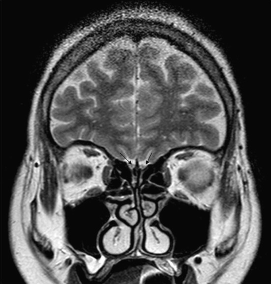
The OB volume was measured using MRI (Fig. 1). All of the measurements were taken from 3-mm consecutive T2-weighted (T2 W) turbo spin echo (TSE) images using the Philips Achieva 1.5-T MRI system (Philips Healthcare, Andover, MA, USA). An experienced radiologist measured OB size by manually tracing the OB on the MRI coronal T2 W sections. The radiologist measured the right and left OB separately and was blinded to the patient and control groups.
Fig. 1.

T2-Weighted coronal image showing reduced olfactory bulb volume (arrows)
Patients were excluded from the study if MRI T2 W gradient echo (GRE) imaging revealed post-traumatic, parenchymal, or meningeal hemosiderin retention in brain tissue. In addition, patients were excluded if the T2 W TSE images revealed other organic brain disorders.
Statistical Analysis
The data were evaluated using MedCalc statistical software v11.1.1. The Wilcoxon signed-rank test was used to compare repeated measures variables and the Mann–Whitney U test was used to test between-group differences. The data are expressed as the mean ± standard deviation. P < 0.05 was deemed to be statistically significant.
Results
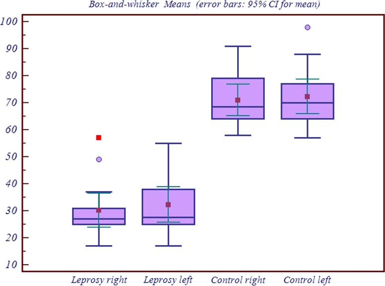
The OB volume varied widely in the patient group. The mean left OB volume was 31.6 ± 11.35 mm3 (range 17–55); the mean right OB volume was 30.26 mm3 ± 10.58 (range 17–57); and the mean total OB volume was 30.93 ± 10.55 mm3 (range 20–55; Table 1).
For the control group, the mean right and left OB volumes were 71.73 ± 10.97 mm3 (range 56–91) and 75.06 ± 13.11 mm3 (range 57–104), respectively, and the mean total OB volume was 73.33 ± 11.17 mm3 (range 60–96; Table 1).
No within-group differences between the right and left OB volumes were detected (patient group, P = 0.4212; control group, P = 0.2524).
The OB volume of the patient group was significantly reduced compared with that of the control group (P < 0.0001; Fig. 2).
Fig. 2.
Box plots showing the distribution of olfactory bulb volume measurements in the patient and control groups
Table 2 summarizes the orthonasal olfactory test results. On a seven-point scale for the butanol threshold and identification test, the leprosy group scored 1.41 ± 0.38 (range 0.75–2.75) and the control group scored 5.73 ± 0.5 (range 4.25–6.75). According to the CCCRC scoring system, the leprosy group was anosmic and the control group was hyposmic.
The orthonasal test detected olfactory dysfunction in all of the patients: 12 were anosmic and three were severely hyposmic. In the control group, two subjects were moderately hyposmic, five were mildly hyposmic, and eight were normal.
Orthonasal olfactory function was significantly reduced in the leprosy group compared with the control group (P < 0.0001). There was a significant cross-correlation between the orthonasal score and OB volumes in both groups (P < 0.0001).
Discussion
The OB is a neuroplastic structure and its size may change in relation the level of afferent neural activity [1]. Although nasal pathology is common in leprosy, few studies have examined changes to the sense of smell in this patient group [1, 16]. Olfactory system dysfunction and anosmia have been observed in all types of leprosy, but no study has investigated the underlying physiopathology.
This study is the first to evaluate olfactory bulb volume changes caused by loss of the sense of smell in patients who have leprosy. Animal studies have shown that one of the most critical effects of olfactory deprivation is a reduction in OB size as a result of hypoplasia [20]. Bulbar neuroplasticity is associated with the stimulation from the olfactory receptor neurons [21].
Chaturvedi et al. [14] observed olfactory loss in 41.7% of 225 patients with leprosy, and Ozturan et al. [16] reported that 91% of their patients had olfactory loss. Mishra et al. [1] reported that all of their patients with leprosy suffered olfactory loss, but that medical treatment improved their olfactory test scores. However, the improvement was smaller in patients who had lepromatous leprosy, the more severe form of leprosy. All of the patients in our study had lepromatous leprosy and our finding that all had severe olfactory dysfunction concurred with that of Mishra et al. [1]. Although other studies have examined olfactory function in patients with leprosy, to our knowledge, our study is the first to investigate involvement at the level of the OB.
Mishra et al. [1] suggested that impairment of the olfactory receptors and OB developed in the early stages of the disease; however, no study was conducted to test this theory. It has been established that the non-myelinated axons of the olfactory receptor cells are the initial target of toxic agents and viruses [22]. M. leprae, which is spread through droplet infection, may infect the olfactory receptors and OB. Other changes affecting olfactory function occur in later stages of the disease. M. lepra causes edema, swelling, ulceration, septic perforation, and collapse at the upper respiratory tract [23]. Peripheral neuron infiltration, motor and sensorial abnormalities, autonomic nerve dysfunction, and ganglion infiltration have been reported in people who have leprosy [24]. Liu and Qiu [25] suggested that the infection reaches the nerves through the blood, lymph, or by direct exposure. Primary atrophic rhinitis is caused by thinning nasal membranes that are the result of the regional effects of leprosy, such as defects in mucosal innervation and olfactory nerve end damage. Furthermore, leprosy is known to cause secondary atrophic rhinitis [1].
Conclusions
To our knowledge, this study is the first to report olfactory bulb volume reduction in patients with leprosy. Olfactory dysfunction and a significant reduction in OB volume were observed in all of our patients. We believe that the OB dysfunction in patients with leprosy is the result of a primary or secondary rhinitis in the upper respiratory tract where the sense of smell originates. The rhinitis causes the peripheral neuropathy that leads to loss of the sense of smell and a subsequent reduction in OB volume.
Acknowledgments
Conflict of Interest
None.
References
- 1.Mishra A, Saito K, Barbash SE, Mishra N, Doty RL. Olfactory dysfunction in leprosy. Laryngoscope. 2006;116:413–416. doi: 10.1097/01.MLG.0000195001.03483.F2. [DOI] [PubMed] [Google Scholar]
- 2.Low WK, Ngo R, Qasim A. Leprosy: otolaryngologist’s perspective. Otorhinolaryngology. 2002;64:281–283. doi: 10.1159/000064133. [DOI] [PubMed] [Google Scholar]
- 3.Nations SP, Katz JS, Lyde CB, Barohn RJ. Leprous neuropathy: an American perspective. Semin Neurol. 1998;18:113–124. doi: 10.1055/s-2008-1040867. [DOI] [PubMed] [Google Scholar]
- 4.Mastro TD, Redd SC, Breiman RF. Imported leprosy in the United States. JAMA. 2000;283:1004–1005. doi: 10.1001/jama.283.8.1004. [DOI] [PMC free article] [PubMed] [Google Scholar]
- 5.Koyuncu M, Celik O, Inan E, Ozturk A. Doppler sonography of vertebral arteries and audiovestibular system investigation in leprosy. Int J Lepr Other Mycobact Dis. 1995;63:23–27. [PubMed] [Google Scholar]
- 6.Yowan P, Danneman K, Koshy S, Richard J, Daniel E. Knowledge and practice of eye-care among leprosy patients. Indian J Lepr. 2002;74(2):129–135. [PubMed] [Google Scholar]
- 7.Soni K. Leprosy of the tongue. Indian J Lepr. 1992;64:325–330. [PubMed] [Google Scholar]
- 8.Ackerstaf AH, Hilgers FJM, Aarson NK, Balm AJM. Communication, functional disorders and lifestyle changes after total laryngectomy. Clin Otolaryngol. 1994;19:295–300. doi: 10.1111/j.1365-2273.1994.tb01234.x. [DOI] [PubMed] [Google Scholar]
- 9.Yousem DM, Geckle RJ, Bilker WB, McKeown DA, Doty RL. MR evaluation in patients with congenital hyposmia or anosmia. Am J Radiol. 1996;166:439–443. doi: 10.2214/ajr.166.2.8553963. [DOI] [PubMed] [Google Scholar]
- 10.Yousem DM, Geckle RJ, Doty RL (1995) Evaluation of olfactory deficits in neurodegenerative disorders. In: Abstract of The Radiological Society of North America Scientific Program. Chicago, 1995
- 11.Yousem DM, Geckle RJ, Bilker WB, Doty RL. Olfactory bulb and tract and temporal lobe volumes: normative data across decades. An N Y Acad Sci. 1998;855:546–555. doi: 10.1111/j.1749-6632.1998.tb10624.x. [DOI] [PubMed] [Google Scholar]
- 12.Buschhüter D, Smitka M, Puschmann S, Gerber JC, Witt M, Abolmaali ND, Hummel T. Correlation between olfactory bulb volume and olfactory function. Neuroimage. 2008;42:498–502. doi: 10.1016/j.neuroimage.2008.05.004. [DOI] [PubMed] [Google Scholar]
- 13.Mueller A, Rodewald A, Reden J, Gerber J, Kummer R, Hummel T. Reduced olfactory bulb volume in posttraumatic and postinfectious olfactory dysfunction. Neuroreport. 2005;16:475–478. doi: 10.1097/00001756-200504040-00011. [DOI] [PubMed] [Google Scholar]
- 14.Özturan O, Saydam L, Gökçe G, Çekkaya S. Leprada olfaktor ve trigeminal sinir fonksiyonlarında bozulma. KBB İhtisas Dergisi. 1994;8:25–29. [Google Scholar]
- 15.Barton RP. Clinical manifestation of leprous rhinitis. Ann Otol Rhinol Laryngol. 1976;85:74–82. doi: 10.1177/000348947608500113. [DOI] [PubMed] [Google Scholar]
- 16.Chaturvedi VN, Rathi SS, Raizada RM, Jain SK. Olfaction in leprosy. Indian J Lepr. 1985;57:814–819. [PubMed] [Google Scholar]
- 17.Cain WS, Gent JF, Goodspeed RB, Leonard G. Evaluation of olfactory dysfunction in the Connecticut Chemosensory Clinical Research Center. Laryngoscope. 1988;98:83–88. doi: 10.1288/00005537-198801000-00017. [DOI] [PubMed] [Google Scholar]
- 18.Cain WS. Testing olfaction in a clinical setting. Ear Nose Throat J. 1989;68:322–328. [PubMed] [Google Scholar]
- 19.Leon EA, Catalanotto FA, Werning JW. Retronasal and orthonasal olfactory ability after laryngectomy. Arch Otolaryngol Head Neck Surg. 2007;133:32–36. doi: 10.1001/archotol.133.1.32. [DOI] [PubMed] [Google Scholar]
- 20.Cummings DM, Knab BR, Brunjes PC. Effects of unilateral olfactory deprivation in the developing opossum, Monodelphis domestica. J Neurobiol. 1997;33:429–438. doi: 10.1002/(SICI)1097-4695(199710)33:4<429::AID-NEU7>3.0.CO;2-C. [DOI] [PubMed] [Google Scholar]
- 21.Lledo PM, Gheusi G. Olfactory processing in a changing brain. Neuroreport. 2003;14:1655–1663. doi: 10.1097/00001756-200309150-00001. [DOI] [PubMed] [Google Scholar]
- 22.Baker H, Genter MB. The olfactory system, the nasal mucosa as portal of entry of viruses, drugs, and other exogenous agents into the brain. In: Doty RL, editor. Handbook of olfaction and gustation. New York: Marcel Dekker; 2003. pp. 549–573. [Google Scholar]
- 23.Srinivisan S, Nehru V, Mann SB, Sharma VK, Bapuraj JR, Das A. Study of ethmoid sinus involvement in multibacillary leprosy. J Laryngol Otol. 1998;112:1038–1041. doi: 10.1017/s0022215100142410. [DOI] [PubMed] [Google Scholar]
- 24.Solomon S, Kurian N, Ramadas P, Rao PS. Incedence of nerve damage in leprosy patients treated with MDT. Int J Lepr Other Mycobact Dis. 1998;66:451–456. [PubMed] [Google Scholar]
- 25.Liu TC, Qui JS. Pathological findings on peripheral nerves, lymph nodes and viceral organs of leprosy. Int J Lepr. 1984;52:377–383. [PubMed] [Google Scholar]


