Abstract
Rare-earth-doped laser materials show strong prospects for quantum information storage and processing, as well as for biological imaging, due to their high-Q 4f↔4f optical transitions. However, the inability to optically detect single rare-earth dopants has prevented these materials from reaching their full potential. Here we detect a single photostable Pr3+ ion in yttrium aluminium garnet nanocrystals with high contrast photon antibunching by using optical upconversion of the excited state population of the 4f↔4f optical transition into ultraviolet fluorescence. We also demonstrate on-demand creation of Pr3+ ions in a bulk yttrium aluminium garnet crystal by patterned ion implantation. Finally, we show generation of local nanophotonic structures and cell death due to photochemical effects caused by upconverted ultraviolet fluorescence of praseodymium-doped yttrium aluminium garnet in the surrounding environment. Our study demonstrates versatile use of rare-earth atomic-size ultraviolet emitters for nanoengineering and biotechnological applications.
 The optical transitions that occur in rare-earth-doped crystals offer promise for quantum information storage and processing. Kolesov et al. report the optical detection of a single praseodymium ion residing in a crystal host by using an excited-state absorption process to enhance its fluorescence yield.
Apart from being the cornerstone of modern laser technology, crystals and glasses doped with rare-earth metals are known to have excellent properties for classical and quantum information storage and for performing quantum computation protocols1,2,3,4,5. They historically set the performance mark for these applications. Examples include demonstrations of optical memory in europium-doped yttrium silicate (Eu:YSO)1, two-qubit gates in Eu:YSO (ref. 2), storage of quantum coherence in Pr3+:YSO for over a second3, single photon storage in Nd3+:YVO4 (ref. 4), preservation of entanglement after photon storage and readout in Nd3+:YSO (ref. 5), etc. Another rapidly developing field of research on rare-earth-doped optical materials is creation of photostable upconverting multicolour fluorescent markers for bioimaging with low background6,7. While quantum applications of rare-earth-doped materials take advantage of the high quantum efficiency of their high quality-factor optical transitions in a solid as well as the numerous optical Raman transitions suitable for spin–photon entanglement, biological applications benefit from the complex energy level structure of rare-earth ions, their exceptional photostability, long lifetimes of the excited electronic states and high flexibility in choosing the dopant species and host material. Long fluorescent lifetimes of 4f ↔ 4f transitions of rare-earth emitters result in their low emission rate and, consequently, inability to detect a single ion. At the same time, such detection would be highly beneficial for both quantum, biological and nanophotonic applications resulting in a fully functional rare-earth-based qubit with exceptionally long coherence lifetime8 and, on the other hand, ultimately small photostable fluorescent biomarker9 and versatile nanoscopic light source for near field microscopy or biomanipulation.
Only a very limited number of indirect observations of a few rare-earth impurities have been reported due to their low fluorescence yield10,11,12. However, the emission rate of rare-earth centres can be dramatically improved by promoting the ions into higher-lying electronic states from which they can emit more efficiently due to much stronger low-Q parity-allowed transitions between 4f n−15d excited and 4f n ground states. In that way, the excitation–emission cycle shortens by orders of magnitude, whereas the fluorescence yield increases by the same factor.
Here we demonstrate optical detection of a single rare-earth ion, namely, trivalent praseodymium, in a crystalline host (in the present case, yttrium aluminium garnet, YAG). We exploit the visible-to-ultraviolet upconversion phenomenon in Pr3+:YAG13 to boost the efficiency of excitation–emission cycle and make a single Pr3+ ion detectable. High-quality photon antibunching measurements prove that the emission originates from a single quantum object, whereas excitation spectra and their polarization dependence prove that the single emitter is indeed a Pr3+ ion. The same approach can be used to detect Pr3+ in other hosts including low-spin YSO14 in which decoherence time of the dopant nuclear spin is the longest8. We also show that the ultraviolet emission originating from Pr3+ in YAG under visible excitation induces photochemical effects in the surrounding environment, such as exposure of photoresists and apoptotic cell death.
Results
Optical properties of Pr3+:YAG
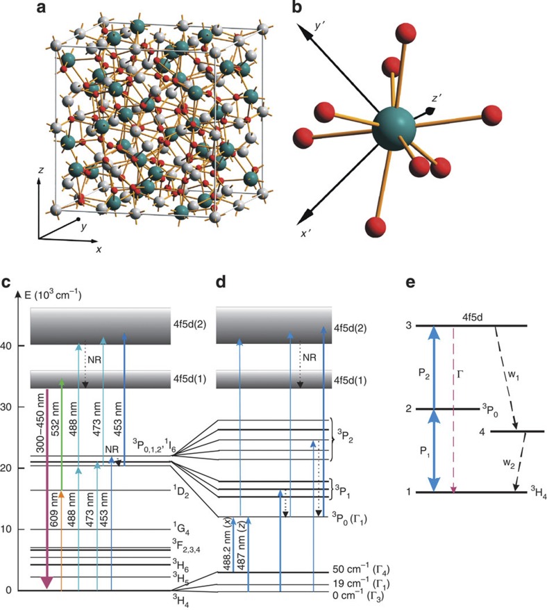
YAG has a complex crystal structure with yttrium ions occupying distorted octahedral sites of D2 symmetry as shown in Fig. 1a and b. Praseodymium dopant ions substitute yttrium in these sites. The electronic level structure of Pr3+ ion in YAG crystal is well studied15. It is represented by a number of 4f2 states of rather low energy and more energetic 4f5d states as depicted in Fig. 1c,d. 4f2 states are formed by the combined action of the central Coulomb potential of the [Xe] core of Pr3+, Coulomb interaction of the unpaird 4f electrons, spin–orbit coupling and crystal field potential. All states belong to either of the four irreducible representations Γ1−4 of the symmetry group D2. The ground state of Pr3+ is the 3H4 multiplet split by crystal field into nine sublevels. 4f2 states of higher energy give rise to spectrally narrow parity forbidden optical transitions in the visible and infrared. Higher lying electronic levels belonging to the 4f5d shell give rise to parity allowed optical transitions to 4f2 states. The 5d electron is not screened from its crystalline environment by outer 5s2 and 5p2 shells as in the case of 4f electrons. Therefore, 4f2 ↔ 4f5d transitions exhibit strong phonon sidebands in absorption and emission similar to singlet–singlet optical transitions in organic dyes or triplet–triplet transitions in nitrogen-vacancy centre in diamond16. Higher lying 4f5d states suffer from radiationless decay into the lowest 4f5d level from where fluorescence emission commences. Emission originating from this level covers a spectral range 300–450 nm17. The quantum efficiency of this ultraviolet transition is close to unity even at room temperature18 with lifetime ≈18 ns. Thus, ultraviolet fluorescence can be used for detecting a single Pr3+ ion in a YAG crystal. It can be excited either by direct ultraviolet (shorter than 300 nm) pumping into the absorption phonon sidebands of 4f5d levels or by a two-step upconversion excitation process involving an intermediate 4f2 state. The second way of exciting Pr3+ is more advantageous for a number of reasons. First of all, excitation with wavelengths shorter than 300 nm would inevitably produce strong fluorescent background from the optical elements along the excitation path (mainly the focusing objective lens) within the detection range (300–450 nm). Second and most importantly, the two-step pumping allows one to exploit the spectrally narrow features of the 4f2↔4f2 transitions in the first pumping step and read out the result of the manipulation with the second pumping step.
Figure 1. Optical properties of Pr3+ ion in YAG.
(a) Unit cell of YAG crystal. Green, white and red sites are occupied by yttrium, aluminium and oxygen, respectively. All together, the unit cell contains eight molecular units Y3Al5O12. Praseodymium occupies yttrium sites substitutionally. (b) The local surrounding of one yttrium site. The orientation is identical to (a). While the local z′-axis is along the y crystal axis, the axes x′ and y′ are rotated with respect to x and z crystal axes by 45°. Other five local symmetries can be obtained from this one by one or two successive 90° rotations around x, y and z crystal axes. (c) Electronic level structure of Pr3+ ion in a YAG crystal. Relevant optical transitions are indicated by arrows. NR, non-radiative decay channels. (d) Fine structure of 3H4→3P0,1,2 transitions giving rise to upconverted ultraviolet fluorescence. Only the lowest three sublevels of 3H4 manifold populated at room temperature are shown. (e) Four-level model used to explain the shape of photon antibunching signal. The metastable trapping state is assumed to be populated via decay of the emitting state 3.
There are a number of processes that can lead to upconverted ultraviolet emission of Pr3+ in YAG as shown in Fig. 1c,d. The first one is excitation of a metastable 1D2 state with an orange laser (611 or 609 nm) followed by the second excitation step at 532 nm. This method was used by us to demonstrate super-resolution microscopy of Pr:YAG nanoparticles19. Even a single colour optical pumping in the orange can be used though this upconversion scheme is much less efficient17. The second and the most straightforward way being exploited in the present work is to use 3H4↔3P0,1,2 transitions as the first upconversion step13. Once promoted into 3P0 state, the electron can be excited further into 4f5d(2) band, nonradiatively decay onto the emitting 4f5d(1) level, and emit an ultraviolet photon. As will be shown below, this results in a number of sharp (>1 nm at room temperature) upconversion resonances whose width is determined by the first excitation step. A simplified level structure diagram showing upconversion process via 3P0 state is given in Fig. 1e. This model will be used later for description of Pr3+ excited-state dynamics.
Localization of a single Pr3+ ion in YAG
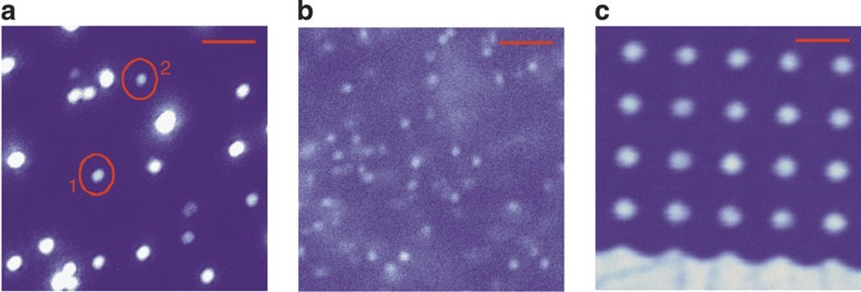
Praseodymium is one of the most abundant impurities in yttrium-based compounds20. To localize a single Pr3+ ion in a bulk YAG crystal, the distance between the neigbouring praseodymium ions must be greater than the resolution of the optical setup. This sets the upper limit of 1 ppb (parts per billion) on the praseodymium dopant concentration relative to yttrium. As we show below, the purest commercially available YAG crystals satisfy this condition. At cryogenic temperatures the requirements on the crystal purity can be significantly relaxed by spectrally selecting a subensemble of ions within an inhomogeneous excitation profile by means of a narrow-band excitation laser21,12. However, a much simpler approach towards isolation of a single Pr3+ impurity even at room temperature is to use Pr-doped YAG nanoparticles finely dispersed on a substrate (see Methods section). Samples prepared in this way were optically studied in a home-built high-resolution fluorescent microscope (see Methods section for details on its construction). A fluorescent image of nanoparticles is shown in Fig. 2a. Most of the bright spots on the image reveal several (up to 20–30) Pr3+ centres and are thought to be clusters of nanoparticles. However, the antibunching measurements performed on the marked nanoparticles revealed g(2) function, indicating single photon emission from those particles.
Figure 2. Upconversion scanning microscopy images of Pr3+:YAG.
(a) Ultraviolet fluorescence image of YAG nanoparticles dispersed on a glass slide. Within the 10×10 μm2 scan two marked spots are showing single emitter antibunching. (b) Fluorescence image of high purity YAG crystal. Bright spots are attributed to individual Pr3+ single ion impurities. (c) Ultraviolet fluorescence image of nominally pure YAG single crystal implanted with Pr3+ ions. The implantation mask contained holes arranged in a square grid. Scale bars in all three panels correspond to 2 μm.
A high purity YAG crystal was also searched for single Pr3+ emitters. A typical fluorescent image of the sample is shown in Fig. 2b. In the present 10×10 μm2 scan around 60 individual ions are seen. With the measured depth resolution being 1 μm, we estimate the density of praseodymium impurities to be 6×1011 cm−3 or 0.04 ppb relative to yttrium. Unlike nanoparticle sample, the out-of-focus Pr3+ impurities caused significant background fluorescence contributing to 35% of the signal. Therefore, the photon antibunching deep corresponding to a single emitting centre had only ≈30% contrast and was rather noisy (Supplementary Fig. S1). Furthermore, the fluorescent signal collected from individual Pr3+ ions in the bulk crystal was 2.5 times lower compared with the nanoparticle case. All experiments described below in this section are thus carried out on single praseodymium ions in YAG nanocrystals.
Spectral properties of a single Pr3+ in YAG
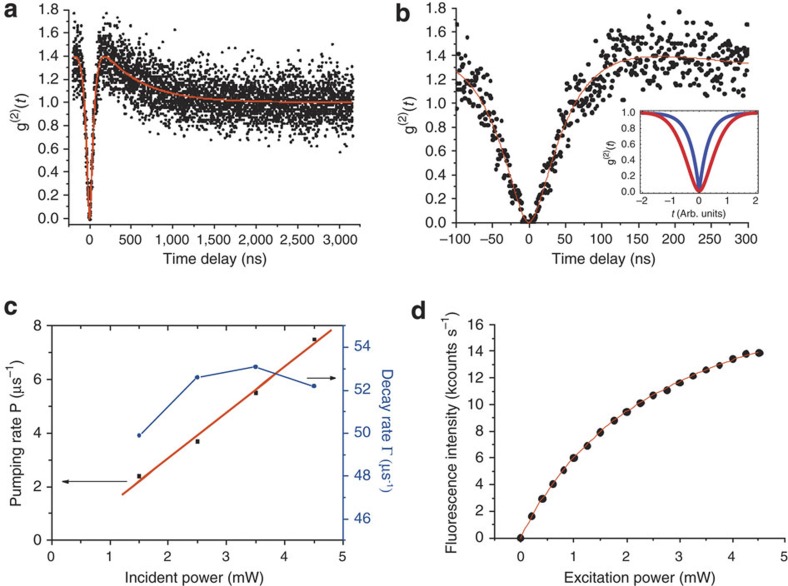
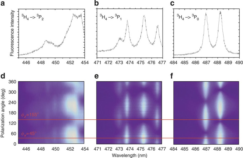
The photon antibunching signal from a single Pr3+ ion is shown in Fig. 3a. Owing to the upconversion process, the fluorescent background (~100 counts s−1) is negligible compared with the signal from the single emitting centre (~1.3×10⁴ counts s−1). Centres are usually photostable at room temperature for many hours of continuous illumination. To prove that fluorescence is indeed from a Pr3+ ion in YAG host, the excitation spectrum of upconverted fluorescence was measured and the results are shown in Fig. 4a–c. The spectra exactly match previously reported experimental results on Pr:YAG visible-to-ultraviolet upconversion13, absorption spectra of Pr3+:YAG (ref. 22), and our own measurements performed on a bulk YAG crystal doped with 0.18% of Pr3+. They consist of a number of sharp (down to <0.5 nm) lines and exemplify the high spectral selectivity of the upconversion process even at room temperature. Further evidence for Pr3+ excitation comes from the polarization selectivity of the first upconversion step. The optical dipoles associated with transitions between any states of Γm and Γn symmetry representations are oriented along the principal axes of the local dodecahedron15 as indicated in Fig. 1b: Γ1↔Γ4 and Γ2↔Γ3 are along x-axis, Γ3↔Γ4 and Γ1↔Γ2 are along y-axis, and Γ1↔Γ3 and Γ2↔Γ4 are along z-axis. The transitions Γn↔Γn between the states of the same group representation are forbidden. At room temperature only 3 out of 9 sublevels of lowest energy belonging to 3H4 manifold, namely, Γ3 (0 cm−1), Γ1 (≈19 cm−1) and Γ4 (≈50 cm−1), are populated. Thus, for example, in the vicinity of 3H4→3P0(Γ1) transition the two allowed optical dipoles, Γ3(3H4)→Γ1(3P0) (487.0 nm) and Γ4(3H4)→Γ1(3P0) (488.2 nm) (corresponding transitions are indicated in Fig. 1d), should have z- and x-orientations, respectively. Same arguments apply to 3H4→3P1,2 excitations. Polarization dependence of the excitation spectra are shown in Fig. 4d–f and reveal the projections of the x and z dipoles onto the plane of the sample. For the ion investigated, we could not identify the direction of the y dipole at room temperature. The probable reason for this is that y-polarized transitions are much weaker than x and z ones. Qualitatively similar results were obtained for other spots indicating single photon emitter behaviour. Overall, 39 single upconverting emitters were found within a 100×100 μm2 area. Statistical analysis of their excitation line positions in the vicinity of 3H4→3P0 transition is shown in Supplementary Fig. S2.
Figure 3. Excited-state dynamics of a single Pr3+ ion.
(a) Normalized g(2) function measured at spot 1 on Fig. 2a with 4.5 mW of excitation at 488.2 nm wavelength. The signal is fitted with the solution of the rate equations according to the four-level model shown in Fig. 1e (red line). (b) Zoom into zero-delay region of the antibunching curve. Flat bottom corresponding to quadratic temporal dependence is clearly seen. The inset compares the theoretical antibunching shapes of the emissions originating from one-step (blue, casp-like) and two-step (red, parabolic at the bottom) excitations. (c) Power dependences of the pumping rate P (black squares) and radiative decay Γ (blue circles) extracted from the fits of antibunching signals taken at 1.5, 2.5, 3.5 and 4.5 mW are shown. Taking the focused laser spot diameter to be ≈300 nm (the size of the nanoparticle seen by our microscope) and specified 85% transmission of the objective lens in the range 480–490 nm, the derived pumping rate gives the ground- and excited-state absorption cross-sections to be 7×10⁻¹⁹ cm2. This value agrees well with ground-state absorption cross-section of 6×10⁻¹⁹ cm2 deduced from the data reported previously33 and with excited-state absorption cross-section of 8×10⁻¹⁹ cm2 (ref. 23) for 532 nm wavelength. (d) Saturation behaviour of ultraviolet fluorescence at spot 1. Saturation power is 2.75 mW, whereas saturated fluorescence count rate is 22.5 kcounts s−1.
Figure 4. Spectral properties of a single Pr3+ ion.
(a–c) Excitation spectrum of ultraviolet emission from centre 1 on Fig. 1a in the vicinity of 3H4→3P2, 3H4→3P1 and 3H4→3P0 transitions, respectively. (d–f) show the dependence of the corresponding spectra on the polarization of the excitation beam.
Excited-state dynamics
The fluorescence lifetime measurements performed on spot 1 with frequency-doubled femtosecond Ti:sapphire laser operating at 906 nm (453 nm for the doubled output) revealed 19 ns in rather good agreement with known value of 18 ns lifetime of the lowest 4f5d state of Pr3+ ion in YAG. The saturation behaviour of Pr3+ fluorescence as a function of excitation intensity at 488.2 nm is shown in Fig. 3d. Three fluorescent regimes are expected: quadratic dependence at very low excitation power; linear dependence once the transition 3H4↔3P0 is saturated while the second step is not; and full saturation. The absorption cross-sections from the ground state 3H4 to the intermediate 3P0 and the one from 3P0 to 4f5d(2) are approximately identical, ~6–8×10⁻¹⁹ cm2 (ref. 23), but the intermediate and the emitting states have significantly different lifetimes, 8 μs and 18 ns, respectively. Only linear and fully saturated regimes were observed experimentally as saturation of the first excitation step occurs at very low excitation intensity at which no upconverted ultraviolet fluorescence was possible to collect.
The shape of the photon correlation signal reveals two distinct features: significant bunching at non-zero time delays and very flat bottom of the antibunching dip, clearly visible in Fig. 3b. The former indicates the existence of a metastable state, which can effectively trap population for rather a long time24. Probable candidates for this metastable state are the 1G4 level whose lifetime is known to be ≈0.4 μs (ref. 25), the very long-lived 1D2 level, which, however, can be repumped by the excitation laser, the 3H4 and 3H5 levels of unknown lifetime or some electron trap within the YAG bandgap. The latter can be populated by ionization of Pr3+ due to excited-state absorption from the emitting 4f5d(1) level23. A simplified 4-level model depicted in Fig. 1e is used to describe the second feature of the g(2)(t) function. The Pr3+ ion is pumped from its ground state 1 (3H4) into an intermediate state 2 (3P0) and back at rate P1. From state 2 it can be promoted further into state 3 (4f5d) at pumping rate P2 from where it can decay either into level 1 at rate Γ emitting an ultraviolet photon or into a metastable state 4 at rate w1. Finally, the metastable state can decay into level 1 at rate w2. After the start photon is detected by the Hanbury–Brown and Twiss setup, the initial populations of the levels are ρ11(0)=1, ρ22(0)=ρ33(0)=ρ44(0)=0. The g(2)(t) function is determined as follows:
 |
1 |
At short time delays between start and stop photons, g(2) can be approximated as:
 |
2 |
The t2 dependence of the intensity correlation function is an indication of probing the population of intermediate state 2 with the second excitation photon and explains the flat bottom of the antibunching dip. It can also be seen that the antibunching width is no longer approaching the lifetime of the emitting state for low pump power as in the case of a single-step excitation, but increases as (P2+2P1)−1. Under the assumption of equal pump rates P1=P2=P (in agreement with known absorption cross-sections) it is possible to fit the experimental antibunching curves with the numerical solutions of the set of rate equations and extract the pumping rates and decay parameters of the Pr3+ ion as a function of pump intensity (Fig. 3c). The measured antibunching curves are shown in Supplementray Fig. S3. As expected, the pumping rate increases linearly whereas the radiative decay rate Γ stays constant around 52 μs−1. The latter value is in good agreement with the measured lifetime of ~19 ns.
Implantation of Pr3+ into YAG crystal
Not only the ability to detect, but also to create fluorescent centres at the desired location in the crystal is a significant ingredient for the technological implementation of single Pr3+. We demonstrate that Pr3+ centres can be created in pure bulk YAG crystals by ion implantation. Some parts of the surface of nominally pure YAG crystal were covered with photoresist with lithographically defined holes. After implantation the photoresist was removed and the sample was annealed at 1,200 °C. Figure 1c shows the upconverted fluorescence scan of the crystal surface subject to such patterned implantation with 30 keV praseodymium ions with an implantation dosage of 1013 cm−2. The grid of the holes in the photoresist is clearly seen. The excitation spectrum taken on the implanted region in the vicinity of 3H4→3P0 transition of Pr3+ was similar to Fig. 4c, indicating that implantation was successful. However, the unimplanted area showed the same excitation spectrum though of 100 times lower brightness. This shows that the nominally pure crystal contained significant amount of optically unresolvable Pr3+ impurities (the crystal used for implantation was much less pure than the one shown in Fig. 2).
Discussion
It is known from optical free-induction decay26, Raman-heterodyne27, and electromagnetically induced transparency3 experiments that Pr3+ ions in various hosts possess nuclear spin–flip optical Raman transitions. Once detected at cryogenic temperature, single fluorescent Pr3+ ions would present a perfect qubit with all-optical access to its nuclear spin states possessing lifetimes of the order of tens of seconds8. In addition, lifetime-limited linewidth of optical transitions28 and their 100% quantum efficiency would allow for efficient dipole–dipole optical coupling between praseodymium ions separated by as far as a few tens of nanometres in the same way as was demonstrated for dye molecules29. In turn, several mutually coupled praseodymium ions would comprise a multi-qubit quantum gate2 while dipolar coupling to other rare-earth species would allow to use Pr3+ as a readout qubit in rare-earth-based quantum computer designs30. However, cryogenic experiments with single Pr3+ ions have to be carefully designed to maximize collection efficiency of ultraviolet fluorescence. In the present case, <2% of the emitted photons are being detected. More details on the estimate of the collection efficiency of the setup are given in Supplementary Methods. Implementation of solid immersion lenses (SILs) and dielectric antennas31 could improve the situation. At the same time, proper identification of the metastable trapping state would show the way towards its repumping and, therefore, fluorescence efficiency increase.
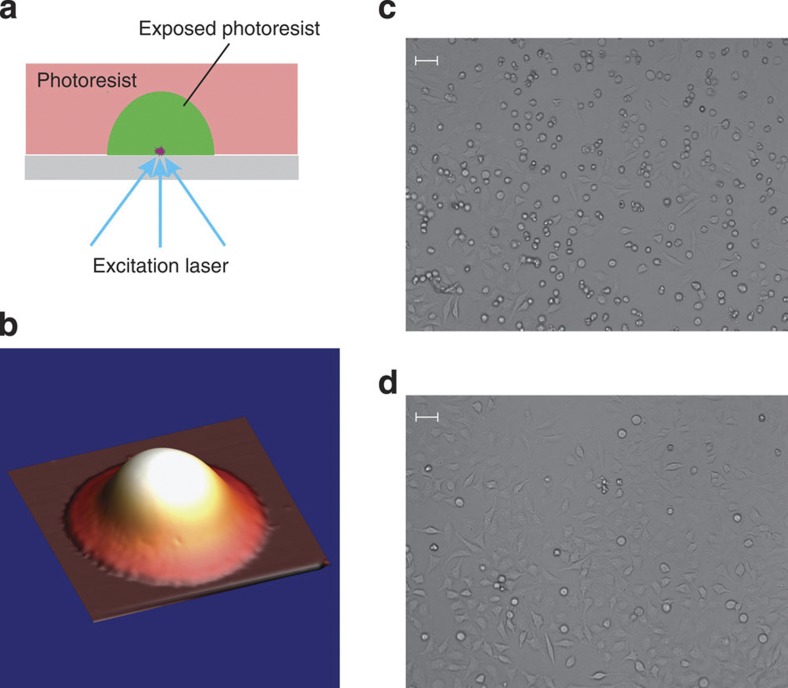
Apart from new opportunities for quantum computing, having a single photostable upconverting ultraviolet emitter at hand enables optical nano-engineering due to ultraviolet-triggered photochemical reactions in the surrounding of the emitter. For example, ultraviolet emission of a Pr:YAG nanoparticle can expose the surrounding photoresist. As the emission of a nanoparticle containing many Pr3+ centres is isotropic, the nanoparticle lying on the substrate and covered with negative-tone photoresist will create a hemispherical shell for itself as illustrated in Fig. 5a. Such a self-assembled SIL will enhance the collection efficiency of the light emitted by this specific nanoparticle as it is situated in the very centre of the hemisphere. Experimental details of creation of a SIL are given in Supplementary Methods. The reconstructed atomic force microscopy image of such a self-assembled SIL is shown in Fig. 5b. The evaluation of SIL performance indicated 5–8 times enhancement in ultraviolet light collection efficiency as indicated in Supplementary Table S1. This example demonstrates applicability of upconverting ultraviolet emission from rare-earth-doped materials for micro- and nanolithography, potentially, at a single-emitter level. This ultraviolet emission can also have biological effects. Although at low excitation power upconverted ultraviolet fluorescence can be used for bioimaging (Supplementary Fig. S4), at higher fluences it can be used for site-specific cell degradation. To demonstrate that we cultured HeLa cells on the surface of bulk Pr:YAG crystal (0.18% Pr3+). Subsequently, part of the crystal was illuminated by scanned focused laser beams of 532 and 609 nm wavelengths (this combination produces upconverted ultraviolet fluorescence according to Fig. 1c). After illumination and incubation of the sample for 1 h, >50% of the cells in the irradiated region died (Fig. 5c), whereas the control region showed <10% of dead cells (Fig. 5d). Pr-doped nanocrystals thus present the opportunity of performing tightly localized (potentially, down to a few nanometers) photochemical reactions in living cells.
Figure 5. Photochemical effects due to upconverted ultraviolet fluorescence.
(a) Self-assembly of a SIL by isotropic exposure of negative-tone photoresist with ultraviolet emission of a nanoparticle. (b) Reconstructed 3D atomic force microscopy (AFM) image of a self-assembled SIL. Lateral size of the image is 6.6×6.6 μm2. (c) Light microscope image of HeLa cells on the surface of Pr:YAG crystal exposed by a combination of 609+532 nm lasers. Most of the cells died (round shaped). (d) Image of the cells exposed by a combination of wavelengths 600+532 nm (no upconverted ultraviolet is produced). Most of the cells are alive. The same fraction of live cells is found in the unexposed region. Scale bars on (c,d) correspond to 50 μm.
Methods
Microscope and laser source construction
Microscopic studies of Pr:YAG nanoparticles spin-coated on a glass slide were performed in a home-built scanning microscope shown schematically in Supplementary Fig. S5a and consisting of a 3D nanopositioning stage, a 425 nm shortpass dichroic beamsplitter, a high NA (1.30) oil immersion objective lens having high transmission in the UV (Zeiss Fluar UV), UV-transmitting filter (UG11 optical glass), UV 50/50 beamsplitter and two UV-sensitive single photon counting PMTs (Hamamatsu) to form a Hanbury–Brown and Twiss setup. Home-made tunable diode lasers with Littmann external cavity operating around 488 nm (3H4↔3P0 transition), 473 nm (3H4→3P1 transition) and 450 nm (3H4↔3P2 transition) were used as excitation sources (all diodes are from Nichia Corporation). The output of the diode lasers was passed through a single-mode optical fiber to assure Gaussian shape of the excitation beam. The illumination of the sample and the fluorescence collection were arranged through the same objective lens. Typical power of the excitation beam at the apperture of the objective lens was several milliwatts.
Laser diode tuning
Laser diodes were tuned by means of Littmann external cavity with 60° prism being a dispersion element as shown in Supplementary Fig. S5b. Single transverse mode laser diodes (488, 473 and 450 nm) were used as manufactured, that is without applying an antireflective coating on their output facets. Laser diode output collimated by an aspheric lens was dispersed by a prism. The first reflection from the dispersion prism was taken as an output beam. The tuning mirror was mounted on a tip-tilt piezo-driven mirror mount for automated wavelength tuning. Typical output power of the diode laser was in the range of a few tens of milliwatts with a possibility of tuning through 7 nm spectral range for 488 and 473 nm diodes and 11 nm spectral range for 450 nm diode. The linewidth of such a tunable laser source was below 0.1 nm.
Nanoparticle preparation and characterization
Pr-doped YAG nanocrystals exploited in the present work were prepared by a sol–gel pyrolysis method according to slightly modified procedure reported in work32. Praseodymium nitrate was added according to required stoichiometry aiming at dopant concentration of 2 ppm. Characterization of nanoparticles by X-ray diffraction measurements resulted in 32 nm average size of single crystalline YAG. Nanoparticles were spin-coated on a 150 μm-thick glass slide out of isopropanol suspension. X-ray diffraction spectrum and SEM image of YAG nanoparticles dried on silicon substrate are shown in Supplementary Figs S6 and S7.
Author contributions
R.K., K.X., R.R., R.S., A.Z., J.M. and P.R.H. performed the experiments; R.K. designed the experiments; R.K. and K.X. analysed the data; R.K., P.R.H. and J.W. wrote the paper; J.W. supervised the project.
Additional information
How to cite this article: Kolesov, R. et al. Optical detection of a single rare-earth ion in a crystal. Nat. Commun. 3:1029 doi: 10.1038/ncomms2034 (2012).
Supplementary Material
Supplementary Figures S1-S7, Supplementary Table S1 and Supplementary Methods
Acknowledgments
We gratefully acknowledge fruitful discussion with Neil Manson, Fedor Jelezko, Peter Siyushev and Ilja Gerhardt. The work was financially supported by ERC SQUTEC, SFB TR21 and DFG FOR 1493.
References
- Yano R., Mitsunaga M. & Uesugi N. Nonlinear laser spectroscopy of Eu3+:Y2SiO5 and its application to time-domain optical memory. J. Opt. Soc. Am. B 9, 992–997 (1992). [Google Scholar]
- Longdell J. J., Sellars M. J. & Manson N. B. Demonstration of conditional quantum phase shift between ions in a solid. Phys. Rev. Lett. 93, 130503 (2004). [DOI] [PubMed] [Google Scholar]
- Longdell J. J., Fraval E., Sellars M. J. & Manson N. B. Stopped light with storage times greater than one second using electromagnetically induced transparency in a solid. Phys. Rev. Lett. 95, 063601 (2005). [DOI] [PubMed] [Google Scholar]
- de Riedmatten H., Afzelius M., Staudt M. U., Simon C. & Gisin N. A solid-state light-matter interface at the single-photon level. Nature 456, 773–777 (2008). [DOI] [PubMed] [Google Scholar]
- Clausen C. et al. Quantum storage of photonic entanglement in a crystal. Nature 469, 508–511 (2011). [DOI] [PubMed] [Google Scholar]
- Wang F. & Liu X. Recent advances in the chemistry of lanthanide-doped upconversion nanocrystals. Chem. Soc. Rev. 38, 976–989 (2009). [DOI] [PubMed] [Google Scholar]
- Wu S. et al. Non-blinking and photostable upconverted luminescence from single lanthanide-doped nanocrystals. Proc. Natl Acad. Sci. USA 106, 10917–10921 (2009). [DOI] [PMC free article] [PubMed] [Google Scholar]
- Fraval E., Sellars M. J. & Longdell J. J. Dynamic decoherence control of a solid-state nuclear-quadrupole qubit. Phys. Rev. Lett. 95, 030506 (2005). [DOI] [PubMed] [Google Scholar]
- Asakura R., Isobe T., Kurokawa K., Takagi T., Aizawa H. & Ohkubo M. Effects of citric acid additive on photoluminescence properties of YAG:Ce3+ nanoparticles synthesized by glycothermal reaction. J. Lumin. 127, 416–422 (2007). [Google Scholar]
- Lange R., Grill W. & Martienssen W. Observation of single impurity ions in a crystal. Europhys. Lett. 6, 499–503 (1988). [Google Scholar]
- Bartko A. P. et al. Observation of dipolar emission patterns from isolated Eu3+:Y2O3 doped nanocrystals: new evidence for single ion luminescence. Chem. Phys. Lett. 358, 459–465 (2002). [Google Scholar]
- Yen W. M. Optical spectroscopy of materials with restricted dimensions Proceedings of the Third International Conference on Trends in Quantum Electronics (eds Prokhorov, A.M. and Ursu, I.) SPIE Proceedings 1033, 183–190 (1989). [Google Scholar]
- Ganem J., Dennis W. M. & Yen W. M. One-color sequential pumping of the 4f5d bands in Pr-doped yttrium aluminum garnet. J. Lumin. 54, 79–87 (1992). [Google Scholar]
- Hu C., Sun C., Li J., Li Z., Zhang H. & Jiang Z. Visible-to-ultraviolet upconversion in Pr3+:Y2SiO5 crystals. Chem. Phys. 325, 563–566 (2006). [Google Scholar]
- Gruber J. B., Hills M. E., Macfarlane R. M., Morrison C. A. & Turner G. A. Symmetry, selection rules, and energy levels of Pr3+:Y3Al5O12. Chem. Phys. 134, 241–257 (1989). [Google Scholar]
- Gruber A. et al. Scanning confocal optical microscopy and magnetic resonance on single defect centers. Science 276, 2012–2014 (1997). [Google Scholar]
- Gayen S. K., Xie B. Q. & Cheung Y. M. Two-photon excitation of the lowest 4f2→4f5d near-ultraviolet transitions in Pr3+:Y3Al5O12. Phys. Rev. B 45, 20–28 (1992). [DOI] [PubMed] [Google Scholar]
- Weber M. J. Nonradiative decay from 5d states of rare earths in crystals. Solid State Commun. 12, 741–744 (1973). [Google Scholar]
- Kolesov R. et al. Super-resolution upconversion microscopy of praseodymium-doped yttrium aluminum garnet nanoparticles. Phys. Rev. B 84, 153413 (2011). [Google Scholar]
- Ozawa L. & Toryu T. Quantitative determination of rare earths in yttrium oxide by spectrophotoluminescence. Anal. Chem. 40, 187–190 (1968). [Google Scholar]
- Moerner W. E. & Carter T. P. Statistical fine structure of inhomogeneously broadened absorption lines. Phys. Rev. Lett. 59, 2705–2708 (1987). [DOI] [PubMed] [Google Scholar]
- van der Ziel J. P., Sturge M. D. & Van Uitert L. G. Optical detection of site selectivity for rare-earth ions in flux-grown yttrium aluminum garnet. Phys. Rev. Lett. 27, 508–511 (1971). [Google Scholar]
- Cheung Y. M. & Gayen S. K. Excited-state absorption in Pr3+:Y3Al5O12. Phys. Rev. B 49, 14827–14835 (1994). [DOI] [PubMed] [Google Scholar]
- Beveratos A., Brouri R., Poizat J.- P. & Grangier P. Bunching and antibunching from single NV color centers in diamond. Preprint at arXiv: 0010044v1 (2000). [DOI] [PubMed]
- Malinowski M., Joubert M. F. & Jacquier B. Dynamics of the IR-to-blue wavelength upconversion in Pr3+-doped yttrium aluminum garnet and LiYF4 crystals. Phys. Rev. B 50, 12367–12374 (1994). [DOI] [PubMed] [Google Scholar]
- Shelby R. M., Tropper A. C., Harley R. T. & Macfarlane R. M. Measurement of the hyperfine structure of Pr3+:YAG by quantum-beat free-induction decay, hole burning, and optically detected nuclear quadrupole resonance. Opt. Lett. 8, 304–306 (1983). [DOI] [PubMed] [Google Scholar]
- Longdell J. J., Sellars M. J. & Manson N. B. Hyperfine interaction in ground and excited states of praseodymium-doped yttrium orthosilicate. Phys. Rev. B 66, 035101 (2002). [Google Scholar]
- Equall R. W., Cone R. L. & Macfarlane R. M. Homogeneous broadening and hyperfine structure of optical transitions in Pr3+:Y2SiO5. Phys. Rev. B 52, 3963–3969 (1995). [DOI] [PubMed] [Google Scholar]
- Hettich C. et al. Nanometer resolution and coherent optical dipole coupling of two individual molecules. Science 298, 385–389 (2002). [DOI] [PubMed] [Google Scholar]
- Wesenberg J. H., Molmer K., Rippe L. & Kröll S. Scalable designs for quantum computing with rare-earth-ion-doped crystals. Phys. Rev. A 75, 012304 (2007). [Google Scholar]
- Lee K. G. et al. A planar dielectric antenna for directional single-photon emission and near-unity collection efficiency. Nat. Photon. 5, 166–169 (2011). [Google Scholar]
- Vaqueiro P. & Lopez-Quintela M. A. Synthesis of yttrium aluminium garnet by the citrate gel process. J. Mater. Chem. 8, 161–163 (1998). [Google Scholar]
- Wittmann G. & Macfarlane R. M. Photon-gated photoconductivity of Pr3+:YAG. Opt. Lett. 21, 426–428 (1996). [DOI] [PubMed] [Google Scholar]
Associated Data
This section collects any data citations, data availability statements, or supplementary materials included in this article.
Supplementary Materials
Supplementary Figures S1-S7, Supplementary Table S1 and Supplementary Methods


