We have recently re-tested the STR fingerprinting data in our paper (Int J Biol Sci 2011; 7:892-901). DNA fingerprinting analysis was performed using genomic DNA from hsNSCs (passage 22); T1 cells (passages <50 and >150) and HeLa cells by GeneMapper ID v3.2 (Applied Biosystems, Foster City, CA) for human STR markers. There are two loci of heterozygosity CSF1PO (9, 10) and D3S1358 (15, 18) in T1 cells (passages <50) compared with the HeLa STR profiles CSF1PO (10) and D3S1358 (18). In addition, four loci variations are seen at CSF1PO (9, 10), D3S1358 (15, 18), TPOX (8, 12) and vWA (18) in T1 cells (passages >150) which are different from those seen in Hela (vWA: 16, 18) and (TPOX: 12). Other than that, the similarity between T1 and Hela was >86%, while the similarity between T1 and hsNSC was <14%. Overall, our recent data suggest that T1 cells may have cross-contaminated by HeLa cells.
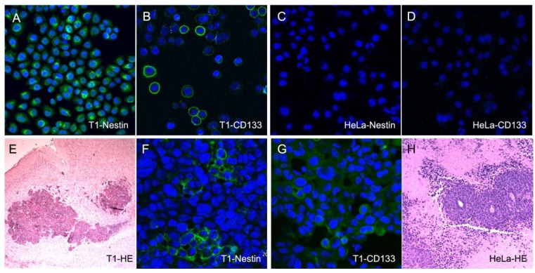
We have also performed additional immunofluorescence assays to show the expression of neural stem cell markers on T1 and Hela cells. Consistent with our previous results, T1 cells expressed both nestin and CD133 (Fig.1A, B). However, HeLa cells were negative for these two markers (Fig. 1C, D).
In separate additional in vivo experiments we have recently completed, Hela and T1 cells were injected into the nude mice subcutaneously and intracranially, respectively. Two weeks later, subcutaneous xenografts were formed in HeLa cell-implanted mice (Fig. 1H). In contrast, T1 cells did not result in xenografts subcutaneously. This is consistent with our previous results we reported in the paper. On the other hand, intracranial neoplasias were found in T1 cell-implanted mice (Fig.1E) after 6 weeks. Immunofluorescence analysis suggested that these tumor cells expressed neural stem cell markers nestin and CD133 (Fig.1F, G). Approximately 10 days after HeLa cells were injected into the mice brain, all the animals died, but no obvious neoplasia was found.
Fig 1.
Immunofluorescence assays showed that the T1 expressed neural stem cell markers nestin (A) and CD133 (B) , but Hela cells were negative (C, D). Intracranial tumorigenicity of T1 cells in immunodeficient nude mice (E, HE staining). Some of these tumor cells were immunoreactive for neural stem cell markers Nestin (F) and CD133 (G). Nuclei were detected using the DAPI staining showed in merged image.
In summary, although the genome of T1 cells was apparently contaminated by HeLa cells; T1 cells are both phenotypically and tumorigenically different from HeLa cells.


