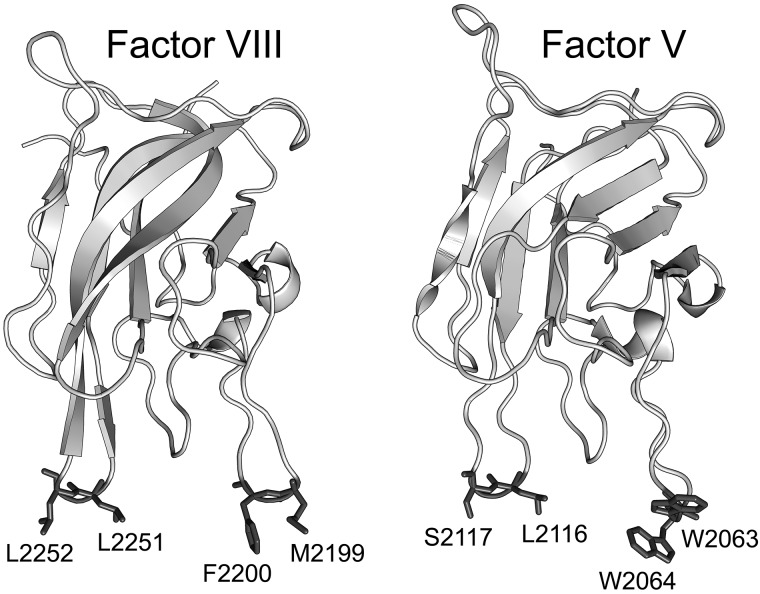
Figure 1.
Crystal structures of the FVIII C2 domain (PDB entry 1D7P) and FV C2 domain (PDB entry 1CZT). The membrane-interactive hydrophobic amino acids for spikes 1 and 3 of FVIII (left) and FV (right) are shown with side chains in dark gray, whereas the rest of the structures show the backbone only in light gray. Residue numbers relate to the full-length proteins.

