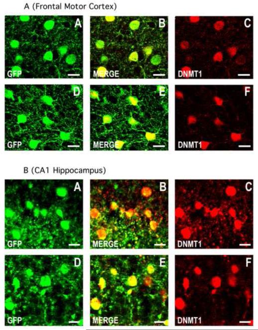
Fig. 2.
DNMT1 is co-expressed with GFP in cortico-limbic GABAergic neurons of the GAD67-GFP knock-in mouse. Confocal double immunofluorescence labeling of GFP (green), DNMT1 (red), and merged images in orange (center panels).
2A) A-C: motor cortex layer II of a 7 day old PRS mouse; D-F: motor cortex layer II of a 7 day old control mouse. Coronal sections correspond roughly to bregma −1.4 mm. 2B) A-C: CA1 field of the hippocampus of the PRS mouse; D-F: CA1 field of the hippocampus of the control mouse. Coronal sections correspond roughly to bregma −2 mm. Scale bars for all panels represent 20 μm.

