Abstract
Auditory verbal hallucinations (AVH) are not only among the most common but also one of the most distressing symptoms of schizophrenia. Despite elaborate research, the underlying brain mechanisms are as yet elusive. Functional MRI studies have associated the experience of AVH with activation of bilateral language-related areas, in particular the right inferior frontal gyrus (rIFG) and the left superior temporal gyrus (lSTG). While these findings helped to understand the neural underpinnings of hearing voices, they provide little information about possible brain mechanisms that predispose a person to experience AVH, i.e. the traits to hallucinate. In this study, we compared resting state connectivity between 49 psychotic patients with chronic AVH and 49 matched controls using the rIFG and the lSTG as seed regions, to identify functional brain systems underlying the predisposition to hallucinate. The right parahippocampal gyrus showed increased connectivity with the rIFG in patients as compared to controls. Reduced connectivity with the rIFG in patients was found for the right dorsolateral prefrontal cortex. Reduced connectivity with the lSTG in patients was identified in the left frontal operculum as well as the parietal opercular area. Connectivity between the lSTG and the left hippocampus was also reduced in patients and showed a negative correlation with the severity of hallucinations. Concluding, we found aberrant connectivity between the seed regions and medial temporal lobe structures which have a prominent role in memory retrieval. Moreover, we found decreased connectivity between language-related areas, indicating aberrant integration in this system potentially including corollary discharge mechanisms.
Introduction
Auditory verbal hallucinations (AVH) are among the most common and distressing symptoms of schizophrenia and also affect persons with affective psychosis and some (e.g., borderline and schizotypic) personality disorders. While AVH are thus a highly prevalent feature of psychiatric illnesses, their underlying brain mechanisms remain elusive. Models proposed for their pathophysiology include the misattribution of inner speech [1], [2], increased top-down processing [3], recollection of verbal memory fragments [4], [5] and sensory-based bottom-up deviations [6], [7]. Functional MRI studies demonstrated activation of inferior-frontal and temporo-parietal areas during the experience of AVH [5], [8], [9] together with deactivation of medial temporal lobe structures at the onset of AVH [5], [10]. However, these symptom-capture studies do not provide information about brain mechanisms predisposing a person to experience AVH, i.e. the trait to hallucinate. Insight into aberrations of neuronal networks underlying such vulnerability, in turn, may be particularly relevant for guiding the development and evaluation of treatment strategies.
It could be hypothesized that a predisposition to AVH may be instantiated by aberrant connectivity between inferior frontal and temporo-parietal areas leading to a misattribution of inner speech [1], [2]. If this were to be the case, we would expect reduced functional connectivity at rest between the inferior frontal and the temporo-parietal areas of both hemispheres, reflecting disturbances within the language network underlying aberrant processing of ambiguous bottom-up signals [11]. This hypothesized reduced couplings between inferior frontal and temporo-parietal areas may render a person vulnerable to hallucinate in the auditory verbal domain. At the structural level, deviations in the fiber-tracts connecting these areas have been demonstrated with Magnetic Transfer Imaging (MTI) in schizophrenia patients [12] and in non-psychotic individuals with frequent hallucinations [13]. However, it is currently unclear if these structural deficiencies also lead to functionally decreased connectivity.
Alternatively, deviant connectivity between speech-related areas and the medial temporal lobe could give rise to spontaneous verbal memory recollection [4], [5]. A predisposition for intrusive memory fragments may be reflected in anomalous functional connectivity between cortical association areas such as the frontal, temporal and parietal cortex and memory related structures such as the hippocampus and parahippocampal gyrus. Such increased vulnerability for intrusive memory fragments may predispose a person for hallucinations per se, as the hippocampal-parahippocampal complex is involved in the recollection of memories from several modalities. Increased vulnerability to memory intrusions could results from either increased connectivity between the cortical and the medial temporal structures, or decreased (inhibitory) connectivity, or a combination of both.
Either of these hypothesized dysconnectivities may become evident in the absence of external tasks, given that the latter may override the endogenous dynamics of AVH. Therefore, aberrations in functional coupling that may predispose a person to hearing voices may become evident by examining resting state connectivity data. Resting state fMRI connectivity measures low-frequency fluctuations in the cerebral haemodynamics (around 0.01–0.1 Hz). Recently, attention has been focused on the possible origins of these slow variations. Various investigations suggested that these signal variations are of neuronal origin and characterize the neuronal baseline activity of the human brain in the absence of externally stimulated neuronal activity.
Resting state connectivity can be assessed by using Regions of Interest (ROIs); areas that are thought to play a major role in the trait under investigation, or without the predefinition of ROIs as in an independent component analysis (ICA). In this paper, we used the first approach as there is good evidence for the essential contribution of certain well-defined areas in AVH. In contrast, the data-driven ICA approach may also pick up other differences in resting state between the groups that may not be related to hallucinations given that lack of focus on a specific system. In this study we aim to investigate the vulnerability to experience AVH in patients who have experienced AVH.
A recent meta-analysis indicated consistent activation of the right inferior frontal gyrus (rIFG) and the left superior temporal gyrus (lSTG) during the experience of AVH [14], these regions therefore provide a strong model to assess dysfunctional connectivity underlying AVH. In the present study we thus compare resting state connectivity of these regions between 49 psychotic patients with chronic AVH and a group of well-matched controls in order to unravel network aberrations underlying the predisposition to AVH. For practical reasons, we selected the Regions of Interest (ROIs) from a single large fMRI study [5], as meta-analytic studies provide rather large ROIs. As the study by Diederen et al. was by far the largest study included in the meta-analysis, results of the meta-analysis are largely in agreement with this study. We selected the two seeds that showed the largest areas of activation, namely the left superior temporal gyrus and the right inferior frontal gryus. These two seeds also have functional significance as the left superior temporal gyrus may be involved in the auditory component of AVH; i.e. the fact that voices are being heard aloud, while the right inferior frontal area may be the place where the words and sentences that are later perceived as voices could be generated [15].
Materials and Methods
Participants
Forty-nine patients with psychosis, as diagnosed according to DSM-IV criteria by an independent psychiatrist using the “Comprehensive Assessment of Symptoms and History (CASH)” [16], who all experienced AVH with a frequency of at least several times a day during at least one year were included in the study (cf. Table 1 for clinical and demographic details). Psychopathology was assessed at the day of scanning with the Positive and Negative Syndrome Scale [17]. For comparison, 49 healthy (as confirmed by the CASH interview) individuals matched for age, sex and handedness were also included. After complete description of the study to the subjects, the participants provided written informed consent into the study, which was approved by the Humans Ethics Committee of the University Medical Center Utrecht.
Table 1. Demographic and clinical description of participants.
| Group | Patients, n = 49 | Control subjects, n = 49 | |
| Age | 38.5 years ±11.72 | 39.5 years ±14.9 | |
| Sex | 20 males, 29 females | 18 males, 31 females | |
| Handedness | 41 right, 8 non-right | 41 right, 7 non-right | |
| Mean time with AVH | 13.9±12.2 years | No AVH | |
| Diagnosis | 43 Schizophrenia, 4 Schizoaffective Disorder, 1 Schizophreniform Disorder, 1 Psychosis NOS | No psychiatric diagnosis | |
| Antipsychotic Medication | 10 clozapine, mean dose 464 mg, 3 flufenazine, mean dose 30 mg, 7 risperidon, mean dose 3.6 mg, 8 olanzapine, mean dose 14.2 mg, 7 quetiapine, mean dose 514 mg, 1 penfluridol 10 mg, 5 haloperidol, mean dose 4 mg, 1 aripriprazol, 15 mg, 7 medication-free | all medication-free | |
| Mean | Positive scale | 16.2±3.7, | |
| PANSS scores | Negative scale | 16.3±5.2, | |
| Total | 63±13.3 | ||
Data acquisition and processing
Resting state scans of eight minutes duration (600 blood-oxygenation-level-dependent (BOLD) fMRI images) were obtained on a Philips Achieva 3 T MRI scanner using the following parameter: 40 (coronal) slices, TR/TE 21.75/32.4 ms, flip angle 10°, FOV 224×256×160, matrix 64×64×40, voxel-size 4 mm isotropic. (PRESTO scans typically have shorter TR than TE times, as the whole head is scanned with each volume in stead of the slab-wise read-out of EPI scans). This scan sequence achieves full brain coverage within 609 ms by combining a 3D-PRESTO pulse sequence with parallel imaging (SENSE) in two directions, using a commercial 8-channel SENSE headcoil [18]. Participants were instructed to lie in the scanner as still as possible with their eyes closed yet stay awake (which was confirmed by post-scan debriefing). After scanning, patients were asked whether or not they had experienced AVH during the resting state scan. Likewise, healthy subjects were asked for AVH, which were denied by all of them.
The resting state scans were first corrected for head movement by affine registration using a two pass procedure. The mean PRESTO image for each subject was then spatially normalized to the MNI single subject template [19] using the unified segmentation approach [20] and the ensuing deformation was applied to the individual PRESTO volumes. Finally, images were smoothed by a 5-mm FWHM Gaussian kernel. Regions of interest (ROIs) used as seeds for the functional connectivity analysis were based on two activation clusters reliably associated with the state of AVH obtained from Diederen et al. [5] (table 2.): the right inferior frontal gyrus, MNI coordinates: 44,16,12 and the left superior temporal gyrus, MNI coordinates −48, 0,0. We selected regions from this large fMRI study rather than using seeds from the meta-analysis [21] because the latter would have provided very large and unspecific clusters.
Table 2. Areas used for seed regions.
| Lobe | Area | MNI Coordinates | T-value | Cluster size | ||
| R Frontal | Inferior Frontal gyrus | 44 | 16 | 12 | 4.31 | 243 |
| L Temporal | Superior Temporal gyrus | −48 | 0 | 0 | 3.46 | 40 |
T-values, cluster sizes and locations of local maxima of activation during AVH in 24 patients, taken from Diederen et al. 2011. Threshold set at p<0.05 whole-brain FDR corrected.
Voxel time courses were extracted for all voxels within a 5 mm sphere around the centre of the particular clusters that were located in the grey matter of the individual subject.
Variance explained by the following nuisance variables was removed from the time series to reduce spurious correlations [22]–[24]: i) motion parameters derived from image realignment and their first derivative; ii) mean grey, white matter and CSF signal intensity per time-point; iii) coherent signal drifts reflected by the first five PCA components on the entire whole-brain data. All nuisance variables entered the model as first and all but the PCA components also as second order terms, as previously described by Behzadi et al. [25] and shown by Chai et al. [26] to increase specificity and sensitivity of the analyses.. Data was then band pass filtered preserving frequencies between 0.01 and 0.08 Hz [27], [28]. The time course of each ROI was then expressed as the first eigenvariate of the processed time series of all voxels associated with that region. A recent study showed that this method is able to detect valid correlation and anti-correlations during rest, which are not an artefact of the preprocessing method, but may reflect valid biological signals [29]. Moreover, it should be emphasized, that the current analysis focussed on differences between the two groups of subjects, which should not be affected by potentially confounds induced by preprocessing steps, even if these were to be present, as long as they do not systematically vary in effect between groups.
Data analysis
For each subject we computed linear (Pearson) correlation coefficients between the time series of the seeds and any other grey matter voxel, which were then transformed into Fisher's Z-scores. Group analysis was then performed on these by an analysis of variance (ANOVA) across subjects using appropriate non-sphericity correction. In the ANOVA, both the main effect of functional connectivity as well as the group-difference between patients and controls were modelled for each seed. Inference on this random-effects analysis was then sought using linear contrasts. First, main effects of connectivity (across both groups) were calculated for both seeds and a conjunction analysis was performed on these to reveal regions showing significant coupling. Subsequently, functional connectivity maps of rIFG and lSTG were assessed for significant differences between patients and controls using unilateral T-tests. These differences in connectivity were evaluated by testing for either significant positive coupling across both groups (main effect) in conjunction with a reduced coupling in patients or significant negative coupling across both groups (main effect) in conjunction with enhanced coupling in patients as compared to controls indicating enhanced and reduced coupling. For all analyses, results were regarded as significant if they passed p<0.05, cluster-level FDR-corrected for multiple comparisons. Anatomical localizations were obtained using the SPM Anatomy Toolbox [30].
Statistical analyses
To test whether the observed differences between patients and controls were additionally related to psychopathology, we extracted individual functional connectivity between the seeds and the peak voxels of the clusters showing significant differences. We then performed a Spearman rank-correlation between these and the individual scores on the “Hallucinatory behaviour” (P3) PANSS item to test for significant (p<0.05) associations. In a second follow-up analysis we divided the patients into those who actually experienced hallucinations during the scan and those who did not. Subsequently, we computed a one-way ANOVA between the groups on the individual functional connectivity scores between the seed and the peak of a significant cluster.
Results
All participants stayed awake during the resting state scan. Thirty-one out of the 49 patients reported the experience of hallucinations during the resting state scan. None of the controls experienced hallucinations during the scan.
The analysis of the main effects for both seeds across both groups is provided in Supplementary Material S1. When resting state connectivity was compared between the patients and the controls, the following differences emerged.
Increased connectivity
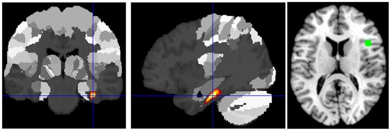
The right parahippocampal gyrus showed significantly increased connectivity with the rIFG (Figure 1) in the patient group.
Figure 1. Increased connectivity with rIFG in patients as compared to controls.
Increased connectivity with rIFG in patients as compared to controls was found in the right parahippocampal gyrus (p<0.05 cluster-level FDR-corrected).
Decreased connectivity
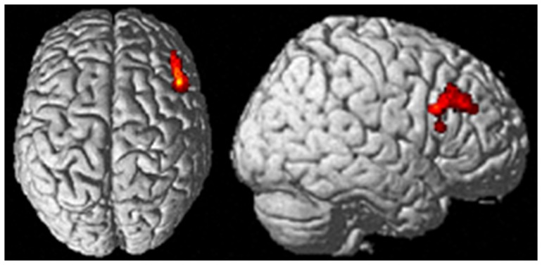
Reduced connectivity with the rIFG in the patients was found for the right dorsolateral prefrontal cortex (DLPFC) just anterior to and slightly encroaching cytoarchitectonically defined Area 45 (Figure 2).
Figure 2. Reduced connectivity with rIFG in patients as compared to controls.
Reduced connectivity with rIFG in patients as compared to controls was found in the right DLPFC/area 45 (p<0.05 cluster-level FDR-corrected).
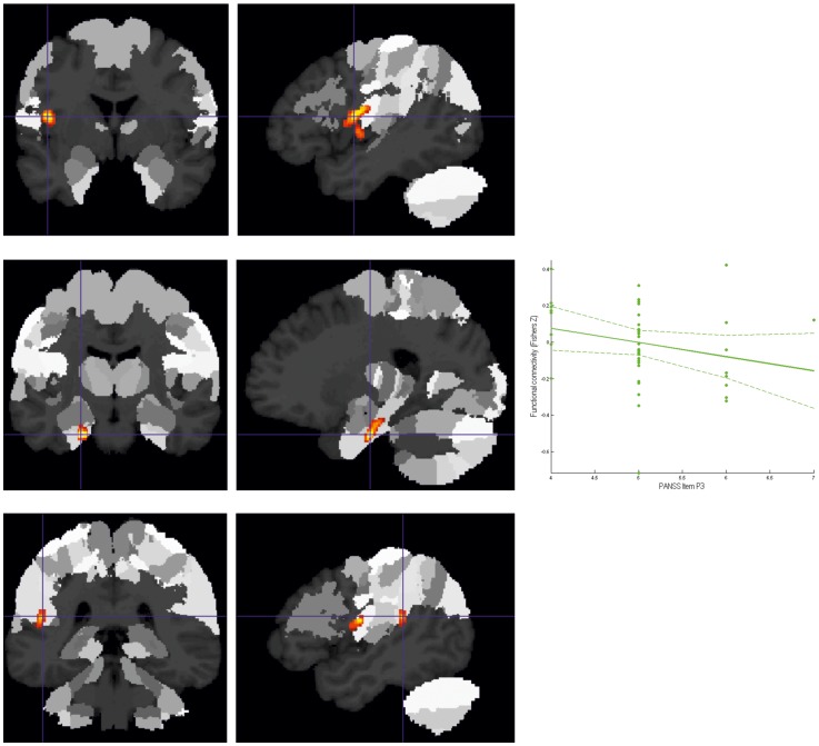
Two regions showed significantly (whole-brain corrected) reduced connectivity to the lSTG in patients (Figure 3). The first was located on the left frontal operculum, extending into the anterior insula areas as well as the parietal opercular areas OP 3 and OP 4. The second was located in the left hippocampus, allocated predominantly to the subiculum and to a smaller extent the entorhinal cortex. Connectivity between the lSTG and the left hippocampus was not only reduced in patients but furthermore showed a significant negative correlation with the item P3 (hallucinations) of the PANSS rating scale.
Figure 3. Decreased connectivity to lSTG in patients as compared to controls.
A. Cluster of significantly (p<0.05 corrected) decreased connectivity with the lSTG in patients in the left frontal operculum/anterior insula shown on the MPM of the SPM Anatomy Toolbox. B. Cluster of significantly (p<0.05 corrected) decreased connectivity with the lSTG in patients in the left hippocampus (subiculum/entorhinal cortex) shown on the MPM of the SPM Anatomy Toolbox. C. Cluster of decreased connectivity (as a strong statistical trend) with the lSTG in patients in the left temporo-parietal operculum/retroinsular cortex.
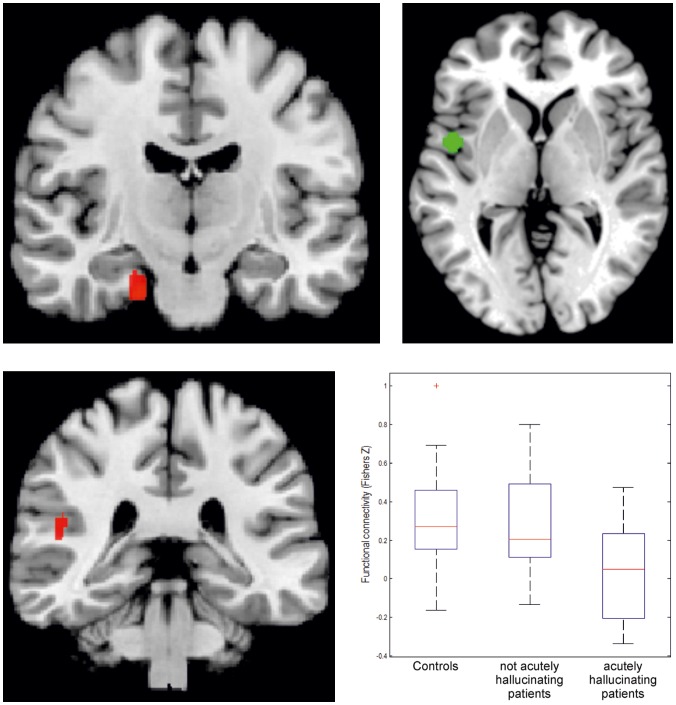
In order to assess state-effects of AVH on functional connectivity with our seed regions, we performed a follow-up analysis for those regions that showed disturbed connectivity in the overall group of trait carriers (i.e., the regions reported above) which assessed differences between controls, trait and state/trait-groups, i.e. individuals with acute AVH during scanning. For this purpose, we divided the patients into one group who hallucinated during the resting state scan, and those who did not. This analysis revealed that the reduction in connectivity between the lSTG and the left hippocampus was significantly stronger in those patients who hallucinated during scanning (Figure 4). That is, in these regions both trait (as demonstrated by the lower connectivity in non-acutely hallucinating patients compared to controls) and state-effects (as evidenced by the further reduction in the acutely hallucinating patients) were evident. For the remaining regions, including those showing abnormal connectivity with the rIFG, no differences were found between acutely hallucinating patients and those showing merely the trait to experience AVH.
Figure 4. Differences in connectivity with lSTG, specified for patients who hallucinated during the scans and those who did not.
A. Aberrations in functional connectivity between lSTG and the left entorhinal cortex gyrus differentiate between healthy controls. Separate findings are shown for patients showing the AVH trait and those acutely experiencing AHV (state). B. Aberrations in functional connectivity between lSTG and the left temporo-parietal operculum/retroinsular cortex differentiate between healthy controls. Separate findings are shown for the patients showing the AVH trait and those patients acutely experiencing AHV during the scan (state). The red crosses in these box plots represent data points outside 1.5 * IQR, i.e. outliers.
Discussion
We compared resting state functional connectivity of 49 psychotic patients experiencing chronic auditory verbal hallucinations (AVH) to 49 healthy controls. Analysis was seeded from the left superior temporal gyrus (lSTG) and the right inferior frontal gyrus (rIFG) as these areas are consistently activated during the state of AVH [5], [21] and are therefore expected to play a crucial role in AVH. In patients, the rIFG showed increased connectivity with the right parahippocampal gyrus and decreased connectivity with the right dorsolateral prefrontal cortex (DLPFC). Decreased connectivity with the lSTG in patients was revealed in the left frontal operculum and the left temporo-parietal operculum. Both opercular areas are considered part of the general neuronal frame work for language [28]. Furthermore, the lSTG showed decreased connectivity with the left hippocampus, this connectivity correlated significantly with the severity of hallucinations as measured in the PANSS interview.
To differentiate between trait and state aspects of AVH we divided the patient group into those who acutely hallucinated during scanning and those who did not. Here, only coupling of the lSTG with the left temporo-parietal operculum and hippocampus was more strongly decreased in the patients who actually hallucinated, indicating that these deviations may result from a combination of state and trait characteristics. Increased connectivity between the rIFG and the parahippocampus was equally strong in both patient groups.
Dys-connectivity with the medial temporal lobe
Our analysis revealed disturbed interactions between both seeds and medial temporal lobe (MTL) structures, including increased connectivity between the rIFG and the right parahippocampal gyrus. This observation resonates strongly with a previous symptom capture study which revealed that cortical activations during AVH were preceded by significant signal changes in the parahippocampal gryus [5]. The MTL structures, especially the hippocampus [31], play important roles in both the consolidation and the retrieval of memories. This latter function may be involved in hallucinations, as hallucinations may be triggered by inappropriate recollections from memory. The parahippocampal gyri have a gatekeeper function as they integrate and transfer information from neocortical association areas to the hippocampus [32]. When this delicate interplay becomes deranged by aberrant connectivity, incidental information from memory may be recollected, giving rise to hallucinations [33]. The finding of both increased and decreased coupling between neocortical and MTL regions represents an important notion about the differentiated organization of the MTL and its interactions, challenging simple notions of “more” or “less integration”. Rather, deregulation of the usually well-balanced interactions between hippocampus, parahippocampal gyrus and neocortical association areas may represent a crucial pathomechanism for hallucinations. Since the hippocampus and parahippocampus regulate retrieval of memories in all modalities, we expect that deregulation between MTL and cortical association areas constitutes a predisposition to hallucinations in general and not specifically for hallucinations in the auditory verbal domain. Indeed, most of these patients who experience chronic AVH also experienced hallucinations in other modalities and can thus be expected prone to hallucinations in general.
Dysconnectivity within the neocortical areas
Secondly, decreased connectivity in the patient group between the lSTG and the left frontal operculum was observed. We also found decreased connectivity between the rIFG and the right DLPFC. These findings may reflect decreased integration between areas mainly associated with the generation (rIFG) and interpretation (lSTG) of language, suggesting misattribution of inner speech as a potential pathomechanism. Furthermore, these regions are often found active during acute hallucinations [5], [21]. Mechanistically, such misattribution may be well reconciled with decreased connectivity by the notion of aberrant corollary discharge [34] failing to suppress the sensory consequences of self-generated actions. A possible mechanism would be that insufficient corollary discharge in the language system as reflected by decreased functional connectivity could result from microstructural alterations in the arcuate fasciculi, the most important fibre bundle between Broca's area and Wernicke's area [35] as observed by de Weijer and co-workers [12], [13]. Flaws or disintegration in the corollary discharge system of language networks may be specifically related to the attribution of internally generated speech to an external origin, which posed a predisposition specifically to hallucinate in the auditory verbal domain.
Comparison to previous functional connectivity studies
Seeding from Wernicke's area (in close vicinity to our lSTG seed), Hoffman and colleagues [36] found increased connectivity in schizophrenia patients with a large subcortical cluster including the thalamus, midbrain and putamen, which is supported by the more localized findings of increased connectivity with the parahippocampal gyrus in the present analysis. Vercammen et al. [37] observed reduced functional connectivity between the left temporo-parietal junction and the right homologue of Broca's area in patients, while Gavrilescu et al. [38] demonstrated reduced connectivity of the bilateral auditory system. These studies thus point to decreased connectivity within the network of language-related areas in patients with AVH, a finding that was confirmed and extended in the present study. Rotarska-Jagiela et al. [39] found that the severity of positive symptoms correlated with functional connectivity of fronto-temporal and auditory networks in schizophrenia patients using an independent component analysis (ICA). In spite of the different approach taken in the present analysis, we could add evidence for the trait-specific aspect of this finding. We thus conclude that the present work is well in line with previous evidence but may provide more specific insights into disturbed circuits by seeding from areas shown to be engaged in AVH, separating state- and trait-dependent dysconnectivity and assessing the association between disturbed coupling and the severity of clinical symptoms.
Limitations and technical considerations
A limitation of this study is the absence of a patient group without the trait to hallucinate. However, to our opinion, all patients with schizophrenia have a predisposition to hallucinate given their psychotic liability. The fact that a small proportion of schizophrenia patients have not experienced hallucinations may reflect adequate pharmacotherapy, rather than an absence of the trait.
The current approach of assessing functional connectivity of regions that have previously been implicated in the pathophysiology of hallucinations allowed for a model-based assessment of dysfunctional circuitry in patients suffering from AVH. This may represent a considerable advantage in terms of functional specificity as compared to purely data-driven approaches such as ICA, as ICA would also identify other differences between the groups unrelated to AVH. In turn, however, analyses based on a-priori information evidently have low sensitivity to aberrations in non-probed circuits.
The removal of noise from resting-state fMRI data as part of the preprocessing has been criticized because this process can introduce artificial anti-correlations [40], [41], [42]. However, component based methods such as PCA have been shown to be less susceptible to this effect while effectively reducing spurious correlations caused by noise [43], [44].
It must also be acknowledged, that the observed differences may not be absolutely specific to hallucinations, as patients and controls also differed in variables such as the presence of other psychotic symptoms, negative symptoms and medication status. While all these factors may have influenced connectivity patterns, the specific, localized aberrations of connectivity with areas activated during AVH should point to a pathophysiological contribution. This is in particular the case for those connections showing a significant state effect or an association with the severity of hallucinations as assessed with the PANSS rating.
In sum, we found two clear deviations in resting state connectivity in the patients with chronic AVH as compared to the controls. The first was aberrant connectivity between the seed regions that are active during AVH and MTL structures which have a prominent role in memory retrieval. The second deviation was decreased connectivity between language-related areas in the neocortex.
Aberrations between neocortical association areas and MTL structures may underlie a liability to experience hallucinations in general. In turn, reduced connectivity within the neocortical language network may indicate deficits in corollary discharge mechanisms as another potentially important pathophysiological mechanism for hallucination in the language domain specifically.
Supporting Information
The analysis of the main effects for both seeds across both groups. The analysis of the main effects for both seeds across both groups demonstrated significant positive coupling between both seed regions with (in particular the left) inferior frontal lobes and anterior insula, dorso-lateral prefrontal cortex, inferior parietal lobules, right anterior temporal lobe and the posterior medial frontal cortex. In turn, both regions were significantly anti-correlated with anterior and posterior cingulated regions, the precuneus, right posterior inferior parietal lobule, bilateral inferior temporal lobe and right cerebellum. Positive coupling, i.e., functional connectivity, with the seeds is shown in green, negative coupling in red. Significantly decreased connectivity with the seeds in patients is superimposed on the main effect in cyan, significantly increased connectivity in patients is superimposed on the main effect in yellow below (all effects significant at p<0.05 cluster-level FDR-corrected).
(TIF)
Funding Statement
This work was supported by NWO/ZonMW (Dutch Scientific Research Organization) Clinical Fellowship [40-00703-97-270] and by NWO/ZonMW Innovation Impulse (VIDI) [017.106.301]. The funders had no role in study design, data collection and analysis, decision to publish, or preparation of the manuscript.
References
- 1. Allen P, Aleman A, McGuire PK (2007) Int Rev Psychiatry. 2007 Aug;19 4:407–15. [DOI] [PubMed] [Google Scholar]
- 2. Waters F, Woodward T, Allen P, Aleman A, Sommer I (2010) Self-recognition Deficits in Schizophrenia Patients With Auditory Hallucinations: A Meta-analysis of the Literature. Schizophr Bull doi: 10.1093/schbul/sbq144. [DOI] [PMC free article] [PubMed] [Google Scholar]
- 3. Vercammen A, Aleman A (2010) Semantic expectations can induce false perceptions in hallucination-prone individuals. Schizophr Bull 36: 151–156. [DOI] [PMC free article] [PubMed] [Google Scholar]
- 4. Copolov DL, Seal ML, Maruff P, Ulusoy R, Wong MTH, et al. (2003) Cortical activation associated with the experience of auditory hallucinations and perception of human speech in schizophrenia: a PET correlation study. Psychiatry Res 122: 139–152. [DOI] [PubMed] [Google Scholar]
- 5. Diederen KM, Neggers SF, Daalman K, Blom JD, Goekoop R, et al. (2010) Deactivation of the parahippocampal gyrus preceding auditory hallucinations in schizophrenia. Am J Psychiatry 167: 427–735. [DOI] [PubMed] [Google Scholar]
- 6. Hunter MD, Eickhoff SB, Pheasant RJ, Douglas MJ, Watts GR, et al. (2010) The state of tranquility: Subjective perception is shaped by contextual modulation of auditory connectivity. Neuro Image 53: 611–618. [DOI] [PubMed] [Google Scholar]
- 7. Hunter MD, Eickhoff SB, Miller TW, Farrow TF, Wilkinson ID, et al. (2006) Neural activity in speech-sensitive auditory cortex during silence. Proc Natl Acad Sci USA 103: 189–194. [DOI] [PMC free article] [PubMed] [Google Scholar]
- 8. Sommer IE, Weijer de AD, Daalman K, Neggers SF, Somers M, et al. (2007) Can fMRI-guidance improve the efficacy of rTMS treatment for auditory verbal hallucinations? Schizophr Res 93: 406–408. [DOI] [PubMed] [Google Scholar]
- 9. Sommer IE, Diederen KM, Blom JD, Willems A, Kushan L, et al. (2008) Auditory verbal hallucinations predominantly activate the right inferior frontal area. Brain 131: 3169–3177. [DOI] [PubMed] [Google Scholar]
- 10. Hoffman RE, Anderson AW, Varanko M, Gore JC, Hampson M (2008) Time course of regional brain activation associated with onset of auditory/verbal hallucinations. Br J Psychiatry 193: 424–425. [DOI] [PMC free article] [PubMed] [Google Scholar]
- 11. Northoff G, Qin P (2011) How can the brain's resting state activity generate hallucinations? A ‘resting state hypothesis’ of auditory verbal hallucinations. Schizophr Res 127: 202–214. [DOI] [PubMed] [Google Scholar]
- 12. Weijer de AD, Neggers SFW, Daalman K, Kahn RS, Sommer IE (2011) Microstructural alterations of the arcuate fasciculus in schizophrenia patients with frequent auditory verbal hallucinations. Schizophr Res 130: 68–77. [DOI] [PubMed] [Google Scholar]
- 13. Weijer de AD, Neggers SFW, Diederen KMJ, Mandl RCW, Kahn RS, et al. (2011) Aberrations in the arcuate fasciculus in psychotic and non-psychotic individuals with auditory verbal hallucinations. Human Brain Mapping doi: 10.1002/hbm.21463. [DOI] [PMC free article] [PubMed] [Google Scholar]
- 14. Bandettini PA, Bullmore E (2008) Endogenous oscillations and networks in functional magnetic resonance imaging. Human Brain Mapping 29: 737–739. [DOI] [PMC free article] [PubMed] [Google Scholar]
- 15. Sommer IE, Selten JP, Diederen KM, Blom JD (2010) Dissecting auditory verbal hallucinations into two components: audibility (Gedankenlautwerden) and alienation (thought insertion). Psychopathology 43 2:137–40. [DOI] [PubMed] [Google Scholar]
- 16. Andreasen NC, Flaum M, Arndt S (1992) The comprehensive assessment of symptoms and history (CASH): An instrument for assessing psychopathology and diagnosis. Arch Gen Psychiatry 49: 615–623. [DOI] [PubMed] [Google Scholar]
- 17. Kay S, Fiszbein A, Opler L (1987) The positive and negative syndrome scale (PANSS) for schizophrenia. Schizophrenia Bulletin 13: 261–276. [DOI] [PubMed] [Google Scholar]
- 18. Neggers SF, Hermans EJ, Ramsey NF (2008) Enhanced sensitivity with fast three-dimensional blood -oxygen-level-dependent functional MRI: comparison of SENSE-PRESTO and 2D-EPI at 3 T. NMR Biomed 21: 663–676. [DOI] [PubMed] [Google Scholar]
- 19. Holmes CJ, Hoge R, Collins L, Woods R, Toga AW, et al. (1998) Enhancement of MR images using registration for signal averaging. J Comput Assist Tomogr 22: 324–333. [DOI] [PubMed] [Google Scholar]
- 20. Ashburner J, Friston KJ (2005) Unified segmentation. Neuroimage 26: 839–851. [DOI] [PubMed] [Google Scholar]
- 21. Jardri R, Pouchet A, Pins D, Thomas P (2011) Cortical activations during auditory verbal hallucinations in schizophrenia: a coordinate-based meta-analysis. Am J Psychiatry 168: 73–81. [DOI] [PubMed] [Google Scholar]
- 22. Fox MD, Zhang D, Snyder AZ, Raichle ME (2009) The global signal and observed anticorrelated resting state brain networks. J of Neurophysiology 101: 3270–3283. [DOI] [PMC free article] [PubMed] [Google Scholar]
- 23. Weissenbacher A, Kasess C, Gerstl F, Lanzenberger R, Moser E, et al. (2009) Correlations and anticorrelations in resting-state functional connectivity MRI: A quantative comparison of preprocessing strategies. Neuro Image 47: 1408–1416. [DOI] [PubMed] [Google Scholar]
- 24. Eickhoff SB, Bzdok D, Laird AR, Roski C, Caspers S, et al. (2011) Co-activation patterns distinguish cortical modules, their connectivity and functional differentiation. Neuro Image 57: 938–949. [DOI] [PMC free article] [PubMed] [Google Scholar]
- 25. Behzadi Y, Restom K, Liau J, Liu TT (2007) A component based noise correction method (CompCor) for BOLD and perfusion based fMRI. Neuroimage 37: 90–101. [DOI] [PMC free article] [PubMed] [Google Scholar]
- 26. Chai XJ, Castañón AN, Öngür D, Whitfield-Gabrieli S (2011) Anticorrelations in resting-state networks without global signal regression. Neuroimage 29: 1420–1428. [DOI] [PMC free article] [PubMed] [Google Scholar]
- 27. Biswal B, Yetkin FZ, Haughton VM, Hyde JS (1995) Functional connectivity in the motor cortex of resting human brain using echo-planar MRI. Magn Reson Med 34: 537–541. [DOI] [PubMed] [Google Scholar]
- 28. Greicius MD, Krasnow B, Reiss AL, Menon V (2003) Functional connectivity in the resting brain: a network analysis of the default mode hypothesis. Proc Natl Acad Sci USA 100: 253–258. [DOI] [PMC free article] [PubMed] [Google Scholar]
- 29. Murphy K, Birn RM, Handwerker DA, Jones TB, Bandettini PA (2009) The impact of global signal regression on resting state correlations: Are anti-correlated networks introduced? Neuro Image 44: 893–905. [DOI] [PMC free article] [PubMed] [Google Scholar]
- 30. Eickhoff SB, Paus T, Caspers S, Grosbras MH, Evans AC, et al. (2007) Assignment of functional activations to probabilistic cytoarchitectonic areas revisited. Neuro Image 36: 511–521. [DOI] [PubMed] [Google Scholar]
- 31. Bartsch T, Döhring J, Rohr A, Jansen O, Deuschl G (2011) CA1 neurons in the human hippocampus are critical for autobiographical memory, mental time travel, and autonoetic consciousness. Proc Natl Acad Sci USA 108: 17562–17567. [DOI] [PMC free article] [PubMed] [Google Scholar]
- 32. Bauer EP, Paz R, Paré D (2007) Gamma oscillations coordinate amygdalo-rhinal interactions during learning. J Neurosci 27: 9369–9379. [DOI] [PMC free article] [PubMed] [Google Scholar]
- 33. Badcock JC, Waters FA, Maybery MT, Michie PT (2005) Auditory hallucinations: failure to inhibit irrelevant memories. Cogn Neuropsychiatry 10: 125–36. [DOI] [PubMed] [Google Scholar]
- 34. Ford JM, Mathalon DH (2005) Corollary discharge dysfunction in schizophrenia: can it explain auditory hallucinations? Int J Psychophysiol 58: 179–189. [DOI] [PubMed] [Google Scholar]
- 35. Lichtheim L (1885) On Aphasia. Brain 7: 433–484. [Google Scholar]
- 36. Hoffman RE, Fernandez T, Pittman B, Hampson M (2011) Elevated functional connectivity along a corticostriatal loop and the mechanism of auditory/verbal hallucinations in patients with schizophrenia. Biol Psychiatry 69: 407–414. [DOI] [PMC free article] [PubMed] [Google Scholar]
- 37. Vercammen A, Knegtering H, den Boer JA, Liemburg EJ, Aleman A (2010) Auditory hallucinations in schizophrenia are associated with reduced functional connectivity of the temporo-parietal area. Biol Psychiatry 67: 912–918. [DOI] [PubMed] [Google Scholar]
- 38. Gavrilescu M, Rossell S, Stuart GW, Shea TL, Innes-Brown H, et al. (2010) Reduced connectivity of the auditory cortex in patients with auditory hallucinations: a resting state functional magnetic resonance imaging study. Psychol Med 40: 1149–1158. [DOI] [PubMed] [Google Scholar]
- 39. Rotarska-Jagiela A, van de Ven V, Oertel-Knöchel V, Uhlhaas PJ, Vogeley K, et al. (2010) Resting-state functional network correlates of psychotic symptoms in schizophrenia. Schizophr Res 117: 21–30. [DOI] [PubMed] [Google Scholar]
- 40. Saad ZS, Gotts SJ, Murphy K, Chen G, Jo HJ, et al. (2012) Trouble at rest: how correlation patterns and group differences become distorted after global signal regression. Brain Connect 2 1:25–32. [DOI] [PMC free article] [PubMed] [Google Scholar]
- 41. Bellec P, Perlbarg V, Jbabdi S, Pelegrini-Issac M, Anton JL, et al. (2006) Identification of large-scale networks in the brain using fMRI. Neuroimage 29: 1231–1243. [DOI] [PubMed] [Google Scholar]
- 42. Birn RM, Diamond JB, Smith MA, Bandettini PA (2006) Separating respiratory-variation-related fluctuations from neuronal-activity-related fluctuations in fMRI. Neuroimage 31 4:1536–48. [DOI] [PubMed] [Google Scholar]
- 43. Weissenbacher A, Kasess C, Gerstl F, Lanzenberger R, Moser E, et al. (2009) Correlations and anticorrelations in resting-state functional connectivity MRI:A quantitative comparison of preprocessing strategies. Neuroimage 47 4:1408–16. [DOI] [PubMed] [Google Scholar]
- 44. Lohmann G, Hoehl S, Brauer J, Danielmeier C, Bornkessel-Schlesewsky I, et al. (2010) Setting the frame: the human brain activates a basic low-frequency network for language processing. Cereb Cortex 20: 1286–1292. [DOI] [PubMed] [Google Scholar]
Associated Data
This section collects any data citations, data availability statements, or supplementary materials included in this article.
Supplementary Materials
The analysis of the main effects for both seeds across both groups. The analysis of the main effects for both seeds across both groups demonstrated significant positive coupling between both seed regions with (in particular the left) inferior frontal lobes and anterior insula, dorso-lateral prefrontal cortex, inferior parietal lobules, right anterior temporal lobe and the posterior medial frontal cortex. In turn, both regions were significantly anti-correlated with anterior and posterior cingulated regions, the precuneus, right posterior inferior parietal lobule, bilateral inferior temporal lobe and right cerebellum. Positive coupling, i.e., functional connectivity, with the seeds is shown in green, negative coupling in red. Significantly decreased connectivity with the seeds in patients is superimposed on the main effect in cyan, significantly increased connectivity in patients is superimposed on the main effect in yellow below (all effects significant at p<0.05 cluster-level FDR-corrected).
(TIF)


