β-thalassemia is the most common inherited disorder characterized by a reduction or absence of β-globin chain synthesis. So far, over 200 mutations have been identified that result in β-thalassemia. Most of the mutations are single nucleotide substitutions or deletions, or insertions in the β-globin gene or its flanking sequences. Heterozygous β-thalassemia usually presents with mild microcytic and hypochromic anemia with a slight increase in hemoglobin A2 (HbA2) levels (3.5–5.5%).1,2 The rare large deletions in the β-globin cluster cause abnormal hemoglobin patterns in heterozygous states; deletions involving δ- and β-globin genes raise fetal hemoglobin (HbF) levels and those involving promoter regions of β-globin gene, without the deletion of δ-globin gene, raise HbA2 levels.2 Identification and characterization of these deletions are important for understanding the molecular mechanisms involved in regulation of globin genes in adults. In this study, we characterized a novel 4056bp deletion of β-globin gene and its promoter causing increased HbA2 in an Indian family. We characterized the deletion using a combination of gene dosage analysis, multiplex ligation-dependent probe amplification (MLPA) and PCR amplification across the breakpoints.
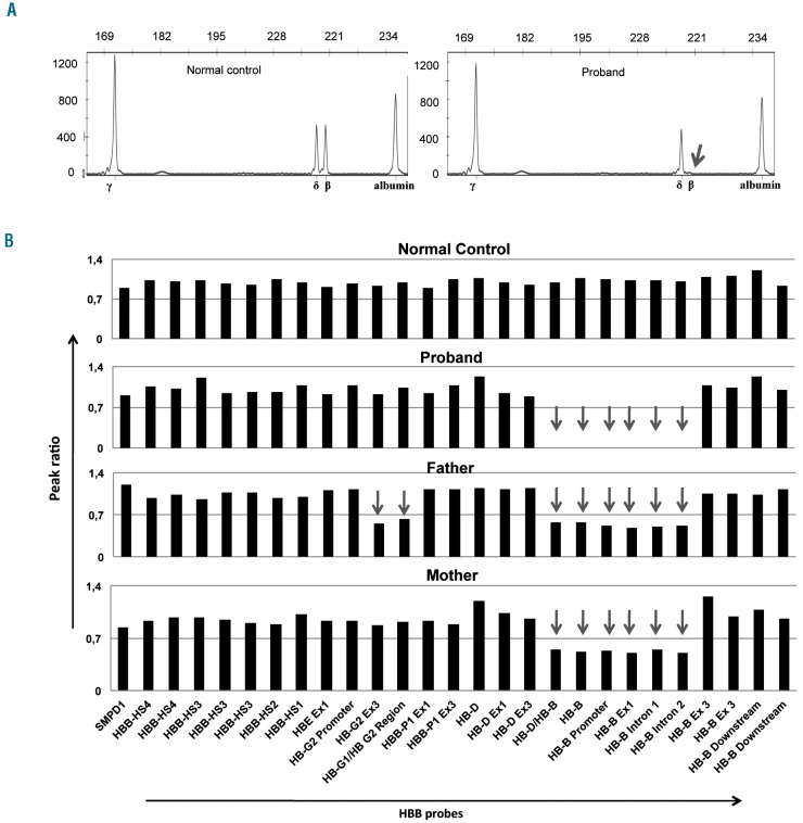
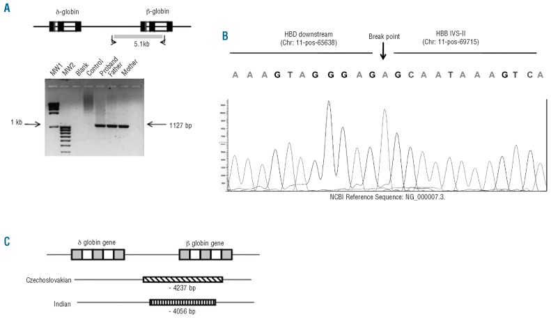
The patient was a 4-year old boy born of a consanguineous marriage from Gujarat, India, who presented with severe anemia requiring frequent blood transfusions from the age of two years. His pre-transfusion hemoglobin level was 5.5 g/dL. He had hepatomegaly but the spleen was not palpable. Complete blood count showed that both the parents had hypochromic microcytic anemia (64.8 and 61.6 fL) and hemoglobin analysis using cation exchange chromatography (Bio-Rad, VARIANT, CA, USA) revealed that they had increased HbA2 (7.7 and 7.1%) with normal HbF (1.1 and 1.4%). Reverse dot blot analysis showed that in this family the common point mutations were absent. To detect possible deletions in the β-globin cluster a multiplex PCR using fluorescently labeled primers was performed for γ, δ- and β-globin genes with the albumin gene as the control. For accurate determination of the copy numbers of the genes in heterozygotes, 200 ng of DNA was used with 0.4 μM primers and the amplification was carried out for 20 cycles. Amplified products were separated by a capillary electrophoresis in an ABI-3130 Genetic Analyzer (Applied Biosystems) and results were analyzed by GeneMapper software version 4.0 (Applied Biosystems). The peak heights obtained for amplified products of the globin genes were divided by those obtained from the albumin gene from each sample, and the ratios obtained for patient and parents were then divided by those obtained from normal individuals. This gene dosage analysis showed absence of amplification from the β-globin gene in the patient in the homozygous state and the parents were heterozygous for a deletion involving the β-globin gene (Figure 1A and Online Supplementary Figure S1). For further identification of the extent of deletion in the β-globin cluster, we performed Multiplex Ligation-dependent Probe Amplification (MLPA) for the β-globin cluster in this family using the SALSA MLPA Kit P102 HBB software version 09 (MRC Holland, Amsterdam, The Netherlands) which contain 29 probes for the 73 kb region of the β-globin gene cluster targeting the locus control region, coding genes in the cluster and the inter-genic sequences.3 The MLPA products were separated by capillary electrophoresis in a Genetic Analyzer and analyzed using GeneMapper software version 4.0. MLPA analysis showed loss of amplification of the probes targeted to the δ-β globin intergenic region and promoter, exons 1 and 2 and introns 1 and 2 confirming the presence of a large deletion of a region from δ-β intergenic region to IVS-2 of β globin gene (Figure 1B). To characterize the breakpoints, we performed PCR with three forward primers that bind to different regions downstream of the δ-globin gene with a constant reverse primer 5′ AGCAGAATGGTAGCTGGATTG 3′ that binds to sequences in β IVS-2. Using a forward primer 5′ CAGGC-CTACTTGAGGGTTGA 3′ that binds at ~2kb downstream of HBD, we obtained an ~1.1 kb amplification product from the patient and the parents while the expected fragment size from a normal individual was 5.1 kb (Figure 2A). DNA sequencing of the amplified product showed that the deletion encompasses 4056 bp region that extends from 2.7 kb downstream of the β-globin gene to IVS-2 of the β-globin gene (Figure 2B).
Figure 1.
(A) Genomic quantitative-PCR for gene dosage analysis to calculate the copy numbers of γ-, δ- and β-globin genes. The peak heights of amplified products of the globin genes and the control albumin gene are shown. (B) MLPA analysis of the β-globin cluster in the family. Arrows indicate the genomic regions that are deleted. Father is heterozygous for an additional deletion of Gγ-globin.
Figure 2.
(A) Location of the primers used for amplification across the break points and agarose electrophoresis of 1127bp amplified products from the patient and the parents. From normal control the expected amplified product 5.1kb was not obtained by the conditions used for amplification. (B) DNA sequencing of the PCR product showing the breakpoints. (C) Comparison of Czechoslovakian and Indian deletions.
MLPA analysis identified an additional deletion present in a heterozygous state in the γ-globin region in the father (Figure 1B) and this is probably due to the Gγ-Aγ fusion gene which is frequently present in this population.4 However, this co-inheritance of a β-globin gene deletion along with the β-globin gene in a single allele does not alter the phenotype in heterozygous β-thalassemia. For a further investigation of the more severe anemia in the mother (Hb 9.1 g/dL, MCV 61.5 fL), we performed multiplex PCR analysis for α-globin genes and found that she was heterozygous for ααα3.7 (ααα3.7/αα) (Online Supplementary Figure S1); it is well known that this genotype is consistent with severe heterozygous β-thalassemia.
So far, 11 deletions, varying from 260bp-67kb in length, of the β-globin gene promoter and its flanking sequence have been reported.5 In an Indian population, a deletion of 10.3 kb that extends from 3011 bp 5′ to the mRNA cap site to an L1 repeat element present downstream of the β-globin gene has been found to cause elevated levels of HbA2 (7.1–7.8%) in a heterozygote state.6 The novel mutation that we identified is similar to the 4237 bp deletion reported in a Czechoslovakian family which caused elevated levels of HbA2 (8.1%–9.0%)7 in a heterozygous state and DNA sequence analysis showed that the 5′ and 3′ breakpoints are a few bases apart (Figure 2C). We used robust, reliable and easier assays to identify the deletion in the β-globin cluster that are useful for appropriate genetic counseling and diagnosis of β-thalassemia, and help predict genotype-phenotype prediction. The exact mechanism for unusual levels of HbA2 in these deletions is not known and it may be hypothesized that it is due to increased availability of transcription factors at the δ-globin promoter when the β-globin gene promoter is deleted, and this promoter competition is not evident between the β- and γ-globin genes.
Footnotes
The online version of this article has a Supplementary Appendix.
The information provided by the authors about contributions from persons listed as authors and in acknowledgments is available with the full text of this paper at www.haematologica.org.
Financial and other disclosures provided by the authors using the ICMJE (www.icmje.org) Uniform Format for Disclosure of Competing Interests are also available at www.haematologica.org.
Funding: this study was supported by funding from the Department of Biotechnology, Government of India.
References
- 1.Weatherall DJ, Clegg JB. The Thalassaemia Syndromes. 4th ed. Oxford, England: Blackwell Science Ltd; 2001. [Google Scholar]
- 2.Databases of Human Hemoglobin Variants and Other resources at the Globin Gene Server. http://globin.cse.psu.edu/hbvar/menu.html. [DOI] [PubMed]
- 3.Harteveld CL, Voskamp A, Phylipsen M, Akkermans N, den Dunnen JT, White SJ, et al. Nine unknown rearrangements in16p13.3 and 11p15.4 causing α- and β-thalassaemia characterized by high resolution multiplex ligation-dependent probe amplification. J Med Genet. 2005;42(12):922–31. doi: 10.1136/jmg.2005.033597. [DOI] [PMC free article] [PubMed] [Google Scholar]
- 4.Sukumaran PK, Nakatsuji T, Gardiner MB, Reese AL, Gilman JG, Huisman THJ. Gamma thalassemia resulting from the deletion of a gamma-globin gene. Nucleic Acids Res. 1983;11(13):4635–43. [PMC free article] [PubMed] [Google Scholar]
- 5.Steinberg MH, Forget BG, Higgs DR, Weatherall DJ. Disorders of Hemoglobin: Genetics Pathophysiology, and Clinical Management. 2nd ed. Cambridge: Cambridge University Press; 2009. [Google Scholar]
- 6.Craig JE, Kelly SJ, Barnetson R, Thein SL. Molecular characterization of a novel 10.3 kb deletion causing β-thalassaemia with unusually high HbA2. Br J Haematol. 1992;82(4):735–44. doi: 10.1111/j.1365-2141.1992.tb06952.x. [DOI] [PubMed] [Google Scholar]
- 7.Popovich BW, Rosenblatt DS, Kendall AG, Nishioka Y. Molecular characterization of an atypical β-thalassemia caused by a large deletion in the 5′ β-globin gene region. Am J Hum Genet. 1986;39(6):797–810. [PMC free article] [PubMed] [Google Scholar]


