

Abstract
Background:
Calabash chalk is a naturally occurring mineral consumed by members of some Nigerian communities for pleasure and by pregnant women as a remedy for morning sickness. The consumption of this geophagic material motivated our interest on the effect of the chalk on the histomorphology of the gastro-oesophageal tract.
Methods:
Twenty-eight young Wistar rats, 4 weeks old, were divided into 4 groups of equal size. Group 1 animals served as controls and received 1 mL of distilled water. Groups 2, 3, and 4 received orally 1 mL of a Calabash chalk suspension containing 40 mg/mL for 14, 21, and 28 days, respectively. Upon completion of the treatments, the animals in groups 2, 3, and 4 were sacrificed on days 15, 22, and 29, respectively, and the control group animals were sacrificed on day 29. All animals were euthanised using chloroform anaesthesia. The oesophagus and the stomach of each animal were dissected out and routinely processed for histological studies.
Results:
There was oedema with haemorrhages in the mucosa of the stomach, and acanthosis, hyperkeratosis, and koilocytic changes were observed in the mucosa of the oesophagus of the groups treated with 40 mg/mL of Calabash chalk suspension.
Conclusion:
Calabash chalk caused histological changes to the stomach and the oesophagus that may lead to other pathophysiological conditions.
Keywords: Calabash chalk, gut, stomach, oesophagus, histology, Wistar rats
Introduction
Geophagia is defined as the practice of eating earth, including soil and chalk (1). People engage in geophagia for a variety of reasons including religious beliefs, medicinal purposes, or as part of a regular diet (2). One geophagic material is Calabash chalk, which is popularly consumed by members of Nigerian and West African communities for pleasure and by pregnant women as a remedy for morning sickness (3,4).
Calabash chalk, also known as Calabar stone, Poto, La craie, Argiles, Mabele, Nzu, and Ndom, is available in a variety of forms including powder, moulded shapes, and blocks. Though native to Africa, it is available in the United Kingdom in ethnic stores and markets. The practice of eating Calabash chalk is also observed among women of African descent in the southern United States, especially in Georgia (4). In addition to the already mentioned uses, Calabash chalk is also used in facial masks and soaps (5). Although this chalk is consumed by both sexes and by many age groups, the prevalence of chalk consumption is greater among women, especially during pregnancy (3).
This chalk is a naturally occurring substance made up of fossilised sea shells but can be prepared artificially (5). It is prepared using a combination of clay and mud, with other ingredients such as sand, wood ash and sometimes salt. This mixture is moulded and then heated to produce the final product (5).
Calabash chalk has the same structure as aluminium silicate hydroxide, which is a member of the kaolin clay group with a possible formula Al2Si2O5(OH)4 (6). Multi-elemental analysis using energy dispersive X-ray fluorescence spectroscopy identified 22 elements in Calabash chalk, including lead and aluminium, and persistent organic pollutants (6).
Another report established the presence of arsenic in Calabash chalk (7). Lead and other toxic elements present in the chalk have been reported to be associated with numerous gastrointestinal disorders including nausea, ulcers, and gastritis. (8). This geophagic material is consumed by the oral route, and several pollutants have been reported to be present in Calabash chalk. It is based on this background that this research was carried out to determine if this chalk has an effect on the oesophagus and the stomach, as these organs serve as the conduit for food entering into the body.
Materials and Methods
Twenty-eight 4-week-old Wistar rats (males and females) were used for the experiment. They were purchased and kept in the Animal House, College of Health Sciences, University of Uyo, Nigeria. The temperature of the room was maintained at 26–28 °C with a 12-hour light/ 12-hour dark cycle.
The animals were cared for according to the international regulations governing the use and care of laboratory animals. They were housed in wooden cages and maintained on standard feeds pellets (growers marsh, Vital Feed, Grand Cereal, Nigeria). Drinking water was allowed ad libitum. The animals were allowed to acclimatise for 1 week. Each animal was weighed prior to the commencement of the experiment and every week thereafter.
Blocks of non-salted Calabash chalk were purchased from a local market in Calabar, Nigeria. The blocks were ground into powder using a manually operated grinder. A 40-g sample of the powder was dissolved in 1000 mL of distilled water, and the mixture was stirred continuously. The chalk was administered as a suspension and was stirred prior to administration. Each millilitre of the suspension contained 40 mg of Calabash chalk.
The rats were weighed and divided into 4 groups of 7 animals each. Group 1 served as the control group and received 1 mL of distilled water for 28 days. Groups 2, 3, and 4 were the test groups and received 1 mL of the Calabash chalk suspension for 14, 21, and 28 days, respectively (Table 1). The rats were humanely sacrificed using chloroform anaesthesia the day after the end of their treatment: rats in groups 2, 3, and 4 were sacrificed on days 15, 22, and 29, respectively, and the controls were sacrificed on day 29. The oesophagus and the stomach from each animal was immediately removed and preserved in 10% buffered formalin for 1 week. These samples were then routinely processed using the haematoxylin and eosin staining method.
Table 1:
The treatment schedule in the experimental groups
| Group | Dosage | Duration (days) |
|---|---|---|
| 1 (control) | 1 mL of distilled water | 28 |
| 2 | 1 mL of suspension (40 mg/mL of Calabash chalk) | 14 |
| 3 | 1 mL of suspension (40 mg/mL of Calabash chalk) | 21 |
| 4 | 1 mL of suspension (40 mg/mL of Calabash chalk) | 28 |
Results
The change in weight relative to the control was smaller in groups 2 and 3 but higher in group 4. The mean weekly weights of the animals in each group are given in Table 2.
Table 2:
Weekly weight of Wistar rats in the control and the test groups treated with 40 mg/mL of Calabash chalk suspension for 14, 21, or 28 days
| Group | Weight (g) | Change in weight | ||||
|---|---|---|---|---|---|---|
| Week 0 | Week 1 | Week 2 | Week 3 | Week 4 | ||
| 1 (control) | 99.26 (6.22) | 120.29 (7.80) | 126.57 (10.00) | 135.14 (11.32) | 136.57 (9.95) | 37.31 |
| 2 | 81.43 (9.92) | 101.57 (10.34) | 109.57 (11.88) | NA | NA | 28.14 |
| 3 | 107.43 (8.36) | 120.86 (12.01) | 129.00 (15.11) | 136.29 (16.72) | NA | 28.86 |
| 4 | 99.14 (10.16) | 118.14 (9.87) | 136.43 (16.46) | 139.14 (14.87) | 142.86 (14.63) | 43.72 |
Results are presented as the mean (SD). Each group consisted of 7 rats. The Week 0 weight represents the weight before the commencement of the experiment. Abbreviation: NA = not applicable.
Histomorphological observations Stomach
The stomach sections of group 1 (control) were normal. A preserved cyto-architecture of the mucosa consisting of simple columnar epithelium forming folds and glands lined by mucus-secreting goblet cells, parietal cells, and chief cells, in addition to endocrine cells having coarse cytoplasm, was observed. The submucosa consisted of a fibro-collagenous stroma, glands, and plexuses. The muscle layer was made up of smooth muscle fibres, and the serosa was composed of mesothelium (Figure 1a).
Figure 1:
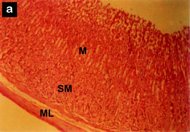
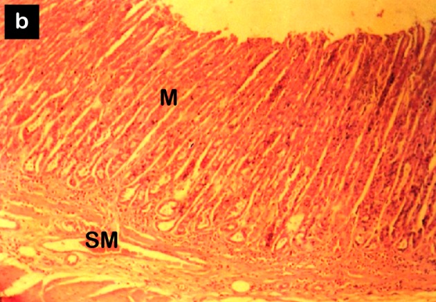
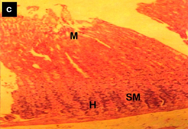
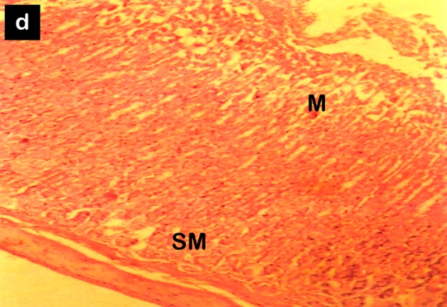
Photomicrograph of the stomach of rats from (a) group 1 (control), (b) group 2 treated with 40 mg/mL of Calabash chalk for 14 days, (c) group 3 treated with 40 mg/mL of Calabash chalk for 21 days, and (d) group 4 treated with 40 mg/mL of Calabash chalk for 28 days (haematoxylin and eosin staining, 100× magnification). Both groups 1 and 2 showed normal mucosal and submucosal layers, whereas groups 3 and 4 showed oedematous morphological changes in the mucosal and submucosal layers. Abbreviation: M = mucosa, SM = submucosa, ML = muscle layer, H = haemorrhage.
Group 2 had normal mucosae with inflammatory cell infiltrates consisting mainly of lymphocytes and plasma cells. The submucosa, muscle layer, and serosa were intact (Figure 1b).
Group 3 had oedematous mucosal linings with haemorrhages and mononuclear inflammatory cell infiltrates consisting mainly of plasma cells and lymphocytes. The submucosa and the serosa were intact, whereas the muscle layer was thickened and hypertrophic compared with that in the control (Figure 1c).
Group 4 had oedematous mucosae close to the basement membrane with haemorrhages and mononuclear inflammatory cell infiltrates consisting mainly of lymphocytes and plasma cells. The submucosa, muscle layer, and serosa were intact (Figure 1d).
Oesophagus
Group 1 (control) had a stratified squamous non-keratinised epithelium overlying loose connective tissue made up of blood vessels, glands, and plexuses. The submucosae contained glands. The muscle layer was made up of smooth muscle fibres, and the serosa was intact (Figure 2a).
Figure 2:
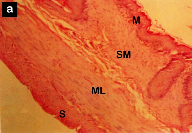
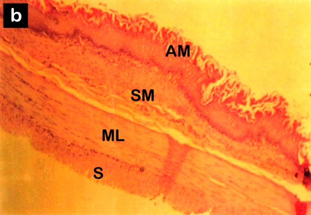

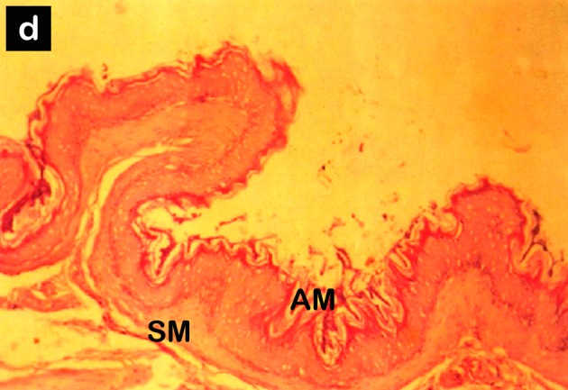
Photomicrograph of the oesophagus of rats from (a) group 1 (control), (b) group 2 treated with 40 mg/mL of Calabash chalk for 14 days, (c) group 3 treated with 40 mg/mL of Calabash chalk for 21 days, and (d) group 4 treated with 40 mg/mL of Calabash chalk for 28 days (haematoxylin and eosin staining, 100× magnification). Group 1 showed no histomorphological changes in the layers of the oesophagus, whereas groups 2, 3, and 4 showed changes in the mucosal layer. Abbreviation: M = mucosa, SM = submucosa, M = muscle layer, S = serosa, AM = acanthosis mucosa.
Group 2 showed keratinised squamous epithelial cells displaying hyperkeratosis, acanthosis, and koilocytosis, and these cells overlaid a loose connective tissue consisting of blood vessels. The submucosa, muscle layer, and serosa were prominent (Figure 2b).
Group 3 showed stratified squamous keratinised epithelial cells displaying acanthosis, hyperkeratosis, and koilocytosis. The granular cell layers were prominent, and the basement membrane was intact. The submucosa, muscle layer, and serosa were unremarkable (Figure 2c).
Group 4 showed a prominent stratified squamous keratinised epithelial cell displaying hyperkeratosis, koilocytosis, and acanthosis. The granular layer was prominent, and few histiocytes and melanocytes were observed. The submucosa, muscle layer, and serosa were unremarkable (Figure 2d).
Discussion
The oesophagus and the stomach are vital organs that serve as conduits and temporary storage spaces for substances that enter the body via the oral cavity. The stomach, which churns and temporarily stores ingested substances, can absorb some substances, such as alcohol (9,10). Substances that are highly toxic may cause morphological changes in the mucosae of both the oesophagus and the stomach.
The results of this study revealed oedema and haemorrhages in the mucosa of the stomach, and acanthosis, hyperkeratosis, and koilocytic changes were found in the mucosae of the oesophagus of the groups treated with 40 mg/mL of Calabash chalk suspension. These findings indicate that the chalk may compromise the integrity of the gastro-oesophageal tract.
Calabash chalk contains poisonous substances such as lead, arsenic, aluminium, and alpha lindane (5,6). Lead intake has been implicated in gastritis, nausea, vomiting, and constipation (7) and has also been found to cause ulcers when large amounts are consumed (11,12). Arsenic has been implicated in gastric distress and stomach upset (13), and aluminium has been shown to be associated with constipation, impaction, colicky pain, anorexia, nausea, and gastrointestinal irritation (14,15).
In addition to the poisonous substances whose adverse effects have so far been reported (5,6), the chalk is reported to be largely composed of kaolin, a substance known to coat the gastrointestinal tract. Kaolin adsorbs drugs and other substances including toxins, which, in addition to reducing their bioavailability, initiates diarrhoeal episodes, thus providing bulk to the stool (16,17). The histological changes in the oesophagus and the stomach observed in this study may also arise as a result of kaolin. The oesophagus is prone to injury (18), and the presence of these substances may increase the susceptibility to injury. This increased susceptibility to injury may lead to further physiological and histomorphological damage to the stomach and the oesophagus due to the increased vulnerability to other toxins that may be consumed.
Superficial injury to the gastric mucosa has been reported to trigger an acute inflammatory response, characterised by an increase in blood flow, plasma exudation, and the recruitment of leukocytes into the mucosa. The objective of this response is thought to be to minimise tissue injury, facilitate the repair of the damaged tissue, and prevent the entry of foreign substances, including microbes and microbial products, into the systemic circulation (19). This inflammatory response is coordinated via the release of an array of soluble mediators from cells such as mucosal mast cells that act as “sentinels” within the mucosa (20). This could have been a reason for the results observed in this study. Morphological changes to the gastrointestinal tract can lead to pseudoneoplasms, a worrisome outcome of the gut’s response mechanisms to injury (21).
A previous study (22) has shown that the Calabash chalk affected the liver, as there was fragmentation of the parenchymal cells, a reduction in the number of hexagonal hepatic lobules and dilated sinusoids when Wistar rats were treated with this substance. Another study (23) found that this chalk alters the normal concentration of haemoglobin, the red blood cell count, the platelet count, and the erythrocyte sedimentation rate in rats.
Conclusion
Calabash chalk caused histomorphological changes to the stomach and the oesophagus, which may lead to other pathophysiological changes and neoplasms of the gastrointestinal tract.
Footnotes
Authors’ contributions
Conception and design, final approval of the article: MBE
Analysis and interpretation of the data: EEJ
Drafting of the article: CCM, EIB
Critical revision of the article: MBE, TB
References
- 1.Halsted JA. Geophagia in man: Its nature and nutritional effects. Am J Clin Nutr. 1968;21(12):1384–1393. doi: 10.1093/ajcn/21.12.1384. [DOI] [PubMed] [Google Scholar]
- 2.Reilly C, Henry J. Geopaghia: Why do humans consume soil? Nutr Bull. 2000;25(2):141–144. [Google Scholar]
- 3.Callahan GN. Eating dirt. [cited 2011 Jun 6];Emerg Infect Dis. 2003 9(8):1016–1021. doi: 10.3201/eid0908.030033. [Internet] Available from: http://www.cdc.gov/ncidod/EID/vol9no8/pdfs/03-0033.pdf. [DOI] [PMC free article] [PubMed] [Google Scholar]
- 4.Grigsby RK, Thyer BA, Waller RJ, Johnston GA Jr. Chalk eating in middle Georgia: a culture-bound syndrome of pica? South Med J. 1999;92(2):190–192. doi: 10.1097/00007611-199902000-00005. [DOI] [PubMed] [Google Scholar]
- 5.Agency warns of the dangers of traditional remedy for morning sickness. London (GB): Food Standards Agency;; 2002. [cited 2011 May 1]. [Internet] Available from: http://tna.europarchive.org/20110116113217/http://www.food.gov.uk/news/pressreleases/2002/oct/calabash. [Google Scholar]
- 6.Dean JR, Deary ME, Gbefa BK, Scott WC. Characterisation and analysis of persistent organic pollutants and major, minor and trace elements in Calabash chalk. Chemosphere. 2004;57(1):21–25. doi: 10.1016/j.chemosphere.2004.05.023. [DOI] [PubMed] [Google Scholar]
- 7.Campbell H. Belfast (IE): Department of Health, Social Service and Public Safety (IE):; 2002. [cited 2011 May 1]. Calabash chalk (calabar stone, la craie, argile, nzu, mabele) [Internet] Available from: http://www.docstoc.com/docs/54253409/calabashchalk-(calabar-stone-La-Argile-Nzu-Mabele) [Google Scholar]
- 8.Health effects of lead. Ontario (CA): Canadian Centre for Occupational Health and Safety;; 2008. [cited 2010 Oct 24]. [Internet] Available from: http://www.ccohs.ca/oshanswers/chemicals/chem_profiles/lead/health_lead.html. [Google Scholar]
- 9.Bode C, Bode JC. Alcohol’s role in gastrointestinal tract disorders. Alcohol Health Res World. 1997;21(1):76–83. [PMC free article] [PubMed] [Google Scholar]
- 10.Snell RS. Clinical anatomy by regions. 8th ed. Philadelphia (PA): Lippincott Williams & Wilkins; 2007. [Google Scholar]
- 11.Marcus S. New York (NY): WebMD; 2011. [cited 2011 May 1]. Lead toxicity in emergency medicine. [Internet] Available from: http://emedicine.medscape.com/article/815399-overview. [Google Scholar]
- 12.Murata K, Iwata T, Dakeishi M, Karita K. Lead toxicity: Does the critical level of lead resulting in adverse effects differ between adults and children. J Occup Health. 2009;51(1):1–12. doi: 10.1539/joh.k8003. [DOI] [PubMed] [Google Scholar]
- 13.Smith AH, Hopenhayn-Rich C, Bates MN, Goeden HM, Hertz-Picciotto I, Duggan HM, et al. Cancer risks from arsenic in drinking water. Environ Health Perspect. 1992;97:259–267. doi: 10.1289/ehp.9297259. [DOI] [PMC free article] [PubMed] [Google Scholar]
- 14.AHFS consumer medication information: Aluminum hydroxide. Bethesda (MD): American Society of Health-System Pharmacists, Inc.; 2008. [cited 2012 Jan 2]. [Internet] Available from: http://www.nlm.nih.gov/medlineplus/druginfo/meds/a699048.html#other-information. [Google Scholar]
- 15.Ganrot PO. Metabolism and possible health effects of aluminum. Environ Health Perspect. 1986;65:363–441. doi: 10.1289/ehp.8665363. [DOI] [PMC free article] [PubMed] [Google Scholar]
- 16.Onyekweli AO, Usifoh CO, Ukonrobo LO, Zuofa JD. Adsorptive property of kaolin in some drug formulations. Trop J Pharm Res. 2003;2(1):155–159. [Google Scholar]
- 17.Kaolin. Place unknown: Wolters Kluwer Health; 2009. [cited 2012 Jan 2]. [Internet] Available from: http://www.drugs.com/npp/kaolin.html#ref9. [Google Scholar]
- 18.Roy S. Drug related lesions of the gastrointestinal tracts. [cited 2011 Apr 6]; [Internet] Available from: http://www.histopathology-india.net/drugGIT.htm. [Google Scholar]
- 19.Wallace JL, Granger DN. The cellular and molecular basis of gastric mucosal defense. FASEB J. 1996;10(7):731–740. doi: 10.1096/fasebj.10.7.8635690. [DOI] [PubMed] [Google Scholar]
- 20.Martin GR, Wallace JL. Gastrointestinal inflammation: A central component of mucosal defense and repair. Exp Biol Med. 2006;231(2):130–137. doi: 10.1177/153537020623100202. [DOI] [PubMed] [Google Scholar]
- 21.De Petris G, Leung ST. Pseudoneoplasms of the gastrointestinal tract. Arch Pathol Lab Med. 2010;134(3):378–392. doi: 10.5858/134.3.378. [DOI] [PubMed] [Google Scholar]
- 22.Ekong MB, Akpantah AO, Ibok OS, Eluwa MA, Ekanem TB. Differentia effects of calabash chalk on the histology of liver of adult Wistar rats. [cited 2010 Feb 8];Internet J Health. 2009 8(2) [Internet J Health] Available from: http://www.ispub.com/journal/the_internet_journal_of_health/volume_8_number_2_12/article/differentia_effects_of_calabash_chalk_on_the_histology_of_the_liver_of_adult_wister_rats.html. [Google Scholar]
- 23.Akpantah AO, Ibok OS, Ekong MB, Eluwa MA, Ekanem TB. The effect of calabash chalk on some haematological parameters in female adult Wistar rats. Turk J Hematol. 27;3(3):177–181. doi: 10.5152/tjh.2010.25. [DOI] [PubMed] [Google Scholar]