Abstract
Objectives
Streptococcus pneumoniae is one of the most common pathogens of otitis media (OM) that exists in biofilm, which enhances the resistance of bacteria against antibiotic killing and diagnosis, compared to the free-floating (planktonic) form. This study evaluated biofilm formation by S. pneumoniae on an abiotic surface and in the middle ear cavity in a rat model of OM.
Methods
In vitro biofilm formation was evaluated by inoculation of a 1:100 diluted S. pneumoniae cell suspension in a 96-well microplate. Adherent cells were quantified spectrophotometrically following staining with crystal violet by measurement of optical density at 570 nm. The ultrastructure of pneumococcal biofilm was assessed by scanning electron microscopy (SEM). For in vitro biofilm study, S. pneumoniae cell suspensions containing 1×10⁷ colony forming units were injected through transtympanic membrane into the middle ear cavity of Sprague Dawley rats. The ultrastructure of middle ear mucus was observed by SEM 1 and 2 weeks post-inoculation.
Results
The in vitro study revealed robust biofilm formation by S. pneumoniae after 12-18 hours of incubation in high glucose medium, independent of exogenously supplied competence stimulating peptide and medium replacement. Adherent cells formed three-dimensional structures approximately 20-30 µm thick. The in vivo study revealed that ciliated epithelium was relatively resistant to biofilm formation and that biofilm formation occurred mainly on non-ciliated epithelium of the middle ear cavity. One week after inoculation, biofilm formation was high in 50% of the treated rats and low in 25% of the rats. After 2 weeks, biofilm formation was high and low in 25% and 37.5% of rats, respectively.
Conclusion
The results imply that glucose level is important for the S. pneumoniae biofilm formation and S. pneumoniae biofilm formation may play important role in the pathophysiology of OM.
Keywords: Otitis media, Streptococcus pneumoniae, Biofilm
INTRODUCTION
Otitis media (OM) is the most common illness for which children visit a physician, receive antibiotics, or undergo surgery world-wide [1, 2]. OM arises in the complex microbial community of the upper respiratory tract. The bacteria most often associated with OM are Streptococcus pneumoniae, Haemophilus influenzae, and Moraxella catarrhalis [3]. S. pneumoniae asymptomatically colonizes up to 50% of children [4, 5] and the colonization of the upper respiratory tract is the first step in infection. Even transient colonization provides an opportunity for S. pneumoniae to invade the middle ear space.
S. pneumoniae can form biofilms in vitro and in vivo [6-8]. The adherent biofilm bacteria are phenotypically and genotypically different from their free-floating (planktonic) counterpart. Within the extracellular exopolysaccharide matrix, biofilm bacteria display reduced metabolic and growth/divisional rates from the planktonic form [9]. These more quiescent bacteria are less affected by antibiotics and can be more difficult to detect by conventional culture-based techniques [10].
Although past studies of OM have focused on planktonic bacteria, biofilms are now recognized to play an important role in the pathophysiology of OM [11-14]. A previous study using chinchillas model of middle ear infection by S. pneumoniae demonstrated the presence of biofilms [8]. Moreover, direct microscopic evidence for the presence of biofilms in OM was recently obtained by analysis of middle ear tissue from OM patients who underwent tympanostomy tube insertion and surface-attached communities of many bacterial species were evident [12]. To elucidate the role of biofilm in the pathophysiology of OM, establishment of a stable in vitro and in vivo biofilm formation models is very important.
In this study, we evaluated in vitro biofilm formation using a microplate method and in vivo biofilm formation by transtympanic injection of S. pneumonia.
MATERIALS AND METHODS
Bacterial strain and growth conditions
S. pneumoniae R6 strain (ATCC BAA-255) used in this study was obtained from the American Type Culture Collection (Manassas, VA, USA). Bacteria were grown on a blood agar plate with 5% sheep blood. A fresh colony was transferred in trypticase soy broth (TSB) and grown at 37℃ for 12 hours in 5% CO2. One microliter of bacterial suspension was diluted in 50 mL of fresh, warm TSB and grown to mid-logarithmic phase (data not shown). The number of colony forming units (CFU) of the bacterial suspension was determined by a serial dilution plate count method [15].
In vitro biofilm formation
In vitro biofilm formation analysis was performed with crystal violet microplate assay by a modification of a previously described procedure [16, 17]. Briefly, biofilm formation was carried out in 96-well (flat-bottom) polystyrene (PST) microplates (BD Falcon; BD, Sparks, MD, USA). S. pneumoniae grown to mid-logarithmic phase containing 1×10⁸ CFU/mL (optical density at 600 nm=0.4) was diluted 1:100 with fresh sterile medium, and 200 µL was inoculated in 96-well PST microplates. Plates were filled with TSB medium supplemented with 0%, 1%, and 2% glucose, and incubated stationary at 37℃ for different times (6, 12, 18, and 24 hours) in 5% CO2. After incubation, the medium was discarded and plates were gently washed three times with 200 µL sterile phosphate buffered saline (PBS). Thereafter, plates were air dried, and stained with 50 µL of 0.1% crystal violet for 15 minutes. Excess stain was decanted-off and plates were washed three times with sterile distilled water. Adherent crystal violet was dissolved in 200 µL of 95% ethanol and optical density was measured at 570 nm a SpectraMax plus 384 automatic spectrophotometer microplate reader (Molecular Devices, Sunnyvale, CA, USA). The experiment was performed in triplicate and the average was calculated. To compensate for background absorbance, values from the sterile medium and crystal violet (CV) were averaged and subtracted.
For SEM analysis, the bacterial suspension of 1×10⁸ CFU/mL was diluted 1:100 with fresh TSB medium and 2 mL were in a 12-well PST microplate (BD Falcon). The plate was incubated stationary at 37℃ for 18 hours and supplied with 5% CO2. After incubation, the medium was removed and the plate was gently washed two times with sterile PBS to remove planktonic cells.
In vivo biofilm formation
The experimental protocol was reviewed and approved by the animal research and care committee at Dongguk University Ilsan Hospital (Goyang, Korea). Twenty four specific pathogen-free, Sprague Dawley (SD) rats weighing 150-200 g (Orient Bio, Seongnam, Korea) were used. Animals were examined by otomicroscopy to document abnormal middle ear status before the experiment and were kept isolated in an infection free zone for 2 weeks. Rats were assigned randomly to groups that received either a bacterial inoculation (n=16) or no procedure (control group, n=8). Fifty microliters of S. pneumoniae cell suspension containing 3×10⁷ CFU of S. pneumoniae was injected into the middle ear cavity through the tympanic membrane of the right ear using a tuberculin syringe and a 27-gauge needle. Animals were sacrificed at 1 week (n=8) and 2 weeks (n=8) after bacterial inoculation, and middle ear bulla was acquired. The tympanic membrane was removed and ears were irrigated to remove planktonic bacteria. The ultrastructure of middle ear mucosa was examined using SEM for determination of the presence or absence of biofilm formation. To measure the degree of biofilm formation at each time point, the sacrificed animals were divided into three groups: no biofilm forming group, low biofilm forming group, and high biofilm forming group.
Scanning electron microscopic analysis
The morphology of pneumococcal biofilms were observed with SEM. Samples were prefixed by immersion in 2% glutaraldehyde in 0.1 M phosphate buffer and post-fixed for 2 hours in 1% osmic acid dissolved in PBS. Samples were treated in a graded series of ethanol and t-butyl alcohol, dried in a model ES-2030 freeze dryer (Hitachi, Tokyo, Japan), platinum-coated using an IB-5 ion coater (Eiko, Kanagawa, Japan), and observed using a model S-4700 FE-SEM (Hitachi).
RESULTS
In vitro biofilm formation
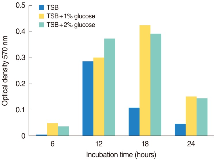
In vitro biofilm formation by S. pneumoniae on a microplate was investigated in TSB medium supplemented with 0%, 1%, and 2% glucose. All three media supported biofilm formation. However, TSB supplied with 1% glucose showed consistently high biofilm formation. The results of biofilm formation in the different concentrations of glucose showed that biofilm formation was closely related with the glucose concentration and maximum biofilm was detected in TSB supplemented with 1% glucose (Fig. 1). To further investigate the influence of incubation time on adherence of pneumococcal cells to PST microplate, 1:100 diluted cell suspension (1×10⁸ CFU) was incubated at 37℃ for different times (6, 12, 18, and 24 hours). The results of the time-course experiment detected biofilm formation after 6 hours following inoculation and reached maximum between 12-18 hours. Thereafter, there was a decline in biofilm formation (Fig. 1). Under optimized conditions, pneumococcal strain R6 consistently showed high in vitro biofilm-forming capacity and optical density values ranged from 0.004-0.424 at 570 nm.
Fig. 1.
In vitro biofilm formation by Streptococcus pneumoniae R6 strain in trypticase soy broth (TSB) medium supplemented with 0%, 1%, and 2% glucose, incubated at 37℃ for 6, 12, 18, and 24 hours in 5% CO2.
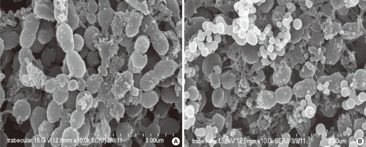
Pneumococcal cells adhered to a surface and adopted a multicellular three-dimensional structure. Adherent cells formed a mat of cells of significant depth (20-30 µm), which were interconnected by small, thin filaments that linked the cells to each other and/or bound the cells to the intercellular matrix (Fig. 2A). In some area, microcolonies were observed revealing non-homogeneous microbial populations, with an irregular and discontinuous surface that coated the cells and contained holes of different sizes, which might represent channels between cell clusters (Fig. 2B).
Fig. 2.
Scanning electron microscopic image of Streptococcus pneumoniae R6 strain biofilm formed on a polystyrene microplate. (A) Filament material linked pneumococcal cells to each other and to the intercellular matrix. (B) Apical view of the irregular surface of a pneumococcal biofilm. Micro-colonies of different sizes revealing non-homogeneous microbial populations are evident.
In vivo biofilm formation
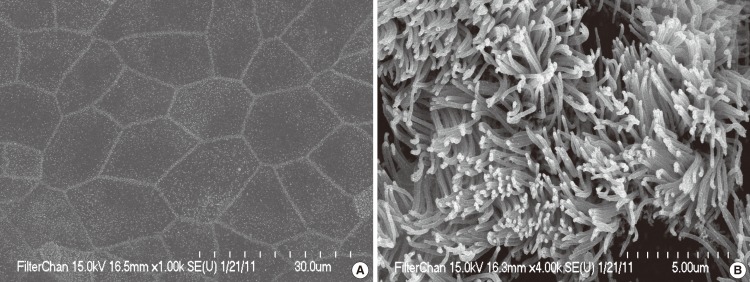
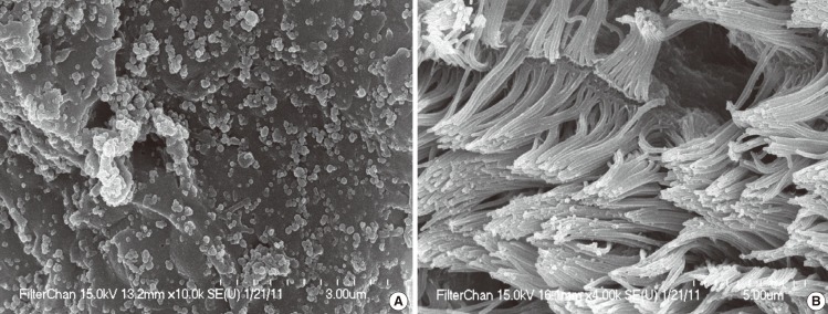
The middle ear mucosa was composed of non-ciliated squamous epithelium and ciliated epithelium. Ciliated epithelium was distributed in the hypotympanum area and Eustachian tube orifice area. The remainder of the middle ear bulla was covered with non-ciliated squamous epithelium. In normal (control) ears, no biofilm formation was detected in either of the areas (Fig. 3). Among eight animals sacrificed after 1 week of bacterial inoculation, two rats (25%) showed no biofilm formation, two rats (25%) showed low biofilm formation and four rats (50%) showed high biofilm formation in middle ear mucosa. In the high biofilm forming group, the non-ciliated epithelium was covered with a thick exopolysaccharide matrix of biofilm of S. pneumoniae, which had a rough and irregular surface (Fig. 4A). Although the cilia of ciliated epithelium were conglomerated, little biofilm was formed in ciliated epithelium region (Fig. 4B). After 2 weeks of bacterial inoculation, eight animals were sacrificed, three rats (37.5%) did not show biofilm formation, three rats (37.5%) showed low biofilm in middle ear, while two rats (25%) showed robust biofilm formation in middle ear mucosa with variably-shaped exopolysaccharide (Fig. 5A, B). Although the degree of biofilm formation was somewhat variable between the animals sacrificed in each group, most of the non-ciliated epithelium in high biofilm forming groups was covered with thick and variably-shaped exopolysaccharide. The ciliated epithelium was resistant to bacterial attachment and biofilm formation. However, the cilia of ciliated epithelium were conglomerated and some exopolysaccharide debris and diplococcal cells were attached to the tip of cilia (Fig. 5C).
Fig. 3.
Scanning electron microscopic image of middle ear mucosa of a control rat. (A) Non-ciliated area of middle ear mucosa. (B) Ciliated area of middle ear mucosa.
Fig. 4.
Scanning electron microscopic image of middle ear mucosa of treated rat after 1 week of bacterial inoculation. (A) High degree of biofilm formation with rough exopolysaccharide matrix. (B) Conglomerated cilia of ciliated epithelium.
Fig. 5.
Scanning electron microscopic image of middle ear mucosa of treated rat after 2 weeks of bacterial inoculation. (A) Thick biofilm with irregular surfaced exopolysaccharide matrix of biofilm. (B) Exopolysaccharide matrix connected with thread like structures. (C) Debris of exopolysaccharide matrix attached to ciliated epithelium.
DISCUSSION
In this study, we evaluated the S. pneumoniae biofilm formation in vitro by using a crystal violet microplate assay without exogenous supplements and in vivo by the transtympanic injection of bacteria into the middle ear cavity of SD rats. The results demonstrated that ciliated epithelium was relatively resistant to biofilm formation and that biofilm formation occurred mainly on non-ciliated epithelium of the middle ear cavity. Also, we showed that S. pneumoniae biofilm formation occurred in vitro in high glucose medium without addition of competence stimulating peptide and media replacement.
S. pneumoniae (pneumococcus) is one of the organisms most frequently isolated from patients with recurrent and persistent OM [18]. In addition to OM, S. pneumoniae is a leading cause of community-acquired pneumonia and meningitis, especially in children and the elderly. Recently, the introduction of the heptavalent pneumococcal capsular conjugate vaccine has clearly reduced the incidence of OM by pneumococcus [19]. However, there was a report that incidence of OM was increased caused by pneumococcal serotypes not included in the heptavalent pneumococcal capsular conjugate vaccine [20].
The culturability of middle ear effusion in OM is variable [21, 22] and recalcitrance to antibiotic treatment is frequent [23]. Although many past studies of OM have focused on planktonic bacteria, biofilm is now recognized to play an important role in the pathophysiology of OM [11, 12]. Though S. pneumoniae is an important pathogen in OM, little is known about the biofilm formation of S. pneumoniae in OM.
In this study, we have used S. pneumoniae R6, which is a well-known biofilm forming strain [24]. Because biofilm is usually resistant to conventional antibiotic treatment, eradication of biofilms is a promising treatment modality [25]. To evaluate the treatment modality, a robust and reliable model is needed. The model reported in this study may be suitable for the evaluation of biofilm formation of S. pneumoniae in vivo and in vitro.
Many studies have supported the biofilm paradigm in OM in animal model [12-14, 26]. In a non-human primate model of chronic suppurative otitis media, biofilm was identified 4 weeks after the inoculation of P. aeruginosa into the middle ear cavity of a monkey via a transtympanic approach [14]. H. influenzae biofilm was visually detected by SEM on the middle ear mucosa after the inoculation of bacteria into the middle ear cavity of chinchillas through the bony bulla and the density of the biofilm increased until at least 96 hours after inoculation [11]. Unlike chinchillas, the middle ear bulla of the rat is relatively deep from the surface and a midline ventral approach is needed to access it; therefore, we adopted a transtympanic approach [27].
In humans, mucosal biofilms were visualized by a confocal laser scanning microscope on 92% of middle ear mucosa from children with OM and recurrent otitis media [12]. In particular, the prevalence of S. pneumoniae was higher than that apparent using polymerase chain reaction [12, 23]. Pneumococcal vaccination would be a promising modality for prevention of pneumococcal infection. However, the effectiveness of vaccination for the prevention of recurrent OM is contentious [28-30].
In vitro biofilm formation was carried out in crystal violet microplate assay, which is easier to perform than continuous culture methods [7]. In addition, the biofilm formation model in this study was independent from exogenously added competence stimulating peptides and medium replacement. We think that it represents the tissue under the pathologic conditions in OM.
However, in this study, maximum biofilm formation occurred between 12 and 18 hours of incubation in high glucose medium. A recent study demonstrated that glucose in medium causes the pH of the medium to drop below 6.0 and inhibits the hydrolytic activity of the pneumococcal autolysin, which is a major autolytic enzyme of pneumococcus [24]. Clinically, hyperglycemia is one of the most important factors in worsening of systemic bacterial infection. We think that our in vitro results may be representative of the clinical biofilm disease common in hyperglycemic patients.
More evidence is emerging to support the biofilm paradigm in OM. However, little is known about the role of biofilm in the pathophysiology of OM and cross-talk between biofilm and middle ear mucosa. In a clinical situation, most infections consist of mixed flora and synergy plays an important role among multiple organisms. Though complex biofilm composed of multiple species is frequently observed in the bacterial infection, little is known about the role of complex biofilm in OM [25]. Further studies are needed to elucidate these issues.
ACKNOWLEDGMENTS
This research was supported by Basic Science Research Program through the National Research Foundation of Korea (NRF) funded by the Ministry of Education, Science and Technology (KRF-2010-0007632).
Footnotes
No potential conflict of interests relevant to this article was reported.
References
- 1.Klein JO. The burden of otitis media. Vaccine. 2000 Dec;19(Suppl 1):S2–S8. doi: 10.1016/s0264-410x(00)00271-1. [DOI] [PubMed] [Google Scholar]
- 2.Hendley JO. Clinical practice: otitis media. N Engl J Med. 2002 Oct;347(15):1169–1174. doi: 10.1056/NEJMcp010944. [DOI] [PubMed] [Google Scholar]
- 3.Revai K, Mamidi D, Chonmaitree T. Association of nasopharyngeal bacterial colonization during upper respiratory tract infection and the development of acute otitis media. Clin Infect Dis. 2008 Feb;46(4):e34–e37. doi: 10.1086/525856. [DOI] [PMC free article] [PubMed] [Google Scholar]
- 4.Bogaert D, De Groot R, Hermans PW. Streptococcus pneumoniae colonisation: the key to pneumococcal disease. Lancet Infect Dis. 2004 Mar;4(3):144–154. doi: 10.1016/S1473-3099(04)00938-7. [DOI] [PubMed] [Google Scholar]
- 5.Syrjanen RK, Kilpi TM, Kaijalainen TH, Herva EE, Takala AK. Nasopharyngeal carriage of Streptococcus pneumoniae in Finnish children younger than 2 years old. J Infect Dis. 2001 Aug;184(4):451–459. doi: 10.1086/322048. [DOI] [PubMed] [Google Scholar]
- 6.Allegrucci M, Hu FZ, Shen K, Hayes J, Ehrlich GD, Post JC, et al. Phenotypic characterization of Streptococcus pneumoniae biofilm development. J Bacteriol. 2006 Apr;188(7):2325–2335. doi: 10.1128/JB.188.7.2325-2335.2006. [DOI] [PMC free article] [PubMed] [Google Scholar]
- 7.Donlan RM, Piede JA, Heyes CD, Sanii L, Murga R, Edmonds P, et al. Model system for growing and quantifying Streptococcus pneumonia biofilms in situ and in real time. Appl Environ Microbiol. 2004 Aug;70(8):4980–4988. doi: 10.1128/AEM.70.8.4980-4988.2004. [DOI] [PMC free article] [PubMed] [Google Scholar]
- 8.Reid SD, Hong W, Dew KE, Winn DR, Pang B, Watt J, et al. Streptococcus pneumoniae forms surface-attached communities in the middle ear of experimentally infected chinchillas. J Infect Dis. 2009 Mar;199(6):786–794. doi: 10.1086/597042. [DOI] [PubMed] [Google Scholar]
- 9.Mah TF, O'Toole GA. Mechanisms of biofilm resistance to antimicrobial agents. Trends Microbiol. 2001 Jan;9(1):34–39. doi: 10.1016/s0966-842x(00)01913-2. [DOI] [PubMed] [Google Scholar]
- 10.Fitzgerald G, Williams LS. Modified penicillin enrichment procedure for the selection of bacterial mutants. J Bacteriol. 1975 Apr;122(1):345–346. doi: 10.1128/jb.122.1.345-346.1975. [DOI] [PMC free article] [PubMed] [Google Scholar]
- 11.Post JC. Direct evidence of bacterial biofilms in otitis media. Laryngoscope. 2001 Dec;111(12):2083–2094. doi: 10.1097/00005537-200112000-00001. [DOI] [PubMed] [Google Scholar]
- 12.Hall-Stoodley L, Hu FZ, Gieseke A, Nistico L, Nguyen D, Hayes J, et al. Direct detection of bacterial biofilms on the middle-ear mucosa of children with chronic otitis media. JAMA. 2006 Jul;296(2):202–211. doi: 10.1001/jama.296.2.202. [DOI] [PMC free article] [PubMed] [Google Scholar]
- 13.Ehrlich GD, Veeh R, Wang X, Costerton JW, Hayes JD, Hu FZ, et al. Mucosal biofilm formation on middle-ear mucosa in the chinchilla model of otitis media. JAMA. 2002 Apr;287(13):1710–1715. doi: 10.1001/jama.287.13.1710. [DOI] [PubMed] [Google Scholar]
- 14.Dohar JE, Hebda PA, Veeh R, Awad M, Costerton JW, Hayes J, et al. Mucosal biofilm formation on middle-ear mucosa in a nonhuman primate model of chronic suppurative otitis media. Laryngoscope. 2005 Aug;115(8):1469–1472. doi: 10.1097/01.mlg.0000172036.82897.d4. [DOI] [PubMed] [Google Scholar]
- 15.Southey-Pillig CJ, Davies DG, Sauer K. Characterization of temporal protein production in Pseudomonas aeruginosa biofilms. J Bacteriol. 2005 Dec;187(23):8114–8126. doi: 10.1128/JB.187.23.8114-8126.2005. [DOI] [PMC free article] [PubMed] [Google Scholar]
- 16.Oggioni MR, Trappetti C, Kadioglu A, Cassone M, Iannelli F, Ricci S, et al. Switch from planktonic to sessile life: a major event in pneumococcal pathogenesis. Mol Microbiol. 2006 Sep;61(5):1196–1210. doi: 10.1111/j.1365-2958.2006.05310.x. [DOI] [PMC free article] [PubMed] [Google Scholar]
- 17.Baldassarri L, Creti R, Recchia S, Imperi M, Facinelli B, Giovanetti E, et al. Therapeutic failures of antibiotics used to treat macrolide-susceptible Streptococcus pyogenes infections may be due to biofilm formation. J Clin Microbiol. 2006 Aug;44(8):2721–2727. doi: 10.1128/JCM.00512-06. [DOI] [PMC free article] [PubMed] [Google Scholar]
- 18.Tonnaer EL, Graamans K, Sanders EA, Curfs JH. Advances in understanding the pathogenesis of pneumococcal otitis media. Pediatr Infect Dis J. 2006 Jun;25(6):546–552. doi: 10.1097/01.inf.0000222402.47887.09. [DOI] [PubMed] [Google Scholar]
- 19.Block SL, Hedrick J, Harrison CJ, Tyler R, Smith A, Findlay R, et al. Community-wide vaccination with the heptavalent pneumococcal conjugate significantly alters the microbiology of acute otitis media. Pediatr Infect Dis J. 2004 Sep;23(9):829–833. doi: 10.1097/01.inf.0000136868.91756.80. [DOI] [PubMed] [Google Scholar]
- 20.McEllistrem MC, Adams J, Mason EO, Wald ER. Epidemiology of acute otitis media caused by Streptococcus pneumoniae before and after licensure of the 7-valent pneumococcal protein conjugate vaccine. J Infect Dis. 2003 Dec;188(11):1679–1684. doi: 10.1086/379665. [DOI] [PubMed] [Google Scholar]
- 21.Diamond C, Sisson PR, Kearns AM, Ingham HR. Bacteriology of chronic otitis media with effusion. J Laryngol Otol. 1989 Apr;103(4):369–371. doi: 10.1017/s0022215100108989. [DOI] [PubMed] [Google Scholar]
- 22.Giebink GS, Juhn SK, Weber ML, Le CT. The bacteriology and cytology of chronic otitis media with effusion. Pediatr Infect Dis. 1982 Mar-Apr;1(2):98–103. doi: 10.1097/00006454-198203000-00007. [DOI] [PubMed] [Google Scholar]
- 23.Rayner MG, Zhang Y, Gorry MC, Chen Y, Post JC, Ehrlich GD. Evidence of bacterial metabolic activity in culture-negative otitis media with effusion. JAMA. 1998 Jan;279(4):296–299. doi: 10.1001/jama.279.4.296. [DOI] [PubMed] [Google Scholar]
- 24.Moscoso M, Garcia E, Lopez R. Biofilm formation by Streptococcus pneumoniae: role of choline, extracellular DNA, and capsular polysaccharide in microbial accretion. J Bacteriol. 2006 Nov;188(22):7785–7795. doi: 10.1128/JB.00673-06. [DOI] [PMC free article] [PubMed] [Google Scholar]
- 25.Smith A, Buchinsky FJ, Post JC. Eradicating chronic ear, nose, and throat infections: a systematically conducted literature review of advances in biofilm treatment. Otolaryngol Head Neck Surg. 2011 Mar;144(3):338–347. doi: 10.1177/0194599810391620. [DOI] [PubMed] [Google Scholar]
- 26.Zuliani G, Carlisle M, Duberstein A, Haupert M, Syamal M, Berk R, et al. Biofilm density in the pediatric nasopharynx: recurrent acute otitis media versus obstructive sleep apnea. Ann Otol Rhinol Laryngol. 2009 Jul;118(7):519–524. doi: 10.1177/000348940911800711. [DOI] [PubMed] [Google Scholar]
- 27.Song JJ, Kown SK, Kim EJ, Lee YS, Kim BY, Chae SW. Mucosal expression of ENaC and AQP in experimental otitis media induced by Eustachian tube obstruction. Int J Pediatr Otorhinolaryngol. 2009 Nov;73(11):1589–1593. doi: 10.1016/j.ijporl.2009.08.011. [DOI] [PubMed] [Google Scholar]
- 28.van Heerbeek N, Straetemans M, Wiertsema SP, Ingels KJ, Rijkers GT, Schilder AG, et al. Effect of combined pneumococcal conjugate and polysaccharide vaccination on recurrent otitis media with effusion. Pediatrics. 2006 Mar;117(3):603–608. doi: 10.1542/peds.2005-0940. [DOI] [PubMed] [Google Scholar]
- 29.Nascimento-Carvalho CM. Pneumococcal conjugate vaccination and otitis media: differences among high-risk and low-risk populations. Expert Rev Vaccines. 2009 Jun;8(6):695–698. doi: 10.1586/erv.09.42. [DOI] [PubMed] [Google Scholar]
- 30.van Kempen MJ, Vermeiren JS, Vaneechoutte M, Claeys G, Veenhoven RH, Rijkers GT, et al. Pneumococcal conjugate vaccination in children with recurrent acute otitis media: a therapeutic alternative? Int J Pediatr Otorhinolaryngol. 2006 Feb;70(2):275–285. doi: 10.1016/j.ijporl.2005.06.022. [DOI] [PubMed] [Google Scholar]


