Abstract
The application of RNA interference-based gene silencing to the airway surface epithelium holds great promise to manipulate host and pathogen gene expression for therapeutic purposes. However, well-differentiated airway epithelia display significant barriers to double-stranded small-interfering RNA (siRNA) delivery despite testing varied classes of nonviral reagents. In well-differentiated primary pig airway epithelia (PAE) or human airway epithelia (HAE) grown at the air–liquid interface (ALI), the delivery of a Dicer-substrate small-interfering RNA (DsiRNA) duplex against hypoxanthine–guanine phosphoribosyltransferase (HPRT) with several nonviral reagents showed minimal uptake and no knockdown of the target. In contrast, poorly differentiated cells (2–5-day post-seeding) exhibited significant oligonucleotide internalization and target knockdown. This finding suggested that during differentiation, the barrier properties of the epithelium are modified to an extent that impedes oligonucleotide uptake. We used two methods to overcome this inefficiency. First, we tested the impact of epidermal growth factor (EGF), a known enhancer of macropinocytosis. Treatment of the cells with EGF improved oligonucleotide uptake resulting in significant but modest levels of target knockdown. Secondly, we used the connectivity map (Cmap) database to correlate gene expression changes during small molecule treatments on various cells types with genes that change upon mucociliary differentiation. Several different drug classes were identified from this correlative assessment. Well-differentiated epithelia treated with DsiRNAs and LY294002, a PI3K inhibitor, significantly improved gene silencing and concomitantly reduced target protein levels. These novel findings reveal that well-differentiated airway epithelia, normally resistant to siRNA delivery, can be pretreated with small molecules to improve uptake of synthetic oligonucleotide and RNA interference (RNAi) responses.
Keywords: Cmap, epithelia, RNAi, siRNA
Introduction
Small-interfering RNA (siRNA)-mediated silencing of genes offers a novel approach for disease treatment. Direct delivery of siRNA to respiratory epithelia is potentially advantageous for pulmonary infections and for chronic diseases like cystic fibrosis, where airway epithelial cells are prominent sites of production and release of proinflammatory cytokines, including interleukin-8 (IL-8).1,2,3 Topical delivery may avoid hepatic or renal clearance and nonspecific accumulation associated with systemic routes. However, due to its high molecular weight and polyanionic nature, siRNAs do not cross the epithelial cell membrane freely. Additionally, mucus presents an intra pulmonary physical barrier to overcome prior to cell entry. Thus, efficient delivery of siRNAs to the airways is challenging due to intracellular and extracellular barriers.
Nonviral siRNA delivery is an attractive approach for achieving gene silencing in airway epithelia. A number of studies report successful delivery of naked siRNA to airways, especially for counteracting viral infections.4,5,6 However, recent reports show that siRNAs delivered intranasally or intratracheally do not enter lung cells and do not elicit RNA interference (RNAi).7 Early reports of siRNA efficacy were likely confounded by the off-target immunostimulatory effects of siRNA constructs.8,9,10,11 Here, we explored further the utility of nonviral based siRNAs in reducing target abundance in well-differentiated primary airway epithelial cells. We show that well-differentiated epithelia are almost completely refractory to entry of siRNA unless subjected to certain treatments.
Results
Dicer-substrate siRNAs (DsiRNA) delivery has no silencing effect in well-differentiated cultures
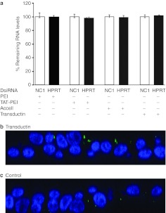
Our overall goal was to test the effectiveness of DsiRNAs in well-differentiated primary airway epithelial cells. Well-differentiated airway epithelia maintained at the air–liquid interface (ALI) mirror to a great extent the in vivo morphology of the airway and thus are an ideal system to test efficacy of inhibitory RNAs in the form of DsiRNAs and nonviral delivery reagents.12 We tested various nonviral reagents or siRNA enhancements, including cationic polymers (PEI or TAT-PEI), modified naked siRNA (Accell), a peptide reagent (PTD-DRBD or Transductin) and a lipid transfection reagent (RNAiMAX). No hypoxanthine–guanine phosphoribosyltransferase (HPRT) silencing was observed after apical transfection of HPRT DsiRNA into pig airway epithelia (PAE) with polymers, Accell or Transductin (Figure 1a). Nor was there a reduction of HPRT mRNA levels in human airway epithelia (HAE) following transfection of siRNA conjugated with RNAiMAX or Transductin (Supplementary Figure S1). However, the DsiRNAs are functional based on their utility to reduce target gene expression (>90%) in PKI cells, a pig kidney cell line grown on plastic (Supplementary Figure S2). Thus, well-differentiated airway epithelia are not amenable to DsiRNA transfection with a broad range of transfection reagents; consequently, RNAi against the targets is not achieved.
Figure 1.
DsiRNA delivery has no silencing effect in well-differentiated PAE cultures. (a) Well-differentiated PAE cultures were transfected with HPRT or NC1 DsiRNA (250 nmol/l) using PEI, Transductin or TAT-PEI, or Accell siRNA at the indicated concentrations. The cells were harvested for RNA after 24 hours, and reverse transcribed into cDNA, which was quantified by quantitative PCR (qPCR). All mRNA levels were normalized to NC1-treated samples. Mean levels (±SD) were calculated from three biological replicates (in triplicate). (b–c) Confocal images (x–z stacks) of epithelia 2 hours after transfection with DIG-HPRT DsiRNA complexed with Transductin (b) or without any transfection reagent (c). Blue, labeled nuclei; green, DIG-labeled oligo.
Well-differentiated cells show minimal uptake of DsiRNA
We next investigated whether failure of silencing is due to limited DsiRNA uptake. We labeled DsiRNA internally with Digoxigenin (DIG) nucleotides and transfected them into cells using Transductin. Subsequently, the cell layers were fixed and processed for immunodetection of DIG label using fluorescent labeled anti-DIG antibodies. Confocal imaging of the cells revealed little to no internalization of DsiRNA in both pig airway cells (Figure 1b,c) and human airway cells (Supplementary Figure S3a,b). The results suggest that the inability of the epithelia to silence gene expression is largely due to a failure of the DsiRNA to enter the cells.
KGF, EGTA, and LLnL treatment of cells prior to siRNA treatment have no effect on target silencing
The decreased susceptibility of well-differentiated cells to DsiRNA prompted us to investigate the effect of interventions known to influence cellular processes including proliferation, permeability, and processing that might influence siRNA transfection and silencing. Keratinocyte growth factor (KGF) is a member of the fibroblast growth factor family. We and others13,14 have shown that KGF stimulates proliferation of differentiated human tracheal and bronchial epithelia. Pretreatment of PAE with KGF before transfection of DsiRNAs with either RNAiMAX or Transductin did not improve gene silencing relative to controls (Supplementary Figure S4a). In addition, we also treated the cells with EGTA, a calcium chelator, which causes a reversible increase in paracellular permeability.15 Similar to results seen with KGF, DsiRNA transfection with either RNAiMAX or Transductin failed to silence HPRT after treatment with EGTA (Supplementary Figure S4b). Lastly, we treated the cells with the proteasome inhibitor [N-acetyl-L-Leucyl-L-Leucyl-L-Norleucinal (LLnL)] before transfection. LLnL is a tripeptide proteasome inhibitor that enhances recombinant adeno-associated virus type 2 transduction from the apical surface of HAE by modulating the intracellular trafficking and processing of the virus.16 Treatment with LLnL had no effect on silencing of HPRT following transfection of specific DsiRNA with either RNAiMAX or Transductin (Supplementary Figure S4c). Thus, well-differentiated cells are refractory to siRNA transfection with physiological manipulations that induce proliferation, changes in paracellular permeability, or proteasome inhibition.
DsiRNA delivery effectively silences targets in poorly differentiated epithelia
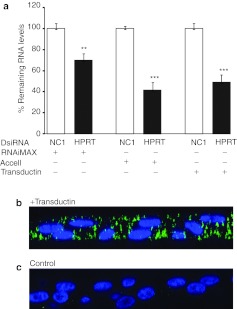
Since well-differentiated cells exhibit significant extracellular barriers to siRNA entry, we hypothesized that poorly differentiated cells, which lack many of the barriers present in mature cells, might be conducive to siRNA entry and knockdown of the target gene. In contrast to well-differentiated cells, poorly differentiated epithelia lack ciliated and goblet cells and do not have a pseudostratified columnar architecture. On transfection of poorly differentiated PAE, also grown at the ALI on culture inserts (2-day-old post-seeding) with DsiRNA against HPRT, mRNA levels were suppressed with all transfection reagents used. HPRT mRNA levels were reduced by 40% with RNAiMAX, 60–70% with Accell, and ~50–60% with Transductin (Figure 2). Similar reductions were found on transfection with DsiRNA against pig IL-8 (Supplementary Figure S5a) or when poorly differentiated human airway cells were transfected with similarly formulated HPRT-targeting DsiRNAs (Supplementary Figure S5b). These results demonstrate that before differentiation airway epithelia are conducive to DsiRNA transfection and target knockdown.
Figure 2.
DsiRNAs effectively silence targets in poorly differentiated PAE cultures. (a) Poorly differentiated cultures, 2-day post-seeding, were transfected with 250 nmol/l of DsiRNA against HPRT with the use of RNAiMAX, Transductin, or Accell siRNA at the indicated concentrations and then processed (RNA isolation, RT-qPCR) 24 hours later. Remaining mRNA levels were normalized to negative control (NC1). Mean levels (±SD) were calculated from three biological replicates (in triplicate) **P < 0.01, ***P < 0.001 (Student's t-test). (b–c) Confocal images (x–z stacks) of epithelia 2 hours after transfection with DIG-HPRT DsiRNA complexed with (b) Transductin or (c) unformulated. Blue, labeled nuclei; green, DIG-labeled oligo.
DsiRNAs enter poorly differentiated transfected cells
We next examined whether DsiRNA internalization was enhanced in poorly differentiated epithelia. Two days after seeding, cells were examined for DsiRNA uptake. In contrast to results in well-differentiated epithelia (Figure 1b), abundant DsiRNA internalization was observed in poorly differentiated epithelia from both pig (Figure 2b,c) and human (Supplementary Figure S3c,d). These data indicate that DsiRNAs can silence targets in poorly differentiated airway epithelia by virtue of their ability to enter the cells.
Airway epithelia become progressively resistant to RNAi when grown at ALI
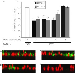
We next investigated how time in culture influenced siRNA entry and target knockdown. Primary airway epithelia, maintained at ALI, take a minimum of 2–4 weeks after seeding to fully differentiate into a pseudostratified columnar epithelium.17 Cells obtained from two pig donors were seeded and batches of cells from 2 to 5 days post-seeding were transfected with HPRT-targeting DsiRNAs complexed with Transductin. Reduction in HPRT mRNA by 20–45% was seen in 2–4-day-old cultures, but there was no silencing in 5-day cultures (Figure 3a). The cultures were also visualized for DsiRNA entry after DIG-DsiRNA transfection. The presence or absence of target knockdown correlated with DIG-DsiRNA internalization (Figure 3b–e). Cells transfected with DsiRNA against IL-8 also showed decreased IL-8 mRNA in 2-day-old, but not in 5-day-old cultures (Supplementary Figure S6a). Similarly, HAE showed no HPRT knockdown when DsiRNAs were applied after 5 days in culture (Supplementary Figure S6b). Thus, silencing of targets is restricted to a narrow time window post-seeding before cell differentiation reaches the point that the cells are refractory to RNAi approaches. Notably, cells undergo differentiation but not extensive cell division during this time frame.
Figure 3.
DsiRNA efficacy as a function of time in culture. (a) PAE cultures that were 2–5 days old were transfected with DsiRNA against HPRT (250 nmol/l) using Transductin. The cells were processed for RNA isolation and cDNA synthesis 24 hours later and then subjected to RT-qPCR for quantification of HPRT mRNA. HPRT levels were normalized to NC1 samples. Mean levels (±SD) were calculated from three biological replicates (in triplicate), **P < 0.01, ***P < 0.001 (Student's t-test). (b–e) Confocal images (x–z stacks) of (b) 2, (c) 3, (d) 4, and (e) 5-day-old cells after transfection of each with DIG-HPRT DsiRNA and then staining 2 hours later for DIG. Red, nuclei; green, DIG-labeled oligo.
EGF pretreatment enhances siRNA entry and silencing
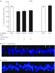
We hypothesized that manipulation of the cells to enhance endocytosis might promote better cellular siRNA uptake and silencing of target genes. Epidermal growth factor (EGF) induces rapid actin filament assembly in the membrane cytoskeleton of many types, which can result in membrane ruffling and macropinocytosis.18,19,20 We reasoned that since the Tat protein, the cell-penetrating domain of Transductin, enter cells through macropinocytosis,21,22 EGF treatment of cells before transfection of DsiRNA–Transductin complexes might enhance DsiRNA cellular entry. Human airway cell cultures from three different donors treated with EGF (100 µg/ml for 15 minutes) before transfection of DsiRNA showed reduced HPRT mRNA levels of 15–20% (Figure 4a). EGF-treated cells (100 µg/ml for 15 minutes) imaged for DsiRNA entry 2 hours after transfection showed increased uptake of the DIG-labeled DsiRNA compared to the mock treated cells (Figure 4b,c). These results show that a transient cellular response to EGF can increase both the uptake of DsiRNA and silencing of target genes.
Figure 4.
EGF treatment of cells before DsiRNA delivery enhances entry and silencing. (a) Well-differentiated airway epithelia from three donors were treated apically with EGF at a concentration of 100 µg/ml for 15 minutes before transfection with DsiRNAs against HPRT (250 nmol/l). RNAs were harvested for quantitation 24 hours later, as above. Mean levels (±SD) were calculated from three biological replicates (in triplicate) *P < 0.02, **P < 0.01 (Student's t-test). (b–c) Well-differentiated human airway epithelia were transfected with a formulation of DIG-HPRT DsiRNA and Transductin with EGF pretreatment (100 µg/ml for 15 minutes) (b) or mock treated (c). Samples were processed as above for confocal microscopy. Blue, nuclei; green, DIG-labeled oligo.
Cmap analysis links gene signatures of mucociliary differentiation to small molecules
We next took advantage of the Connectivity map (Cmap) to identify pathways that, when manipulated, could overcome the resistance of well-differentiated airway epithelia to RNAi. In brief, the Cmap provides users with a dataset of transcriptional changes that occur in a number of cell lines in response to a drug treatment.23 Investigators merge this dataset with expression data from a system of interest, in which gain-of-function and loss-of-function expression studies have been done.24,25 Here, we used previously published data sets representing immature airway epithelia (which are receptive to siRNA delivery and activity) and well-differentiated airway epithelia (which are resistant to siRNA delivery and activity). The rationale is that gene expression changes which underlie the state of differentiation in some way contribute to the emerging resistance of these cells to RNAi. Drugs known to induce pathways active in the more de-differentiated physiological state may provide a “permissive environment,” and allow for siRNA activity even though the cells remain as a mature epithelium. To accomplish this, we used the data set previously reported by Ross et al.,26 which identified genes involved in the differentiation of human bronchial epithelial cells grown at the ALI over 28 days. We found several small molecules which positively or negatively correlated to the reference signatures. Because we were interested in agents that might induce cell physiological changes which mimicked pathways active in poorly differentiated cells, we focused on drugs which correlated negatively with the reference signatures. Four agents were selected that ranked highly with a strong anti-correlation score (connectivity score of –1 or close to –1). These included LY294002, ergocalciferol, paclitaxel, and nifedipine. Two additional agents that were highly ranked in the results according to their P values were also selected: naltrexone and fasudil. As a negative control in our experiments, we selected the drugs monensin and meclofenamic acid; each had strong positive correlation scores (connectivity score of +1 or close to +1).
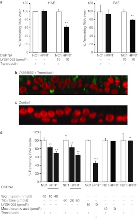
We next treated primary airway epithelia with the selected small-molecule agents. Cells were treated for 6 hours, then transfected with DsiRNA–Transductin complex for 24 hours. When pig or human epithelia were transfected with the HPRT-targeting DsiRNA, we observed no reduction in mRNA levels in epithelia treated with ergocalciferol, paclitaxel, nifedipine, naltrexone, or fasudil (data not shown). In contrast, following LY294002 treatment (10 µmol/l), human and pig epithelia showed ~40% and ~20% HPRT silencing (Figure 5a), respectively, compared to that of control DsiRNA. Results from cells of several donors showed a range of silencing effects, 10–55% inhibition in HAE and 10–20% in PAE (data not shown). The DsiRNA silencing effect was a result of improved DsiRNA uptake, as shown by images of cells treated with LY294002 and transfected with DIG-DsiRNA (Figure 5b,c). LY294002 inhibits four classes of PI3K isoforms in the PI3K pathway and has been one of the most widely used pathway inhibitors.27,28,29,30 Activation of PI3K results in a range of downstream events. To test whether the enhanced silencing following LY294002 treatment is due to PI3K pathway inhibition or another mode of action, we treated cells with the PI3K pathway inhibitors wortmannin and triciribine. Wortmannin, unlike LY294002, is an irreversible inhibitor of the PI3K pathway and triciribine is an inhibitor of AKT, a downstream effector in the PI3K pathway. As HAE showed the most robust knockdown, they were used in these follow-up studies. Pretreatment of epithelia with increasing doses of wortmannin and triciribine before DsiRNA transfection resulted in dose-dependent improvements in RNAi efficacy (Figure 5d). Treatment of cells with 10 and 40 nmol/l of wortmannin decreased HPRT levels by 20 and 30%, respectively, compared to controls. Treatment with 20 and 60 µmol/l triciribine resulted in 20 and 35% decrease, respectively in HPRT mRNA levels compared to controls (Figure 5d). These findings suggest that the improved silencing of the siRNA target is a consequence of cellular changes that occur in response to PI3K pathway inhibitors. Finally, we also tested the effects of LY294002 treatment on two other DsiRNA gene targets, cystic fibrosis transmembrane conductance regulator (CFTR) and SIN3A. Pretreatment of human airway cells with 10 µmol/l of LY294002 before transfection with these specific DsiRNAs resulted in 35 and 25% reduction in CFTR and SIN3A mRNA levels, respectively, when compared to control (Supplementary Figure S7a,b).
Figure 5.
Small molecule treatments prior to DsiRNA delivery improve gene silencing. (a) PAE or HAE, respectively were treated apically with LY294002 at a concentration of 10 µmol/l for 6 hours or mock treated before transfection with HPRT or NC1 DsiRNAs. Data show HPRT mRNA levels (mean ± SD; three biological replicates (in triplicate), **P < 0.01, Student's t-test). (b–c) Confocal imaging (x–z stacks) of HAE (b) with or (c) without pretreatment with LY294002 (10 µmol/l for 2 hours) and transfection with DIG-HPRT DsiRNA with Transductin. Red, nuclei; green, DIG-labeled oligo. (d)Human airway cells treated with PI3K pathway inhibitors wortmannin and triciribine, or with the negative control meclofenamic acid prior to DsiRNA application. Data show HPRT mRNA levels normalized to NC1. Mean levels (±SD) were calculated from three biological replicates (in triplicate), **P < 0.01, ***P < 0.001 (Student's t-test).
LY294002 pretreatment decreases CFTR protein levels in epithelia transfected with CFTR DsiRNA
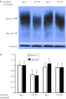
We next tested whether this level of gene silencing was sufficient to reduce the expression of a protein important in lung cell physiology, the CFTR protein. CFTR encodes an anion channel in airway epithelia and other cells.31,32 Loss of CFTR function, caused by mutations in the CFTR gene causes cystic fibrosis, an important chronic disease characterized by progressive pulmonary infection and inflammation.33 Treatment of HAE with LY294002 before transfection of cells with CFTR-targeting DsiRNA formulated in Transductin resulted in 35% reduction in CFTR mRNA levels as shown in Supplementary Figure S7b. Analysis of CFTR protein by western blot showed decreased protein levels in transfected cells treated with LY294002 compared to controls (Figure 6). Both band B and mature, fully glycosylated CFTR band C were significantly decreased in drug-treated cells. The decrease in protein levels was observed in cells from three separate donors tested (data not shown). Thus, well-differentiated airway epithelia, normally refractory to siRNA delivery, are amenable if pretreated with LY294002, allowing for silencing of target mRNA and protein levels.
Figure 6.
LY294002 treatment prior to CFTR siRNA transfection decreases CFTR protein expression in well-differentiated HAE cultures. (a) HAE were treated apically with LY294002 at a concentration of 10 µmol/l for 6 hours or mock treated before transfecting them with CFTR or negative control DsiRNA. Analysis of protein expression was done by Western blot using CFTR antibody. Protein loading was visualized by probing for β-tubulin. (b) Band density detection was performed using densitometry. Protein levels were compared to that of LY294002-treated negative control. Mean levels were calculated from the results of three different transfections, each performed with a different donor from duplicate wells. Mean ± SD;***P < 0.003 (Student's t-test).
Discussion
siRNA-mediated silencing of gene expression holds great promise for disease treatment. While there has been much progress in siRNA design to avoid immunogenicity, toxicity, and off-target effects, one of the remaining hurdles is delivery.34 Unlike many organs, the lung permits direct access of delivery materials to the target cells via topical administration through intranasal, intratracheal, and aerosol routes. Thus, many lung diseases which have epithelial cell pathologies, namely cystic fibrosis, chronic obstructive pulmonary disease, pulmonary fibrosis, and respiratory viral diseases, are ideal targets for siRNA treatment. In this study, we tested DsiRNA-nonviral reagent combinations for effective silencing of gene targets by applying various formulations to the apical surface of primary pig or HAE cells. We used DsiRNAs previously shown to evoke potent RNAi.35
Primary airway epithelia from either trachea or bronchi and grown in an ALI culture become well-differentiated over time, form a pseudostratified columnar epithelium with tight junctions, develop cilia, and produce mucin.12,17 Previous studies showed a strong overall correlation in gene expression between cells grown at ALI and cells obtained directly from in vivo bronchial brushings,12 supporting this culture system as an excellent model for studies of lung epithelia biology. One prior report noted that well-differentiated epithelia transfected apically with siRNA formulated with Oligofectamine or jetSI did not reduce target gene expression.36 And, although siRNA entry was described as limiting, no data were presented. Here, we found that none of our DsiRNA formulations were successful in gene silencing to well-differentiated epithelia, irrespective of the dose or time of transfection, and this correlated strongly with limited oligonucleotide entry.
Thus, successful delivery of DsiRNA to the airways depends on the capacity of the formulations to overcome a variety of barriers. One of these is the mucus biopolymer network, which causes steric obstruction and thereby hinders diffusion of lipid or polymer nanoparticles.37,38 In addition, macromolecules such as antibodies may interact with nonviral nanoparticles37,38 and cause entrapment, aggregation or release of the RNA. However, in our experiments, repeated washings of the apical surface with phosphate-buffered saline, which removes most of the mucus, did not improve RNAi efficacy.
Several different formulations have been investigated for in vivo siRNA delivery to the lungs. Therapeutic efficacies were reported from intranasal or intratracheal administration of naked siRNA, including reduction in the titers of airway-tropic viruses, or reducing transcripts pivotal in airway disease.4,5,6,9,10,39,40,41,42,43 However, misinterpretation may have arisen from the use of control sequences that were less immunostimulatory than the siRNA sequences targeting the gene of interest.8,9,10,11 Additional work indicated that direct intratracheal administration of naked siRNA into mouse lung did not achieve target knockdown.7 Instead, as early as 15 minutes after administration renal excretion was observed, indicating rapid absorption across the respiratory epithelium without access to the cytoplasmic compartments needed to function as triggers of RNAi.
We found that target gene silencing was effective in poorly differentiated pig and HAE as a function of improved entry. Primary cultures of different ages are distinct in their makeup of cell types, average apical membrane surface area, and overall architecture. Also, tight junctions that regulate paracellular passage of molecules are present in epithelia within a day of seeding. This suggests that poor DsiRNA entry is not related to the formation of tight junctions. This interpretation was supported by studies with EGTA of well-differentiated epithelia. Previous studies showed that EGTA treatment of airway epithelia in vitro and in vivo enhanced gene transfer with retroviruses and adenovirus by providing access to the basolateral surface.15 EGTA treatments did not enhance target silencing with DsiRNA formulations, however.
We also considered that actively proliferating cells would enhance entry, since cells early post-seeding are still dividing (albeit slowly) and they were more susceptible to RNAi. However, inducing well-differentiated epithelia to divide and proliferate did not improve gene silencing. In spite of this result, we cannot conclude that cell division has no impact on entry. Another consideration is that the cellular makeup within the epithelia changes as a function of differentiation.
The failure of siRNA to enter cells efficiently after just 5 days post-seeding implies that barrier properties arise before complete differentiation of the epithelia. This suggests that physiological and structural differences associated with cell growth and division may have a bearing on siRNA entry. Cell-specific differences in transfection efficiency have been attributed to effects of not only cell cycle and cell division frequency, but also to endocytic capacity.44,45,46 It has been established that endocytosis acts as a major path of entry for many polyplexes-containing nucleic acid.45 Lipoplexes or PEI polyplexes are internalized by various endocytic pathways, including macropinocytosis, clathrin-mediated endocytosis, and nonclathrin-mediated mechanisms with entry via caveolae.45 Two studies show that the protein transduction domain Tat, which is the cell-penetrating domain of the peptide agent Transductin, enters cells by macropinocytosis.21,22 For these reasons, we tested EGF. EGF acts through a tyrosine kinase receptor (EGFR) to rapidly stimulate actin reorganization at the cell membrane resulting in membrane ruffling and macropinocytosis.18,19,20 EGFR can be activated in the airways and has been implicated in epithelial cell repair and mucin production.46 Additionally, matrix metalloprotease-9, which is abundant in various chronic airway disorders and involved in lung remodeling, induces release of membrane bound EGF-like proligands at the apical surface that then subsequently bind to EGFR.46 We hypothesized that treatment of EGF could enhance internalization and endocytosis of the siRNA cargo, especially those formulated with Transductin since its entry is shown to occur by macropinocytosis. EGF treatment of human primary airway epithelia prior to transfection of DsiRNA with Transductin increased knockdown from 0 to 15–20%. Consistent with this, confocal imaging revealed increased internalization of the DIG-labeled DsiRNA. It will be interesting to test how further modulation of this pathway may impact siRNA delivery to airway.
A resource that provided insight regarding the differences between poorly- and well-differentiated airway epithelia was the molecular atlas of gene expression changes during mucociliary differentiation.26 Many of the functional categories of the genes correlated well with the transformation of these cells into a pseudostratified ciliated epithelium that shares many properties of the in vivo airway epithelium. We used three gene signatures (0 versus 4 day; 0 versus 8 day; 4 versus 8 day) from this dataset to query the Cmap database in an attempt to link genes associated with differentiation to candidate small molecules.23 Treatment with five of the six compounds selected did not enhance DsiRNA delivery or activity in well-differentiated airway epithelia. One of the small molecules, LY294002, a PI3K inhibitor, improved knockdown in pig airway and was therefore also tested in HAE. There its impact to the formulation was more robust. Of note, the Cmap data was generated in human cell lines and thus the resource may be less useful for predicting small molecules that impact gene expression changes in other species.
We also show that manipulation of well-differentiated epithelia with LY294002 facilitates knockdown of disparate DsiRNA targets (SIN3A and CFTR). Silencing of CFTR mRNA by this approach also reduced levels of the CFTR protein. Thus, the drugs we have identified in this study (the PI3K pathway inhibitors) provide a permissive environment for siRNA activity to function in a system that has proved previously to be extremely difficult to access for nucleic acid delivery by nonviral methods.
PI3Ks have key regulatory roles in many cellular processes, including cell survival, proliferation, and differentiation.28 PI3Ks are downstream effectors of receptor tyrosine kinases and G protein-coupled receptors and transduce signals from growth factors and cytokines. These signals activate serine-threonine protein kinase AKT and other downstream effector pathways. Many components of the pathway are mutated in human cancers. LY294002 was the first synthetic drug-like small molecule inhibitor capable of reversibly targeting PI3K family members in the micromolar range.27,28,29,30 The observed enhancement of LY294002 on airway DsiRNA silencing would suggest that treatment with other PI3K pathway inhibitors might have similar effects. We tested two compounds outside of the Cmap database, wortmannin (a PI3K inhibitor) and triciribine (an Akt inhibitor) and observed a dose-dependent silencing effect in HAE with both compounds, suggesting that PI3K pathway inhibition is playing a role. All three drugs inhibit tumorigenesis. However, how these molecules increase siRNA uptake and silencing remains obscure.
In conclusion, we discovered that well-differentiated airway epithelia are refractory to treatment with commonly used siRNA and DsiRNA formulations. This barrier occurs at least in part at the level of cargo entry. Importantly, manipulation of epithelia can induce effective DsiRNA uptake. Further refinements and studies of the mechanism behind these barrier changes may aid in developing additional successful strategies for gene silencing in well-differentiated epithelia, and translating RNAi to in vivo applications.
Materials and Methods
Culture of primary epithelial cells and cell lines. PAE cells were obtained from trachea of lungs removed from euthanized pigs. The studies were approved by the institutional review board at the University of Iowa. HAE cells were obtained from trachea and bronchi of lungs removed for organ donation from non-CF individuals. Cells were isolated by enzyme digestion as previously described.35,47 Following enzymatic dispersion, cells were seeded at a density of 5 × 10⁵ cells/cm2 onto collagen-coated, 0.6-cm2 semipermeable membrane filters (Millipore polycarbonate filters; Millipore, Bedford, MA). The cells were maintained at 37 °C in a humidified atmosphere of 5% CO2 air. Twenty-four hours after plating, the apical media was removed and the cells were maintained at an ALI to allow differentiation of the epithelium. The culture medium consisted of 1:1 ratio mix of Dulbecco's modified Eagle's medium/Ham's F12, 5% Ultroser G (Biosepra SA, Cedex, France), 100 U/ml penicillin, 100 µg/ml streptomycin, 1 % nonessential amino acids, and 0.12 U/ml insulin. Studies were performed on well-differentiated epithelia ~4–6 weeks after initiation of the ALI cultures conditions. Pig kidney cells (PK1) were cultured in Medium 199 (Life Technologies, Carlsbad, CA) supplemented with 3% fetal bovine serum.
DsiRNA oligonucleotides. The 27-mer DsiRNAs were designed using algorithms developed by Integrated DNA Technologies (IDT, Coralville, IA) and synthesized and high-performance liquid chromatography purified by IDT. The protocol for DsiRNA design and manufacture has been described in detail.35,48,49 The DIG-labeled DsiRNA was also designed and synthesized by IDT. The DIG label was internally coupled to an amino-dT base in a 2-O′ methyl modified pig-specific DsiRNA against HPRT. The DsiRNAs used in this study are listed in Supplemental Table S1.
RNAi reagents. Lipofectamine RNAiMAX was purchased from Invitrogen (Life Technologies). The cationic polymers, PEI, and TAT-PEI were synthesized and conjugated with DsiRNA into appropriate NP concentrations. Accell siRNA (Thermo Scientific, Lafayette, CO), which are chemically modified to increase stability and enhance uptake by cells, were custom synthesized as 21-nucleotide siRNA with a sequence for control NC1 and a sequence against pig HPRT. The PTD-DRBD peptide delivery reagent was purchased as Transductin from IDT.
Transfection. For airway epithelia that were either well or poorly differentiated, transfection was performed mostly according to the manufacturer's protocol. In all cases, the apical surface of epithelia was washed twice with phosphate-buffered saline before adding the transfection mixture to the apical surface. The mixture was then left on the cells for 24 hours for all transfection reagents. For PK1 cells, reverse transfection was performed in a 48-well plate by first adding the DsiRNA-RNAiMAX reagent mixture onto the plate and then adding the dispersed cells (40,000 cells), allowing the cells to adhere and establish growth for 24 hours. Before some transfections, cells were pretreated with various chemicals: KGF (Prospec, East Brunswick, NJ) 50 ng/ml added both apically and basolaterally for 24 hours; EGTA 6 mmol/l added apically for 30 minutes LLnL (ICN Biochemicals, Costa Mesa, CA) 40 µmol/l added both apically and basolaterally for 24 hours; human EGF (Sigma-Aldrich, St Louis, MO) 100 µg/ml, added apically for 15 minutes. For all experiments, a minimum of three biological replicates, in triplicate, were done.
RNA isolation and quantitative real-time PCR. Total RNA was isolated using SV96 RNA isolation kit (Promega, Madison, WI), according to manufacturer's protocol. Total RNA (250 ng) were reverse transcribed using oligo (dT) (Roche Biochemicals, Indianapolis, IN) and random hexamers (Life Technologies) and Superscript II (Life Technologies) according to manufacturer's instructions. One-fifteenth of the cDNA was then amplified and analyzed by Taqman assay in the 7900 Real-Time PCR System (Applied Biosystems, Foster city, CA) using synthesized primer-probe pairs (IDT), reaction buffer and Immolase DNA polymerase (Bioline, Taunton, MA). All the primers and probes used in the study are listed in Supplemental Table S2. The reaction mix was contained in a total volume of 10 µl and the reaction condition was an initial cycle of 95 °C for 10 minutes, then 40 cycles of 95 °C for 15 seconds and 60 °C for 1 minutes. All data were normalized to the internal standard, RPL4 mRNA for pig airway samples and SFRS9 mRNA for human airway samples. Absolute quantification of an mRNA target sequence within an unknown sample was determined by reference to a standard curve. The standard curve was based on the real-time PCR amplification of standard amounts of the specific gene in a control NC1 treatment cDNA. PCR efficiency for all reactions was within the acceptable margin of 90–110%. All results of the samples were presented as remaining target mRNA level in comparison to the mRNA level in the control samples (NC1), which was normalized to 100%.
Confocal imaging. The primary HAE grown at ALI were transfected with the DIG-labeled DsiRNA at a concentration of 250 nmol/l after complexing with Transductin. The transfection mixture was left on the apical surface for 2 hours. At the end of this period, the cells were fixed in 2% formaldehyde, permeabilized in 0.2 % Triton-x-100 and blocked in 1% bovine serum albumin for 1 hour. The cells were then stained with mouse antibody to DIG (Roche Biochemicals) for 1 hour followed by Alexa 488-labeled goat anti-mouse secondary antibody for 1 hour and then Alexa 488-labeled rabbit anti-mouse tertiary antibody (Life Technologies). In some cases Alexa 564 goat anti-mouse secondary antibody was used. The cells were finally stained with nuclear stain, ToPro 3 for 10 minutes. The filter, containing the cells, was removed from the culture insert by cutting the edges with razor blade, mounted with Vectashield (Vector Laboratories, Burlingame, CA). The cells were visualized by confocal microscopy (Bio-Rad Radience 2100MP Multiphoton/Confocal Microscope). For each experiment, epithelia from a minimum of three donors were analyzed, with biological replicates done in duplicate.
SDS-PAGE and immunoblotting. Human primary culture was washed with phosphate-buffered saline and lysed in freshly prepared lysis buffer (1% Triton, 25 mmol/l Tris pH 7.4, 150 mmol/l NaCl, protease inhibitors (cOmplete; mini, EDTA-free; Roche Biochemicals) for 30 minutes at 4 oC. The lysates were centrifuged at 14,000 rpm for 20 minutes at 4 oC, and the supernatant quantified by BCA Protein Assay kit (Pierce, Rockford, IL). Fifty nanogram of protein per lane was separated on a 7% SDS-PAGE gel for western blot analysis. CFTR antibody (R-769) was procured from Cystic Fibrosis Foundation and used at a dilution of 1:2000; α-tubulin antibody was purchased from Sigma (1:10,000 dilution). Protein abundance was quantified by densitometry using an AlphaInnotechFluorochem Imager (Alpha Innotech, Santa Clara, CA). Band B and C of CFTR protein were quantified separately. Polyvinylidene difluoride membrane was first probed with the R-769 anti-CFTR antibody, imaged and then stripped and reprobed as follows. The membrane was stripped with Restore Western Blot Stripping Buffer (Thermo Scientific) for 15 minutes, washed in Tris-buffered saline-Tween (TBS-T) and blocked in 5% bovine serum albumin (Pierce) for 1 hour. The membrane was then reprobed for α-tubulin.
Cmap analysis. The Cmap is a public database of human cell line gene expression data sets representing responses to drug treatments that can be queried using input gene expression signatures to identify small molecules that share similar gene expression patterns.23 We used input query signatures derived from published microarray data26 of gene expression changes associated with in vitro mucociliary differentiation of human bronchial epithelial cells. Specifically, genes with more than threefold expression change from 0 to 4 days culture or 0 to 8 days were used as input signatures (probe ID defined by the Affymetrix GeneChip Human Genome U133A array). Each reference signature in the database was compared with the input signature and given a score termed the “connectivity score” based on the extent of similarity between the two. Scores ranged from +1 (+ correlation), 0 (no correlation), and –1 (reverse correlation). We selected candidate agents with connectivity scores approximating –1).
Small molecules. Wortmannin was purchased from Sigma as a ready to use solution. LY294002, triciribine hydrate, and meclofenamic acid were purchased from Sigma and were dissolved in DMSO. Monensin was purchased from Sigma and was dissolved in water. For experiments with drugs, the epithelia were pretreated with drugs for 6 hours at concentrations used in the Cmap23 study or available in the published literature. After drug treatment, the epithelia were transfected with DsiRNA as described before.
SUPPLEMENTARY MATERIAL Figure S1. DsiRNA delivery has no silencing effect in well-differentiated HAE cultures. Figure S2. Dicer-substrate DsiRNA silence gene expression in PK1 cells. Figure S3. Confocal imaging of human airway epithelial cells transfected with DIG-HPRT DsiRNA. Figure S4. DsiRNA delivery has no silencing effect in well-differentiated PAE cultures treated with EGTA, LLnL, or KGF. Figure S5. DsiRNAs effectively silence targets in poorly differentiated epithelial cultures. Figure S6. Effective silencing of targets in 2-day-old cultures. Figure S7. Small molecule treatment of epithelia prior to DsiRNA delivery improves target silencing. Table S1. DsiRNAs. Table S2. qPCR primers and probes.
Acknowledgments
This work was supported by HL51670, and DK54759, the Roy J. Carver Charitable Trust, and the University of Iowa Cystic Fibrosis Foundation RDP. The authors also acknowledge the Central Microscopy Research Facilities for support, the Davidson and McCray labs for helpful discussions.
Supplementary Material
DsiRNA delivery has no silencing effect in well-differentiated HAE cultures.
Dicer-substrate DsiRNA silence gene expression in PK1 cells.
Confocal imaging of human airway epithelial cells transfected with DIG-HPRT DsiRNA.
DsiRNA delivery has no silencing effect in well-differentiated PAE cultures treated with EGTA, LLnL, or KGF.
DsiRNAs effectively silence targets in poorly differentiated epithelial cultures.
Effective silencing of targets in 2-day-old cultures.
Small molecule treatment of epithelia prior to DsiRNA delivery improves target silencing.
DsiRNAs.
qPCR primers and probes.
REFERENCES
- Durcan N, Murphy C., and, Cryan SA. Inhalable siRNA: potential as a therapeutic agent in the lungs. Mol Pharm. 2008;5:559–566. doi: 10.1021/mp070048k. [DOI] [PubMed] [Google Scholar]
- Thomas M, Lu JJ, Chen J., and, Klibanov AM. Non-viral siRNA delivery to the lung. Adv Drug Deliv Rev. 2007;59:124–133. doi: 10.1016/j.addr.2007.03.003. [DOI] [PMC free article] [PubMed] [Google Scholar]
- Starner TD, Barker CK, Jia HP, Kang Y., and, McCray PB., Jr CCL20 is an inducible product of human airway epithelia with innate immune properties. Am J Respir Cell Mol Biol. 2003;29:627–633. doi: 10.1165/rcmb.2002-0272OC. [DOI] [PubMed] [Google Scholar]
- Bitko V, Musiyenko A, Shulyayeva O., and, Barik S. Inhibition of respiratory viruses by nasally administered siRNA. Nat Med. 2005;11:50–55. doi: 10.1038/nm1164. [DOI] [PubMed] [Google Scholar]
- Alvarez R, Elbashir S, Borland T, Toudjarska I, Hadwiger P, John M.et al. (2009RNA interference-mediated silencing of the respiratory syncytial virus nucleocapsid defines a potent antiviral strategy Antimicrob Agents Chemother 533952–3962. [DOI] [PMC free article] [PubMed] [Google Scholar]
- DeVincenzo J, Lambkin-Williams R, Wilkinson T, Cehelsky J, Nochur S, Walsh E.et al. (2010A randomized, double-blind, placebo-controlled study of an RNAi-based therapy directed against respiratory syncytial virus Proc Natl Acad Sci USA 1078800–8805. [DOI] [PMC free article] [PubMed] [Google Scholar]
- Moschos SA, Frick M, Taylor B, Turnpenny P, Graves H, Spink KG.et al. (2011Uptake, efficacy, and systemic distribution of naked, inhaled short interfering RNA (siRNA) and locked nucleic acid (LNA) antisense Mol Ther 192163–2168. [DOI] [PMC free article] [PubMed] [Google Scholar]
- Robbins M, Judge A, Ambegia E, Choi C, Yaworski E, Palmer L.et al. (2008Misinterpreting the therapeutic effects of small interfering RNA caused by immune stimulation Hum Gene Ther 19991–999. [DOI] [PubMed] [Google Scholar]
- Lomas-Neira JL, Chung CS, Wesche DE, Perl M., and, Ayala A. In vivo gene silencing (with siRNA) of pulmonary expression of MIP-2 versus KC results in divergent effects on hemorrhage-induced, neutrophil-mediated septic acute lung injury. J Leukoc Biol. 2005;77:846–853. doi: 10.1189/jlb.1004617. [DOI] [PubMed] [Google Scholar]
- Perl M, Chung CS, Lomas-Neira J, Rachel TM, Biffl WL, Cioffi WG.et al. (2005Silencing of Fas, but not caspase-8, in lung epithelial cells ameliorates pulmonary apoptosis, inflammation, and neutrophil influx after hemorrhagic shock and sepsis Am J Pathol 1671545–1559. [DOI] [PMC free article] [PubMed] [Google Scholar]
- Ge Q, Filip L, Bai A, Nguyen T, Eisen HN., and, Chen J. Inhibition of influenza virus production in virus-infected mice by RNA interference. Proc Natl Acad Sci USA. 2004;101:8676–8681. doi: 10.1073/pnas.0402486101. [DOI] [PMC free article] [PubMed] [Google Scholar]
- Pezzulo AA, Starner TD, Scheetz TE, Traver GL, Tilley AE, Harvey BG.et al. (2011The air-liquid interface and use of primary cell cultures are important to recapitulate the transcriptional profile of in vivo airway epithelia Am J Physiol Lung Cell Mol Physiol 300L25–L31. [DOI] [PMC free article] [PubMed] [Google Scholar]
- Wang G, Davidson BL, Melchert P, Slepushkin VA, van Es HH, Bodner M.et al. (1998Influence of cell polarity on retrovirus-mediated gene transfer to differentiated human airway epithelia J Virol 729818–9826. [DOI] [PMC free article] [PubMed] [Google Scholar]
- Michelson PH, Tigue M, Panos RJ., and, Sporn PH. Keratinocyte growth factor stimulates bronchial epithelial cell proliferation in vitro and in vivo. Am J Physiol. 1999;277 4 Pt 1:L737–L742. doi: 10.1152/ajplung.1999.277.4.L737. [DOI] [PubMed] [Google Scholar]
- Wang G, Zabner J, Deering C, Launspach J, Shao J, Bodner M.et al. (2000Increasing epithelial junction permeability enhances gene transfer to airway epithelia In vivo Am J Respir Cell Mol Biol 22129–138. [DOI] [PubMed] [Google Scholar]
- Ding W, Yan Z, Zak R, Saavedra M, Rodman DM., and, Engelhardt JF. Second-strand genome conversion of adeno-associated virus type 2 (AAV-2) and AAV-5 is not rate limiting following apical infection of polarized human airway epithelia. J Virol. 2003;77:7361–7366. doi: 10.1128/JVI.77.13.7361-7366.2003. [DOI] [PMC free article] [PubMed] [Google Scholar]
- Karp PH, Moninger TO, Weber SP, Nesselhauf TS, Launspach JL, Zabner J.et al. (2002An in vitro model of differentiated human airway epithelia. Methods for establishing primary cultures Methods Mol Biol 188115–137. [DOI] [PubMed] [Google Scholar]
- Roepstorff K, Grøvdal L, Grandal M, Lerdrup M., and, van Deurs B. Endocytic downregulation of ErbB receptors: mechanisms and relevance in cancer. Histochem Cell Biol. 2008;129:563–578. doi: 10.1007/s00418-008-0401-3. [DOI] [PMC free article] [PubMed] [Google Scholar]
- Ridley AJ, Paterson HF, Johnston CL, Diekmann D., and, Hall A. The small GTP-binding protein rac regulates growth factor-induced membrane ruffling. Cell. 1992;70:401–410. doi: 10.1016/0092-8674(92)90164-8. [DOI] [PubMed] [Google Scholar]
- Kurokawa K, Itoh RE, Yoshizaki H, Nakamura YO., and, Matsuda M. Coactivation of Rac1 and Cdc42 at lamellipodia and membrane ruffles induced by epidermal growth factor. Mol Biol Cell. 2004;15:1003–1010. doi: 10.1091/mbc.E03-08-0609. [DOI] [PMC free article] [PubMed] [Google Scholar]
- Kaplan IM, Wadia JS., and, Dowdy SF. Cationic TAT peptide transduction domain enters cells by macropinocytosis. J Control Release. 2005;102:247–253. doi: 10.1016/j.jconrel.2004.10.018. [DOI] [PubMed] [Google Scholar]
- Imamura J, Suzuki Y, Gonda K, Roy CN, Gatanaga H, Ohuchi N.et al. (2011Single particle tracking confirms that multivalent Tat protein transduction domain-induced heparan sulfate proteoglycan cross-linkage activates Rac1 for internalization J Biol Chem 28610581–10592. [DOI] [PMC free article] [PubMed] [Google Scholar]
- Lamb J, Crawford ED, Peck D, Modell JW, Blat IC, Wrobel MJ.et al. (2006The Connectivity Map: using gene-expression signatures to connect small molecules, genes, and disease Science 3131929–1935. [DOI] [PubMed] [Google Scholar]
- Hieronymus H, Lamb J, Ross KN, Peng XP, Clement C, Rodina A.et al. (2006Gene expression signature-based chemical genomic prediction identifies a novel class of HSP90 pathway modulators Cancer Cell 10321–330. [DOI] [PubMed] [Google Scholar]
- Kunkel SD, Suneja M, Ebert SM, Bongers KS, Fox DK, Malmberg SE.et al. (2011mRNA expression signatures of human skeletal muscle atrophy identify a natural compound that increases muscle mass Cell Metab 13627–638. [DOI] [PMC free article] [PubMed] [Google Scholar]
- Ross AJ, Dailey LA, Brighton LE., and, Devlin RB. Transcriptional profiling of mucociliary differentiation in human airway epithelial cells. Am J Respir Cell Mol Biol. 2007;37:169–185. doi: 10.1165/rcmb.2006-0466OC. [DOI] [PubMed] [Google Scholar]
- Maira SM, Stauffer F, Schnell C., and, García-Echeverría C. PI3K inhibitors for cancer treatment: where do we stand. Biochem Soc Trans. 2009;37 Pt 1:265–272. doi: 10.1042/BST0370265. [DOI] [PubMed] [Google Scholar]
- Katso R, Okkenhaug K, Ahmadi K, White S, Timms J., and, Waterfield MD. Cellular function of phosphoinositide 3-kinases: implications for development, homeostasis, and cancer. Annu Rev Cell Dev Biol. 2001;17:615–675. doi: 10.1146/annurev.cellbio.17.1.615. [DOI] [PubMed] [Google Scholar]
- Liu P, Cheng H, Roberts TM., and, Zhao JJ. Targeting the phosphoinositide 3-kinase pathway in cancer. Nat Rev Drug Discov. 2009;8:627–644. doi: 10.1038/nrd2926. [DOI] [PMC free article] [PubMed] [Google Scholar]
- Gharbi SI, Zvelebil MJ, Shuttleworth SJ, Hancox T, Saghir N, Timms JF.et al. (2007Exploring the specificity of the PI3K family inhibitor LY294002 Biochem J 40415–21. [DOI] [PMC free article] [PubMed] [Google Scholar]
- Rowe SM, Miller S., and, Sorscher EJ. Cystic fibrosis. N Engl J Med. 2005;352:1992–2001. doi: 10.1056/NEJMra043184. [DOI] [PubMed] [Google Scholar]
- Riordan JR, Rommens JM, Kerem B, Alon N, Rozmahel R, Grzelczak Z.et al. (1989Identification of the cystic fibrosis gene: cloning and characterization of complementary DNA Science 2451066–1073. [DOI] [PubMed] [Google Scholar]
- Kerem B, Rommens JM, Buchanan JA, Markiewicz D, Cox TK, Chakravarti A.et al. (1989Identification of the cystic fibrosis gene: genetic analysis Science 2451073–1080. [DOI] [PubMed] [Google Scholar]
- Davidson BL., and, McCray PB., Jr Current prospects for RNA interference-based therapies. Nat Rev Genet. 2011;12:329–340. doi: 10.1038/nrg2968. [DOI] [PMC free article] [PubMed] [Google Scholar]
- Collingwood MA, Rose SD, Huang L, Hillier C, Amarzguioui M, Wiiger MT.et al. (2008Chemical modification patterns compatible with high potency dicer-substrate small interfering RNAs Oligonucleotides 18187–200. [DOI] [PMC free article] [PubMed] [Google Scholar]
- Platz J, Pinkenburg O, Beisswenger C, Püchner A, Damm T., and, Bals R. Application of small interfering RNA (siRNA) for modulation of airway epithelial gene expression. Oligonucleotides. 2005;15:132–138. doi: 10.1089/oli.2005.15.132. [DOI] [PubMed] [Google Scholar]
- Zuhorn IS, Engberts JB., and, Hoekstra D. Gene delivery by cationic lipid vectors: overcoming cellular barriers. Eur Biophys J. 2007;36:349–362. doi: 10.1007/s00249-006-0092-4. [DOI] [PubMed] [Google Scholar]
- Sanders N, Rudolph C, Braeckmans K, De Smedt SC., and, Demeester J. Extracellular barriers in respiratory gene therapy. Adv Drug Deliv Rev. 2009;61:115–127. doi: 10.1016/j.addr.2008.09.011. [DOI] [PMC free article] [PubMed] [Google Scholar]
- Merkel OM, Zheng M, Debus H., and, Kissel T. Pulmonary gene delivery using polymeric nonviral vectors. Bioconjug Chem. 2012;23:3–20. doi: 10.1021/bc200296q. [DOI] [PubMed] [Google Scholar]
- Rosas-Taraco AG, Higgins DM, Sánchez-Campillo J, Lee EJ, Orme IM., and, González-Juarrero M. Intrapulmonary delivery of XCL1-targeting small interfering RNA in mice chronically infected with Mycobacterium tuberculosis. Am J Respir Cell Mol Biol. 2009;41:136–145. doi: 10.1165/rcmb.2008-0363OC. [DOI] [PMC free article] [PubMed] [Google Scholar]
- Zhang X, Shan P, Jiang D, Noble PW, Abraham NG, Kappas A.et al. (2004Small interfering RNA targeting heme oxygenase-1 enhances ischemia-reperfusion-induced lung apoptosis J Biol Chem 27910677–10684. [DOI] [PubMed] [Google Scholar]
- Kim TH, Kim SH, Seo JY, Chung H, Kwak HJ, Lee SK.et al.2011Blockade of the Wnt/beta-catenin pathway attenuates bleomycin-induced pulmonary fibrosis Tohoku J Exp Med 22345–54. [DOI] [PubMed] [Google Scholar]
- Senoo T, Hattori N, Tanimoto T, Furonaka M, Ishikawa N, Fujitaka K.et al. (2010Suppression of plasminogen activator inhibitor-1 by RNA interference attenuates pulmonary fibrosis Thorax 65334–340. [DOI] [PubMed] [Google Scholar]
- Elouahabi A., and, Ruysschaert JM. Formation and intracellular trafficking of lipoplexes and polyplexes. Mol Ther. 2005;11:336–347. doi: 10.1016/j.ymthe.2004.12.006. [DOI] [PubMed] [Google Scholar]
- Khalil IA, Kogure K, Akita H., and, Harashima H. Uptake pathways and subsequent intracellular trafficking in nonviral gene delivery. Pharmacol Rev. 2006;58:32–45. doi: 10.1124/pr.58.1.8. [DOI] [PubMed] [Google Scholar]
- Burgel PR., and, Nadel JA. Roles of epidermal growth factor receptor activation in epithelial cell repair and mucin production in airway epithelium. Thorax. 2004;59:992–996. doi: 10.1136/thx.2003.018879. [DOI] [PMC free article] [PubMed] [Google Scholar]
- Yamaya M, Finkbeiner WE, Chun SY., and, Widdicombe JH. Differentiated structure and function of cultures from human tracheal epithelium. Am J Physiol. 1992;262 6 Pt 1:L713–L724. doi: 10.1152/ajplung.1992.262.6.L713. [DOI] [PubMed] [Google Scholar]
- Amarzguioui M, Lundberg P, Cantin E, Hagstrom J, Behlke MA., and, Rossi JJ. Rational design and in vitro and in vivo delivery of Dicer substrate siRNA. Nat Protoc. 2006;1:508–517. doi: 10.1038/nprot.2006.72. [DOI] [PubMed] [Google Scholar]
- Reynolds A, Leake D, Boese Q, Scaringe S, Marshall WS., and, Khvorova A. Rational siRNA design for RNA interference. Nat Biotechnol. 2004;22:326–330. doi: 10.1038/nbt936. [DOI] [PubMed] [Google Scholar]
Associated Data
This section collects any data citations, data availability statements, or supplementary materials included in this article.
Supplementary Materials
DsiRNA delivery has no silencing effect in well-differentiated HAE cultures.
Dicer-substrate DsiRNA silence gene expression in PK1 cells.
Confocal imaging of human airway epithelial cells transfected with DIG-HPRT DsiRNA.
DsiRNA delivery has no silencing effect in well-differentiated PAE cultures treated with EGTA, LLnL, or KGF.
DsiRNAs effectively silence targets in poorly differentiated epithelial cultures.
Effective silencing of targets in 2-day-old cultures.
Small molecule treatment of epithelia prior to DsiRNA delivery improves target silencing.
DsiRNAs.
qPCR primers and probes.


