Abstract
The prognostic and predictive value of pre-treatment serum levels of carcinoembryonic antigen (CEA) and cytokeratin-19 fragments (CYFRA 21-1) were assessed in advanced non-small cell lung cancer (NSCLC) patients treated with gefitinib or erlotinib. Pre-treatment CEA and CYFRA 21-1 levels were measured in 123 advanced NSCLC patients receiving gefitinib or erlotinib. High CEA levels (h-CEA) were significantly associated with females, patients with adenocarcinoma and non-smokers. Low CYFRA 21-1 levels (l-CYFRA 21-1) were significantly associated with a good performance status (ECOG PS 0-1). The overall response rate (RR) was 27.6%, and a higher RR was associated with adenocarcinoma, h-CEA, and epidermal growth factor receptor (EGFR) mutation. Patients with h-CEA had significantly longer progression-free survival (PFS) (P=0.021). Patients with l-CYFRA 21-1 had significantly longer PFS and overall survival (OS) (P=0.006 and P<0.001, respectively). Notably, h-CEA and l-CYFRA 21-1 levels were associated with good prognosis in patients with unknown EGFR mutation status or patients with squamous cell carcinoma (P=0.021 and P=015, respectively). A good ECOG PS (HR=0.45, P=0.017), h-CEA (HR=0.41, P=0.007), l-CYFRA 21-1 (HR=0.52, P=0.025), and an EGFR mutation (HR=0.22, P<0.001) were independently predictive of a longer PFS. A good ECOG PS (HR=0.52, P=0.018), l-CYFRA 21-1 (HR=0.36, P=0.004), and EGFR mutation (HR=0.53, P=0.051) were independently predictive of longer OS. h-CEA and l-CYFRA 21-1 may be prognostic and predictive serum markers for higher response and longer survival in patients with advanced NSCLC receiving gefitinib or erlotinib, particularly in patients with unknown EGFR mutation status or patients with squamous cell carcinoma.
Keywords: carcinoma, non-small cell lung cancer, biological markers, carcinoembryonic antigen, cytokeratin-19 fragments, tyrosine kinase inhibitor
Introduction
Lung cancer is the leading cause of cancer-related mortality in the world. Non-small cell lung cancer (NSCLC) accounts for approximately 85% of all lung cancer cases (1). The oral small molecule epidermal growth factor receptor (EGFR) tyrosine kinase inhibitors (TKIs), such as gefitinib and erlotinib, promote responses in 10–18% of patients who had a failed response to prior chemotherapy. Erlotinib was found to have a 2-month median survival advantage over a placebo (2), and gefitinib did not exhibit an inferior efficacy when compared with docetaxel (3).
Treatment with an EGFR TKI is effective in women, Asians, non-smokers, and patients with adenocarcinoma. An EGFR mutation was found to be the most important predictive factor for patient response to an EGFR TKI (4). However, acquiring adequate tissue for an EGFR mutational analysis is often not feasible, particularly in patients with advanced disease (2–4). Therefore, the identification of clinical parameters that can serve as surrogates markers for an EGFR mutation may prove useful when mutational analysis is not feasible. A recent study reported that the molecular analysis of circulating tumor cells from the peripheral blood of patients with lung cancer was useful in monitoring changes in epithelial tumor genotypes during the course of treatment (5). However, this molecular analysis may prove to be difficult as a specific microfluidic-based device, the CTC chip, is required.
Therefore, a marker that is easily analyzed and predicts the responses to EGFR TKI treatment is needed. Several serum markers have been considered potentially prognostic and predictive in NSCLC. Among these NSCLC markers, carcinoembryonic antigen (CEA) and cytokeratin-19 fragments (CYFRA 21-1) have been considered sensitive and valuable tumor markers for diagnosis, prognosis, and the monitoring of therapy (6–10). According to recent reports, CEA and CYFRA 21-1 were significant predictors of sensitivity and survival in patients treated with gefitinib (11–13). Therefore, we investigated the clinical significance of the pre-treatment serum levels of CEA and CYFRA 21-1 in advanced NSCLC patients who were treated with gefitinib or erlotinib.
Materials and methods
We retrospectively collected clinical data on 123 NSCLC patients whose pre-treatment levels of CEA and CYFRA 21-1 had been measured and who received gefitinib or erlotinib treatment at Severance Hospital, Yonsei University Health System, Seoul, Korea, from January 2006 to December 2008. Variables used in the pre-treatment analysis were age, gender, clinical stage, Eastern Cooperative Oncology Group (ECOG) performance status (PS), histological type, smoking history, number of prior chemotherapy regimens, and EGFR mutation if possible. Serum CEA (normal range, 0–5 ng/ ml) and CYFRA 21-1 (normal range, 0–3.3 ng/ml) were measured using a chemiluminescence enzyme immunoassay kit (Beckman Coulter, MN, USA) and an electrochemiluminescence immunoassay on an automatic analyzer (Elecsys 200; Roche Diagnostics Mannheim, Basel, Switzerland), respectively, before TKI treatment. Histological analysis of the tumors was based on the WHO classification of cell types (14). The clinical response to the drug was defined according to the response evaluation criteria of RECIST 1.0 for patients with measurable disease (15). Nucleotide sequencing of the kinase domain of EGFR (exons 18 to 21) was performed using nested polymerase chain reaction (PCR) amplification of individual exons. The details of sequencing have been described previously (16). This study was approved by the Institutional Review Board of the Yonsei University Health System (approval no. 4-2009-0700).
Statistical methods
The association between pre-treatment levels of CEA and CYFRA 21-1 and other categorical clinical variables were compared using the Pearson's Chi-square test. Progression-free survival (PFS) was defined as the time from the start day of TKI treatment until the date of tumor progression or death. Overall survival (OS) was measured from the date of diagnosis to the date of death or final follow-up. The survival data were estimated using a Kaplan-Meier curve and compared using the log-rank test. Multivariate analyses were performed to find prognostic markers using Cox's proportional hazards model. A P-value <0.05 was considered statistically significant.
Results
Patient characteristics
The clinicopathological characteristics of the 123 patients are summarized in Table I. Notably, a a high serum CEA (h-CEA) level (≥5 ng/ml) was observed in 70 (56.9%) patients, and was significantly more frequent in females, patients with adenocarcinoma and patients without a history of smoking. On the other hand, 64 (52%) patients had an elevated serum CYFRA 21-1 (h-CYFRA 21-1) level (≥3.3 ng/ml), which was significantly more frequent in patients with a poor ECOG PS (P=0.017) and in those with a history of smoking (P=0.072). There was no difference in either CEA or CYFRA 21-1 levels in terms of EGFR mutation status.
Table I.
Comparison of pre-treatment clinicopathological characteristics according to CEA and CYFRA 21-1 levels.
| CEA, n (%)
|
CYFRA 21-1, n (%)
|
||||||
|---|---|---|---|---|---|---|---|
| Patient characteristics | n (%) | <5 ng/ml | ≥5 ng/ml | P-value | <3.3 ng/ml | ≥3.3 ng/ml | P-value |
| Total | 123 (100) | 53 (43.1) | 70 (56.9) | 59 (48) | 64 (52) | ||
| Age (years) | |||||||
| Median (range) | 55 (34–88) | 0.265 | 0.451 | ||||
| <65 | 81 (65.9) | 32 (39.5) | 49 (60.5) | 41 (50.6) | 40 (49.4) | ||
| ≥65 | 42 (34.1) | 21 (50.0) | 21 (50.0) | 18 (42.9) | 24 (57.1) | ||
| Gender | <0.001 | 0.192 | |||||
| Male | 70 (56.9) | 40 (57.2) | 30 (42.9) | 30 (42.9) | 40 (57.1) | ||
| Female | 53 (43.1) | 13 (24.5) | 40 (75.5) | 29 (54.7) | 24 (45.3) | ||
| Histologic type | 0.043 | 0.995 | |||||
| Adenocarcinoma | 73 (59.3) | 26 (35.6) | 47 (64.4) | 35 (47.9) | 38 (52.1) | ||
| Non-adenocarcinoma | 50 (40.7) | 27 (54.0) | 23 (46.0) | 24 (48.0) | 52 (52.0) | ||
| Clinical stage | 0.439 | 0.628 | |||||
| IIIB | 35 (18.5) | 17 (48.6) | 18 (51.4) | 18 (51.4) | 17 (48.6) | ||
| IV | 88 (71.5) | 36 (40.9) | 52 (59.1) | 41 (46.6) | 47 (53.4) | ||
| Performance status | 0.100 | 0.017 | |||||
| 0–1 | 83 (67.5) | 40 (48.2) | 43 (51.8) | 46 (55.4) | 37 (44.6) | ||
| 2 | 40 (32.5) | 13 (32.5) | 27 (67.5) | 13 (32.5) | 27 (67.5) | ||
| Smoking history | 0.036 | 0.072 | |||||
| None | 59 (47.5) | 19 (32.8) | 39 (67.2) | 33 (56.9) | 25 (43.1) | ||
| Current + former | 64 (52.5) | 33 (51.6) | 31 (48.2) | 26 (40.6) | 38 (59.4) | ||
| No. of prior regimens | 0.631 | 0.485 | |||||
| ≤1 | 40 (32.5) | 16 (40.0) | 24 (60.0) | 21 (52.5) | 19 (47.5) | ||
| ≥2 | 83 (67.5) | 37 (44.6) | 46 (55.4) | 38 (45.8) | 45 (54.2) | ||
| TKI | 0.669 | 0.203 | |||||
| Gefitinib | 72 (58.5) | 29 (40.3) | 43 (61.4) | 37 (51.4) | 35 (48.6) | ||
| Erlotinib | 51 (41.5) | 24 (47.1) | 27 (52.9) | 22 (43.1) | 29 (56.9) | ||
| EGFR mutation (n=84) | 0.418 | 0.789 | |||||
| Negative | 47 (38.2) | 19 (40.4) | 28 (59.6) | 23 (48.9) | 24 (51.1) | ||
| Positive | 37 (30.1) | 14 (37.8) | 23 (62.2) | 19 (51.4) | 18 (48.6) | ||
| Unknown | 39 (31.7) | 20 (51.3) | 19 (48.7) | 17 (43.6) | 22 (56.4) | ||
CEA, carcinoembryonic antigen; CYFRA 21-1, cytokeratin-19 fragments; TKI, tyrosine kinase inhibitor; EGFR, epidermal growth factor receptor
Association of serum markers with responses to EGFR TKIs
The median follow-up duration was 9.0 months (range, 0.2–43 months). The median PFS was 5.0 months (95% CI, 3.3–6.7 months), and the median OS was 16.0 months (95% CI, 8.7–23.3 months). Responses were not assessable in 7 patients; 4 patients died and 3 patients refused treatment before response evaluation. Thirty-two of the evaluable 116 (27.6%) patients showed partial responses. The response rate to EGFR TKIs was significantly higher in patients with adenocarcinoma, an EGFR mutation, and a h-CEA (≥5 ng/ ml) serum level. The disease control rate in the patients with h-CEA levels was significantly higher than those with low CEA (l-CEA) levels (75 vs. 51.9%, P=0.034). There were no differences in the response rates according to gender, smoking history, or the number of prior chemotherapy regimens. There was a trend towards a better response rate in patients with low CYFRA 21-1 (l-CYFRA) levels (P=0.104). To evaluate whether the combination of CEA and CYFRA 21-1 levels improved the prediction accuracy, patients were divided into three groups according to their CEA and CYFRA 21-1 levels. Patients with a l-CEA and a h-CYFRA 21-1 level were defined as group A (CEA <5 ng/ml and CYFRA 21-1 ≥3.3 ng/ ml, n=24), while those with l-CEA and l-CYFRA 21-1 levels or h-CEA and h-CYFRA 21-1 levels were considered group B (CEA <5 ng/ml and CYFRA 21-1 <3.3 ng/ml, or CEA ≥5 ng/ ml and CYFRA 21-1 ≥3.3 ng/ml, n=66). Finally, patients with h-CEA and l-CYFRA 21-1 levels were defined as group C (CEA ≥5 ng/ml and CYFRA 21-1 <3.3 ng/ml, n=26). The three groups showed significantly different response rates, with the most favorable responses noted in group C (42.3 vs. 25.8 vs. 16.7%, P=0.005, for groups C, B and A, respectively) (Table II).
Table II.
Comparison of pretreatment clinicopathological characteristics according to EGFR TKI responses.
| Patient characteristics | PR, n (%) | SD, n (%) | PD, n (%) | P-value |
|---|---|---|---|---|
| Total (n=116) | 32 (27.6) | 43 (37.1) | 41 (35.3) | |
| Age (years) | ||||
| <65 | 20 (25.6) | 31 (39.8) | 27 (34.6) | 0.663 |
| ≥65 | 12 (31.6) | 12 (31.6) | 14 (36.8) | |
| Gender | ||||
| Male | 16 (24.2) | 28 (42.4) | 22 (33.4) | 0.371 |
| Female | 16 (32.0) | 15 (30.0) | 19 (38.0) | |
| Histologic type | ||||
| Adenocarcinoma | 25 (35.2) | 19 (26.8) | 27 (38.0) | 0.009 |
| Non-adenocarcinoma | 7 (15.6) | 24 (53.3) | 14 (31.1) | |
| Performance status | ||||
| 0–1 | 26 (32.9) | 30 (38.0) | 23 (29.1) | 0.07 |
| 2 | 6 (16.2) | 13 (35.2) | 18 (48.6) | |
| Smoking history | ||||
| None | 17 (30.9) | 18 (32.7) | 20 (36.4) | 0.673 |
| Current + former | 15 (25.0) | 24 (40.0) | 21 (35.0) | |
| No. of prior regimens | ||||
| ≤1 | 13 (35.1) | 13 (35.1) | 11 (29.8) | 0.436 |
| ≥2 | 19 (24.1) | 30 (38.0) | 30 (38.0) | |
| Serum CEA level (ng/ml) | ||||
| <5 | 11 (21.1) | 16 (30.8) | 25 (48.1) | 0.034 |
| ≥5 | 21 (32.8) | 27 (42.2) | 16 (25.0) | |
| Serum CYFRA 21-1 level (ng/ml) | ||||
| <3.3 | 18 (32.7) | 23 (41.8) | 14 (25.5) | 0.104 |
| ≥3.3 | 14 (23.0) | 20 (32.8) | 27 (44.2) | |
| Combination of CEA and CYFRA 21-1 | ||||
| Group C | 11 (42.3) | 12 (46.2) | 3 (11.5) | 0.005 |
| Group B | 17 (25.8) | 26 (39.4) | 23 (34.8) | |
| Group A | 4 (16.7) | 5 (20.8) | 15 (62.5) | |
| EGFR mutation (n=84) | ||||
| Negative | 7 (15.6) | 12 (26.7) | 26 (57.8) | <0.001 |
| Positive | 18 (52.9) | 10 (29.4) | 6 (17.6) |
Group A, CEA <5 and CYFRA 21-1 ≥3.3 ng/ml. Group B, CEA <5 and CYFRA 21-1 <3.3 or CEA ≥5 and CYFRA 21-1 ≥3.3 ng/ml. Group C, CEA ≥5 and CYFRA 21-1 <3.3 ng/ml. PR, partial remission; SD, stable disease; PD, progression of disease; CEA, carcinoembryonic antigen; CYFRA 21-1, cytokeratin-19 fragments; TKI, tyrosine kinase inhibitor; EGFR, epidermal growth factor receptor.
Association of serum markers with survival
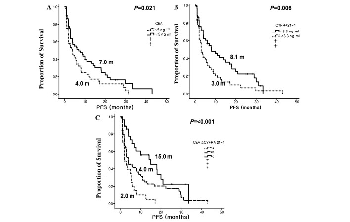
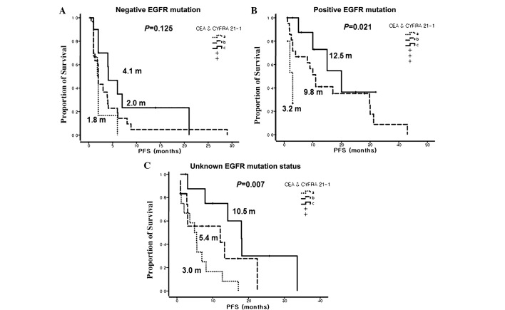
Patients with a h-CEA level had significantly better PFS than those with a l-CEA level (7.0 vs. 4.0 months, P=0.021). In contrast, patients with a l-CYFRA 21-1 level also had significantly better PFS than those with h-CYFRA 21-1 (8.1 vs. 3.0 months, P=0.006). When subgrouped by combined CEA and CYFRA 21-1 levels, the three groups showed significantly different PFS, and group C showed the longest PFS among the three groups (15.0 vs. 4.0 vs. 2.0 months, P<0.001, for groups C, B and A, respectively) (Fig. 1). Particularly, group C had the longest PFS among the patients with squamous cell carcinoma (Fig. 2). In addition, a h-CEA and a l-CYFRA 21-1 level was a significant prognostic marker, not only in patients with EGFR-mutant tumors, but in patients with an unknown EGFR mutation status (Fig. 3). Finally, univariate analysis revealed several significant factors for PFS including good ECOG PS (6.1 vs. 3.0 months, P=0.016) and positive EGFR mutation status (11.0 vs. 2.0 months, P<0.001) (Table III).
Figure 1.
Progression-free survival (PFS) curves according to pre-treatment serum levels of carcinoembryonic antigen (CEA) and cytokeratin-19 fragments (CYFRA 21-1). (A) CEA. (B) CYFRA 21-1. (C) Combinations of CEA and CYFRA 21-1 by group: (a) patients with l-CEA and h-CYFRA 21-1, (b) patients with l-CEA and l-CEA or h-CEA and h-CYFRA 21-1 and (c) patients with a h-CEA and l-CYFRA 21-1.
Figure 2.
Progression-free survival (PFS) curves according to pre-treatment serum levels of a combination of carcinoembryonic antigen (CEA) and cytokeratin-19 fragments (CYFRA 21-1) according to histologic type. (a) Patients with l-CEA and h-CYFRA 21-1, (b) patients with l-CEA and l-CYFRA 21-1 or h-CEA and h-CYFRA 21-1 and (c) patients with h-CEA and l-CYFRA 21-1. (A) Patients with adenocarcinoma. (B) Patients with squamous cell carcinoma.
Figure 3.
Progression-free survival (PFS) curves according to pre-treatment serum levels of a combination of carcinoembryonic antigen (CEA) and cytokeratin-19 fragments (CYFRA 21-1) according to epidermal growth factor receptor (EGFR) mutation status. (a) Patients with l-CEA and h-CYFRA 21-1, (b) patients with l-CEA and l-CYFRA 21-1 or h-CEA and h-CYFRA 21-1 and (c) patients with h-CEA and l-CYFRA 21-1. (A) Patients with a negative EGFR mutation. (B) Patients with a positive EGFR mutation. (C) Patients with unknown EGFR mutation status.
Table III.
Univariate predictions of survival.
| PFS
|
OS
|
|||||
|---|---|---|---|---|---|---|
| Category | Median (months) | 95% CI | P-value | Median (months) | 95% CI | P-value |
| Total | 5.0 | 3.3–6.7 | 16.0 | 8.7–23.3 | ||
| Age (years) | ||||||
| <65 | 4.1 | 2.0–6.2 | 0.982 | 15.1 | 10.5–19.7 | 0.843 |
| ≥65 | 5.6 | 2.8–8.4 | 22.0 | 5.5-38-5 | ||
| Gender | ||||||
| Male | 4.1 | 2.7–5.6 | 0.985 | 15.1 | 9.3–20.9 | 0.902 |
| Female | 6.0 | 2.5–9.50 | 18.1 | 5.7–30.6 | ||
| Histologic type | ||||||
| Adenocarcinoma | 5.6 | 3.3–7.8 | 0.942 | 18.1 | 0.0–36.3 | 0.716 |
| Non-adenocarcinoma | 4.1 | 1.2–7.0 | 16.0 | 8.3–23.7 | ||
| Performance status | ||||||
| 0–1 | 6.1 | 3.2–9.0 | 0.016 | 29.6 | 19.9–39.4 | <0.001 |
| 2 | 3.0 | 1.0–5.0 | 6.1 | 1.7–10.6 | ||
| Smoking history | ||||||
| None | 5.0 | 2.8–7.2 | 0.331 | 16.0 | 7.0–25.0 | 0.780 |
| Current + former | 4.9 | 3.1–6.7 | 14.1 | 1.7–26.5 | ||
| No. of prior regimens | ||||||
| 0–1 | 8.1 | 3.2–13.0 | 0.176 | 29.6 | 3.3–55.3 | 0.447 |
| ≥ 2 | 4.0 | 2.0–6.0 | 15.0 | 9.1–20.9 | ||
| TKI | ||||||
| Gefitinib | 5.6 | 3.6–7.5 | 0.679 | 16.0 | 8.9–23.1 | 0.935 |
| Erlotinib | 3.9 | 1.5–6.3 | 24.0 | 13.2–34.9 | ||
| Serum CEA level (ng/ml) | ||||||
| <5 | 4.0 | 1.7–6.3 | 0.021 | 14.0 | 2.2–25.8 | 0.505 |
| ≥5 | 7.0 | 2.5–11.5 | 18.0 | 10.3–25.7 | ||
| Serum CYFRA 21-1 level (ng/ml) | ||||||
| <3.3 | 8.1 | 2.9–13.3 | 0.006 | NR | <0.001 | |
| ≥3.3 | 3.0 | 2.0–4.0 | 8.0 | 5.2–10.8 | ||
| Combination of CEA and CYFRA 21-1 (ng/ml) | ||||||
| CEA ≥5 and CYFRA 21-1 <3.3 | 15.0 | 5.7–24.3 | <0.001 | NR | 0.002 | |
| CEA <5 and CYFRA 21-1 <3.3 or | 4.0 | 3.1–4.9 | 14.1 | 5.0–23.2 | ||
| CEA ≥5 and CYFRA 21-1 ≥3.3 | ||||||
| CEA <5 and CYFRA 21-1 >3.3 | 2.0 | 0.9–3.1 | 8.0 | 4.8–11.2 | ||
| EGFR mutation (n=84) | ||||||
| Negative | 2.0 | 1.4–2.7 | <0.001 | 7.1 | 3.9–10.3 | 0.038 |
| Positive | 11.0 | 5.3–16.7 | 22.0 | 13.1–31.0 | ||
PFS, progression-free survival; OS, overall survival; TKI, tyrosine kinase inhibitor; CEA, carcinoembryonic antigen; CYFRA 21-1, cytokeratin-19 fragments; NR, not reached; EGFR, epidermal growth factor receptor.
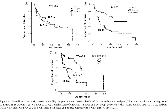
Patients with good ECOG PS and a positive EGFR mutation status also had significantly longer OS than those who had a poor ECOG PS and a negative EGFR mutation status (ECOG PS, 29.6 vs. 6.1 months, P<0.001; EGFR mutation status, 22.0 vs. 7.1 months, P=0.038, respectively). However, OS did not differ according to pre-treatment CEA levels. Patients with a l-CYFRA 21-1 level had a longer OS than those with h-CYFRA 21-1 (not reached vs. 8.0 months, P<0.001). Patients in group C also had the longest OS among the three groups (Table IV and Fig. 4).
Table IV.
Multivariate predictions of survival.
| PFS
|
OS
|
|||||
|---|---|---|---|---|---|---|
| Category | Hazard ratio | 95% CI | P-value | Hazard ratio | 95% CI | P-value |
| Age (years) | ||||||
| <65 vs. ≥65 | 1.23 | 0.67–2.28 | 0.506 | 1.18 | 0.64–2.29 | 0.633 |
| Gender | ||||||
| Female vs. male | 2.86 | 0.75–10.89 | 0.124 | 1.24 | 0.27–6.75 | 0.808 |
| Histologic type | ||||||
| Non-adeno vs. adeno | 0.80 | 0.41–1.56 | 0.521 | 1.59 | 0.81–3.14 | 0.182 |
| Clinical stage | ||||||
| IV vs. III | 0.64 | 0.35–1.15 | 0.134 | 0.80 | 0.40–1.60 | 0.534 |
| Performance status | ||||||
| 0–1 vs. 2 | 2.02 | 1.13–3.61 | 0.017 | 2.13 | 1.14–3.98 | 0.018 |
| Smoking history | ||||||
| None vs. current + former | 1.48 | 0.39–5.56 | 0.57 | 1.38 | 0.25–7.61 | 0.706 |
| Serum CEA level (ng/ml) | ||||||
| <5 vs. ≥5 | 0.41 | 0.24–0.78 | 0.007 | 0.55 | 0.25–1.21 | 0.554 |
| Serum CYFRA 21-1 level (ng/ml) | ||||||
| <3.3 vs. ≥3.3 | 1.93 | 1.09–3.44 | 0.025 | 2.76 | 1.38–5.53 | 0.004 |
| EGFR mutation (n=84) | ||||||
| Negative vs. positive | 0.22 | 0.11–0.42 | <0.001 | 0.53 | 0.28–1.004 | 0.051 |
PFS, progression-free survival; OS, overall survival; TKI, tyrosine kinase inhibitor; CEA, carcinoembryonic antigen; CYFRA 21-1, cytokeratin-19 fragments; EGFR, epidermal growth factor receptor.
Figure 4.
Overall survival (OS) curves according to pre-treatment serum levels of carcinoembryonic antigen (CEA) and cytokeratin-19 fragments (CYFRA 21-1). (A) CEA. (B) CYFRA 21-1. (C) Combinations of CEA and CYFRA 21-1 by group: (a) patients with l-CEA and h-CYFRA 21-1, (b) patients with l-CEA and l-CYFRA 21-2 or h-CEA and h-CYFRA 21-1 and (c) patients with h-CEA and l-CYFRA 21-1.
Multivariate analysis using a Cox proportional hazards model indicated that a good ECOG PS, positive EGFR mutation status, high pre-treatment CEA levels, and low pre-treatment CYFRA 21-1 levels are independent predictive factors for PFS. Meanwhile, predictive factors for OS included a good ECOG PS, positive EGFR mutation status and l-CYFRA 21-1, but not h-CEA (Table IV).
Discussion
Detection of a mutation in the EGFR gene in NSCLC patients treated with an EGFR TKI is the most important factor for the prediction of a good response to these drugs (4). However, the detection of an EGFR mutation may be difficult due to the limited amount of available tissue (2–4). Therefore, a surrogate biomarker that can improve the prediction of response to these targeted drugs is needed.
CEA was first described by Gold and Freedman in 1965 as an antigen expressed by gastrointestinal carcinoma cells (17). Although CEA was often falsely elevated in smokers and in patients with restrictive or obstructive pulmonary disease (18–20), abnormally elevated CEA levels were reported in 30–70% of patients with NSCLC and were most frequently observed in patients with adenocarcinoma and advanced stage carcinoma (21). In addition, several studies have shown that h-CEA is a potential marker of poor prognosis in NSCLC regardless of treatment (7,21).
On the contrary, Okamoto et al (11) reported that patients treated with EGFR TKIs with high pre-treatment levels of CEA had a longer survival and a better response than those with l-CEA. They attributed this to a possible anti-apoptotic signal of the mutant EGFR pathway that may elevate the expression level of CEA protein. Our data are similar to the data of Okamato et al (11). Shoji et al (22) reported that the rate of the EGFR gene mutation significantly increased as the serum CEA level increased (for serum CEA levels of <5, ≥5 but <20, and ≥20, the rate of the EGFR gene mutation was 35, 55 and 87.5%, respectively; P=0.040). However, our data showed that the status of the EGFR mutation made no difference in the CEA levels. Based on previous reports, the function of CEA has not been elucidated but may include the following: i) CEA is a cell surface adhesion protein and may play a role in cell-to-cell adhesion (23); ii) overexpression of CEA is thought to play a role in tumorigenesis (24); iii) CEA has a dominant effect in blocking differentiation, and it also cooperates with Myc and Bcl-2 in cellular transformation (25); and iv) it can inhibit cell death induced by a loss of anchorage to the extracellular matrix (anoikis) (26). Although these findings suggest that CEA has anti-apoptotic effects in cancer cells, a direct relationship between h-CEA and response to EGFR TKIs has not yet been established.
CYFRA 21-1, a fragment of cytokeratin subunit 19, was first identified in 1993 as a valuable marker in lung cancer patients (27). CYFRA 21-1 was found to be associated with TNM stage and ECOG PS, reflecting an unfavorable prognosis for NSCLC patients regardless of treatment (8,21,28–30). In our study, patients with a poor ECOG PS had a higher CYFRA 21-1 level than patients with a good ECOG PS (6.4 vs. 3.0 ng/ml; P=0.03). Patients with h-CYFRA 21-1 levels were more likely to have a history of smoking; however, this association was not significant (P=0.072). Previous studies have also reported that smoking has no effect on serum CYFRA 21-1 levels (31,32). Univariate and multivariate analyses demonstrated that CYFRA 21-1 levels higher than 3.3 ng/ ml had an independent negative impact on PFS (HR=1.93, 95% CI 1.09–3.44; P=0.025) and OS (HR=2.76, 95% CI 1.38–5.53; P=0.0004). Therefore, CYFRA 21-1 is an independent marker for poor prognosis in NSCLC patients receiving an EGFR TKI, which is consistent with a previous study (12).
We demonstrated that pre-treatment levels of CEA and CYFRA 21-1 serve as prognostic and predictive markers in NSCLC patients treated with gefitinib or erlotinib. Patients with a high pre-treatment CEA level showed better responses and longer PFS, and patients with a low pre-treatment CYFRA 21-1 level showed longer PFS and OS. In addition, the prediction accuracy of the EGFR TKI response and prognosis improved when all patients were divided into three groups according to combined levels of CEA and CYFRA 21-1.
It is difficult to predict high efficacy of EGFR TKIs when they are used in patients with non-adenocarcinoma histology since the incidence of EGFR mutation is extremely rare in these tumors (33). However, the present study revealed that CEA and CYFRA 21-1 levels can also be prognostic markers in patients with squamous cell carcinoma or patients with unknown EGFR mutation status (Figs. 2 and 3).
In conclusion, pre-treatment serum levels of CEA and CYFRA 21-1 are simple and easy to detect, and can serve as predictive and prognostic factors for advanced NSCLC patients being treated with EGFR TKIs, particularly in patients with squamous cell carcinoma or patients with an unknown EGFR mutation status.
Acknowledgments
This study was supported by a faculty research grant of Yonsei University College of Medicine for 6-2007-0194.
References
- 1.Jemal A, Siegel R, Ward E, et al. Cancer statistics, 2008. CA Cancer J Clin. 2008;58:71–96. doi: 10.3322/CA.2007.0010. [DOI] [PubMed] [Google Scholar]
- 2.Shepherd FA, Rodrigues Pereira J, Ciuleanu T, et al. Erlotinib in previously treated non-small-cell lung cancer. N Engl J Med. 2005;353:123–132. doi: 10.1056/NEJMoa050753. [DOI] [PubMed] [Google Scholar]
- 3.Kim ES, Hirsh V, Mok T, et al. Gefitinib versus docetaxel in previously treated non-small-cell lung cancer (INTEREST): a randomised phase III trial. Lancet. 2008;372:1809–1818. doi: 10.1016/S0140-6736(08)61758-4. [DOI] [PubMed] [Google Scholar]
- 4.Mok TS, Wu Y, Thongprasert S, et al. Gefitinib or carboplatin-paclitaxel in pulmonary adenocarcinoma. N Engl J Med. 2009;361:947–957. doi: 10.1056/NEJMoa0810699. [DOI] [PubMed] [Google Scholar]
- 5.Maheswaran S, Sequist LV, Nagrath S, et al. Detection of mutations in EGFR in circulating lung-cancer cells. N Engl J Med. 2008;359:366–377. doi: 10.1056/NEJMoa0800668. [DOI] [PMC free article] [PubMed] [Google Scholar]
- 6.Molina R, Auge JM, Escudero JM, et al. Mucins CA 125, CA 19.9, CA 15.3 and TAG-72.3 as tumor markers in patients with lung cancer: comparison with CYFRA 21-1, CEA, SCC and NSE. Tumour Biol. 2008;29:371–380. doi: 10.1159/000181180. [DOI] [PubMed] [Google Scholar]
- 7.Okada M, Nishio W, Sakamoto T, et al. Prognostic significance of perioperative serum carcinoembryonic antigen in non-small cell lung cancer: analysis of 1,000 consecutive resections for clinical stage I disease. Ann Thorac Surg. 2004;78:216–221. doi: 10.1016/j.athoracsur.2004.02.009. [DOI] [PubMed] [Google Scholar]
- 8.Pujol JL, Boher JM, Grenier J, Quantin X. CYFRA 21-1, neuron specific enolase and prognosis of non-small cell lung cancer: prospective study in 621 patients. Lung Cancer. 2001;31:221–231. doi: 10.1016/s0169-5002(00)00186-0. [DOI] [PubMed] [Google Scholar]
- 9.Holdenrieder S, von Pawel J, Dankelmann E, et al. Nucleosomes and CYFRA 21-1 indicate tumor response after one cycle of chemotherapy in recurrent non-small cell lung cancer. Lung Cancer. 2009;63:128–135. doi: 10.1016/j.lungcan.2008.05.001. [DOI] [PubMed] [Google Scholar]
- 10.Ardizzoni A, Cafferata MA, Tiseo M, et al. Decline in serum carcinoembryonic antigen and cytokeratin 19 fragment during chemotherapy predicts objective response and survival in patients with advanced non-small cell lung cancer. Cancer. 2006;107:2842–2849. doi: 10.1002/cncr.22330. [DOI] [PubMed] [Google Scholar]
- 11.Okamoto T, Nakamura T, Ikeda J, et al. Serum carcinoembryonic antigen as a predictive marker for sensitivity to gefitinib in advanced non-small cell lung cancer. Eur J Cancer. 2005;41:1286–1290. doi: 10.1016/j.ejca.2005.03.011. [DOI] [PubMed] [Google Scholar]
- 12.Barlsi F, Tchouhadjian C, Doddoli C, Torre JP, Astoul P, Kleisbauer JP. CYFRA 21-1 level predicts survival in non-small cell lung cancer patients receiving gefitinib as third-line therapy. Br J Cancer. 2005;92:13–14. doi: 10.1038/sj.bjc.6602296. [DOI] [PMC free article] [PubMed] [Google Scholar]
- 13.Chiu C, Shih Y, Tsai C, Liou J, Chen Y, Perng R. Serum tumor markers as predictors for survival in advanced non-small cell lung cancer patients treated with gefitinib. Lung Cancer. 2007;57:213–221. doi: 10.1016/j.lungcan.2007.02.016. [DOI] [PubMed] [Google Scholar]
- 14.Travis WD, Colby TV, Corrin B, Shimosato Y, Brambilla E, editors. Histological Typing of Lung and Pleural Tumours. 3rd edition. Springer-Verlag; Berlin: 1999. [Google Scholar]
- 15.Therasse P, Arbuck SG, Eisenhauer EA, et al. New guidelines to evaluate the response to treatment in solid tumors. European Organization for Research and Treatment of Cancer, National Cancer Institute of the United States, National Cancer Institute of Canada. J Natl Cancer Inst. 2000;92:205–216. doi: 10.1093/jnci/92.3.205. [DOI] [PubMed] [Google Scholar]
- 16.Han S, Kim T, Hwang PG, et al. Predictive and prognostic impact of epidermal growth factor receptor mutation in non-small-cell lung cancer patients treated with gefitinib. J Clin Oncol. 2005;23:2493–2501. doi: 10.1200/JCO.2005.01.388. [DOI] [PubMed] [Google Scholar]
- 17.Gold P, Freedman SO. Demonstration of tumor-specific antigens in human colonic carcinomata by immunological tolerance and absorption techniques. J Exp Med. 1965;121:439–462. doi: 10.1084/jem.121.3.439. [DOI] [PMC free article] [PubMed] [Google Scholar]
- 18.Fujishima T, Honda Y, Shijubo N, Takahashi H, Abe S. Increased carcinoembryonic antigen concentrations in sera and bronchoalveolar lavage fluids of patients with pulmonary alveolar proteinosis. Respiration. 1995;62:317–321. doi: 10.1159/000196473. [DOI] [PubMed] [Google Scholar]
- 19.Rule AH, Straus E, Vandevoorde J, Janowitz HD. Tumor-associated (CEA-reacting) antigen in patients with inflammatory bowel disease. N Engl J Med. 1972;287:24–26. doi: 10.1056/NEJM197207062870106. [DOI] [PubMed] [Google Scholar]
- 20.Okada M, Nishio W, Sakamoto T, et al. Effect of histologic type and smoking status on interpretation of serum carcinoembryonic antigen value in non-small cell lung carcinoma. Ann Thorac Surg. 2004;78:1004–1009. doi: 10.1016/j.athoracsur.2004.03.019. [DOI] [PubMed] [Google Scholar]
- 21.Matsuoka K, Sumitomo S, Nakashima N, Nakajima D, Misaki N. Prognostic value of carcinoembryonic antigen and CYFRA21-1 in patients with pathological stage I non-small cell lung cancer. Eur J Cardiothorac Surg. 2007;32:435–439. doi: 10.1016/j.ejcts.2007.05.014. [DOI] [PubMed] [Google Scholar]
- 22.Shoji F, Yoshino I, Yano T, et al. Serum carcinoembryonic antigen level is associated with epidermal growth factor receptor mutations in recurrent lung adenocarcinomas. Cancer. 2007;110:2793–2798. doi: 10.1002/cncr.23101. [DOI] [PubMed] [Google Scholar]
- 23.Benchimol S, Fuks A, Jothy S, Beauchemin N, Shirota K, Stanners CP. Carcinoembryonic antigen, a human tumor marker, functions as an intercellular adhesion molecule. Cell. 1989;57:327–334. doi: 10.1016/0092-8674(89)90970-7. [DOI] [PubMed] [Google Scholar]
- 24.Hammarström S. The carcinoembryonic antigen (CEA) family: structures, suggested functions and expression in normal and malignant tissues. Semin Cancer Biol. 1999;9:67–81. doi: 10.1006/scbi.1998.0119. [DOI] [PubMed] [Google Scholar]
- 25.Screaton RA, Penn LZ, Stanners CP. Carcinoembryonic antigen, a human tumor marker, cooperates with Myc and Bcl-2 in cellular transformation. J Cell Biol. 1997;137:939–952. doi: 10.1083/jcb.137.4.939. [DOI] [PMC free article] [PubMed] [Google Scholar]
- 26.Ordoez C, Screaton RA, Ilantzis C, Stanners CP. Human carcinoembryonic antigen functions as a general inhibitor of anoikis. Cancer Res. 2000;60:3419–3424. [PubMed] [Google Scholar]
- 27.Stieber P, Bodenmller H, Banauch D, et al. Cytokeratin 19 fragments: a new marker for non-small cell lung cancer. Clin Biochem. 1993;26:301–304. doi: 10.1016/0009-9120(93)90129-t. [DOI] [PubMed] [Google Scholar]
- 28.Nisman B, Lafair J, Heching N, et al. Evaluation of tissue polypeptide-specific antigen, CYFRA 21-1, and carcinoembryonic antigen in nonsmall cell lung carcinoma: Does the combined use of cytokeratin markers give any additional information? Cancer. 1998;82:1850–1859. doi: 10.1002/(sici)1097-0142(19980515)82:10<1850::aid-cncr6>3.0.co;2-r. [DOI] [PubMed] [Google Scholar]
- 29.Barlsi F, Gimenez C, Torre J, et al. Prognostic value of combination of CYFRA 21-1, CEA and NSE in patients with advanced non-small cell lung cancer. Respir Med. 2004;98:357–362. doi: 10.1016/j.rmed.2003.11.003. [DOI] [PubMed] [Google Scholar]
- 30.Pujol JL, Molinier O, Ebert W, et al. CYFRA 21-1 is a prognostic determinant in non-small-cell lung cancer: results of a meta-analysis in 2063 patients. Br J Cancer. 2004;90:2097–2105. doi: 10.1038/sj.bjc.6601851. [DOI] [PMC free article] [PubMed] [Google Scholar]
- 31.Kao CH, Hsieh JF, Ho YJ, Tsai SC, Lee JK. Cytokeratin fragment 19 (CYFRA 21-1) in healthy smokers. Anticancer Res. 1999;19:4545–4546. [PubMed] [Google Scholar]
- 32.Molina R, Filella X, Aug JM, et al. Tumor markers (CEA, CA 125, CYFRA 21-1, SCC and NSE) in patients with non-small cell lung cancer as an aid in histological diagnosis and prognosis. Comparison with the main clinical and pathological prognostic factors. Tumour Biol. 2003;24:209–218. doi: 10.1159/000074432. [DOI] [PubMed] [Google Scholar]
- 33.Shigematsu H, Lin L, Takahashi T, et al. Clinical and biological features associated with epidermal growth factor receptor gene mutations in lung cancers. J Natl Cancer Inst. 2005;97:339–346. doi: 10.1093/jnci/dji055. [DOI] [PubMed] [Google Scholar]


