

Abstract
Multilocular thymic cysts with follicular hyperplasia are uncommon masses that occur in Human Immunodeficiency Virus (HIV) positive individuals. These cysts mostly present in HIV positive children. Here we report a rare case of multilocular thymic cyst in an HIV positive adult female. In this case report, the radiologic findings of multilocular thymic cyst, management and prognosis are discussed.
Keywords: AIDS, follicular hyperplasia, HIV, multilocular thymic cyst, thymus
INTRODUCTION

Xiao Shi, MD
Thymic cysts account for 1% to 3% of anterior mediastinal masses and can be divided into two types - congenital cysts and acquired cysts.[1] Acquired thymic cysts, unlike congenital cysts, are multilocular and arise as a reactive process secondary to inflammation or malignancy.[1] Multilocular thymic cysts (MTCs) have been described in association with Human Immunodeficiency Virus (HIV), however majority of cases described involve the pediatric population.[2–6] The few adult cases that have been reported involve only male patients.[6,7] Here we report a rare case of multilocular thymic cyst with follicular hyperplasia in an HIV positive adult female, with its imaging features and differential diagnosis.
CASE REPORT
A 47-year-old female who has been seropositive for HIV for 19 years, presented with a five-day history of cough, chest pain, and asymptomatic bilateral swelling of the cheek. On physical examination, patient was afebrile, and vital signs were within normal limits. There were mild bibasilar crackles in the lungs, bilateral parotid enlargement, and a palpable left superior cervical lymph node approximately 3 centimeters in size. Laboratory evaluation revealed CD4 count of 551 K/mm[3], and HIV-1 RNA load of 16784 copies/ ml. Chest X-ray demonstrated an abnormal contour along the right cardiomediastinal border [Figure 1]. Subsequent computed tomography (CT) of the chest with contrast revealed a 7.1 × 2.7 × 8.8 cm lobulated low-attenuation mass with heterogeneous enhancement in the anterior mediastinum [Figures 2, 3]. The mediastinal lesion did not show any evidence of invasion into adjacent mediastinal structures. The mass was made up of non-enhancing cysts separated by enhancing septations. The average density of the mass on contrast CT was 14 HU (Hounsfield units) in the cystic regions and 43 HU in the septations. Surgical pathology following thymectomy revealed a multilocular, predominantly cystic structure, filled with turbid yellow to hemorrhagic colloidal material [Figure 4]. Walls of the cystic mass contained abundant lymphoid tissue with follicular hyperplasia and a prominent reactive germinal center, leading to the diagnosis of multilocular thymic cyst (MTC) with follicular hyperplasia [Figure 5]. CT scan of the neck revealed bilateral enlargement of the parotid glands with innumerable cystic areas [Figure 6]. Given the known association with HIV, the parotid lesions were diagnosed clinically as benign lymphoepithelial cysts of the parotid gland, and biopsy was deferred.
Figure 1.
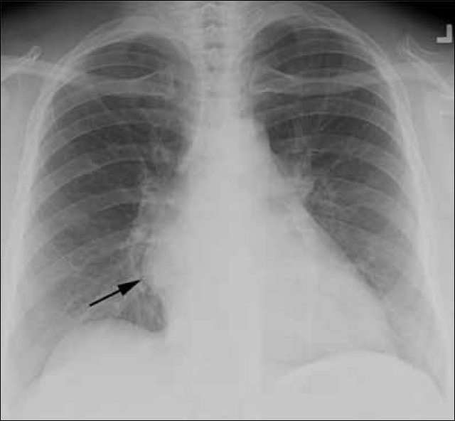
Multilocular thymic cyst with follicular hyperplasia in a 47- year-old HIV+ female with cough and chest pain. Postero-anterior chest radiograph demonstrates an abnormal contour along the right cardiomediastinal border (arrow).
Figure 2.
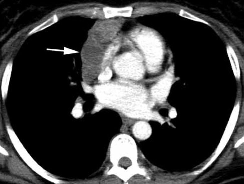
Multilocular thymic cyst with follicular hyperplasia in a 47 year-old HIV+ female with an anterior mediastinal mass. Axial contrast enhanced chest CT at the level of the heart shows a 7.1 × 2.7 × 8.8 cm lobular lowattenuation mass with heterogeneous enhancement draped across the anterior mediastinum (arrow).
Figure 3.
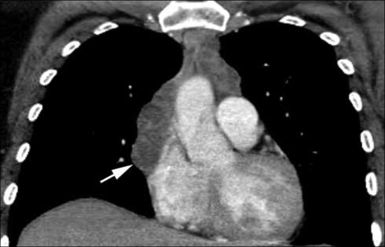
Multilocular thymic cyst with follicular hyperplasia in a 47 year-old HIV+ female with an anterior mediastinal mass. Coronal contrast enhanced chest CT shows low-attenuation cystic areas and enhancing septations (arrow).
Figure 4.
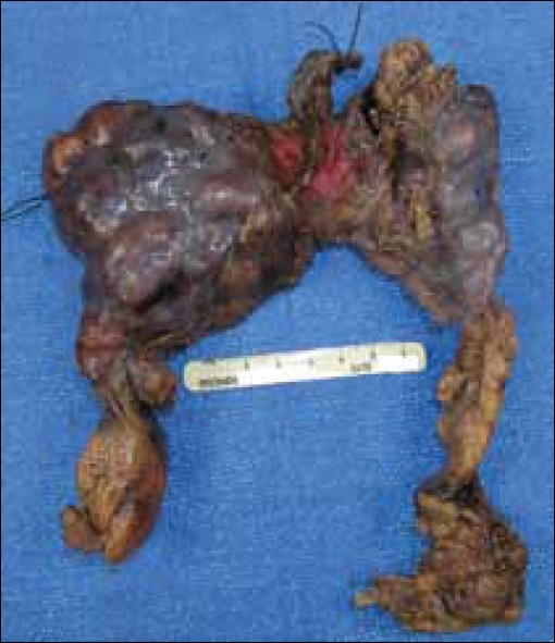
Multilocular thymic cyst with follicular hyperplasia. Gross thymectomy specimen from a 47- year-old HIV+ female weighs 180 gram and measures 14.5 cm from medial to lateral, 15 cm from superior to inferior and up to 3.5 cm from anterior to posterior. The gland is very lobulated in appearance with a moderate amount of attached adipose tissue. The gland appears encapsulated with a smooth and glistening pink-purple surface.
Figure 5.
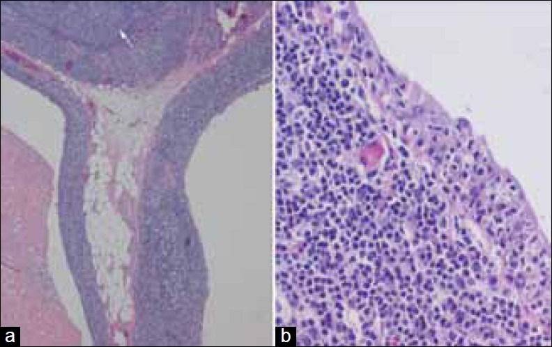
Multilocular thymic cyst with follicular hyperplasia. Hematoxylin and Eosin staining of thymectomy specimen from a 47 year-old HIV+ female shows (a) Two adjacent cysts (left and right) and a large lymphoid follicle with a germinal center (arrow) (2×). (b) Cyst wall with columnar epithelium heavily infiltrated by lymphocytes (20×).
Figure 6.
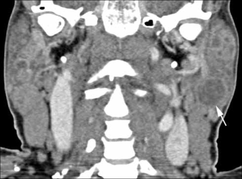
Benign lymphoepithelial cysts in a 47- year-old HIV+ female with bilaterally enlarged parotid glands. Coronal contrast enhanced neck CT demonstrates innumerable hypo dense foci, the largest one measuring 1.8 cm in the tail of the left parotid (arrow).
DISCUSSION
Multilocular thymic cysts with follicular hyperplasia are an uncommon reactive process to HIV infection found in children, with few earlier case reported in adults[2–4,6,7] Furthermore, previous case reports and case series note a male predominance when describing MTCs in patients with HIV.[2–9] MTCs are mostly benign lesions in HIV positive individuals, and malignant transformation is rare. Foci of thymoma and thymic carcinoma have been found in resected MTCs in HIV negative individuals,[1] but in HIV positive patients, only one recent case report has noted a thymic carcinoma in a resolving multilocular thymic cyst.[8] MTCs can exhibit spontaneous reduction in volume, although no long-term follow-up studies have been performed to see if these masses fully resolve.[2,3] Surgical excision is curative, and while MTCs in HIV negative patients have been shown to recur after excision, cyst recurrence has not been noted in HIV positive individuals.[1–9]
In HIV positive patients, multilocular thymic cysts are generally found incidentally on chest radiographs, and are better characterized on CT. Maximum diameter of this multiloculated entity ranges from 8 cm to 17 cm, with individual cysts that can range in size from 1 cm to 6 cm.[3,6,7,9] The mass is difficult to assess on ultrasound due to the lack of an appropriate acoustic window.[3] On magnetic resonance imaging (MRI), the septa demonstrate enhancement on post-gadolinium T1 weighted imaging, and are hypointense relative to the cystic regions on T2 weighted spin echo and Short TI Inversion Recovery (STIR) sequences.[3]
On histopathology, multilocular thymic cyst walls are lined with squamous, cuboidal or columnar epithelium (sometimes ciliated), and the septa contain numerous normal Hassall corpuscles and hyperplastic lymphoid follicles with well-formed germinal centers.[1] Suster and Rosai note the histologic appearance of MTCs strongly resemble that of benign lymphoepithelial cysts seen in parotid glands of HIV positive individuals,[1] and Leonidas et al., subsequently report a case series of three HIV positive MTC patients in which all patients had prior history of parotid gland enlargement.[6]
In our patient, CT scan of the neck demonstrated bilateral enlargement of the parotid glands with innumerable hypodense foci [Figure 6]. Parotid masses were clinically diagnosed as benign lymphoepithelial cysts, given the known association with HIV, and biopsy of the parotid lesion was not performed. The likely presence of the benign lymphoepithelial cysts in the parotid gland supports the proposed link between benign lymphoepithelial cysts of the parotid and MTCs with follicular hyperplasia.
The differential diagnosis of an anterior mediastinal mass in an HIV positive individual generally consists of infectious and neoplastic etiologies. In immunosuppressed individuals with CD4 counts less than 200 K/mm[3], tuberculosis will commonly present with low attenuation mediastinal lymphadenopathy characterized by peripheral rim enhancement on contrast enhanced CT.[10] However, the CD4 count was 410 K/mm[3] in our patient. Mycobacterium avium complex can have a similar presentation, but the chest is rarely the sole site of disease for this entity, and it is also associated with CD4 counts less than 100.[10] Bartonella causes a marked increase in vascularity leading to enlarged lymph nodes that enhance with contrast on CT examination, but in general will not appear cystic in nature and will usually be associated with pulmonary nodules.[10] In the neoplastic category, non-Hodgkin's lymphoma (typically a high grade B-cell lymphoma) occurs at an increased frequency in HIV individuals and can be associated with mediastinal lymphadenopathy. However, generally non-Hodgkin's lymphoma in HIV patients presents as an extra nodal disease when the chest is involved.[10] Kaposi's sarcoma can affect mediastinal lymph nodes as well, although it more commonly presents as ill-defined pulmonary nodules.[10]
In conclusion, a low-attenuation anterior mediastinal mass in an HIV positive adult should raise the suspicion of multilocular thymic cyst with follicular hyperplasia, even though this entity is more common in the pediatric population.
Footnotes
Available FREE in open access from: http://www.clinicalimagingscience.org/text.asp?2012/2/1/55/100379
Source of Support: Nil
Conflict of Interest: None declared.
REFERENCES
- 1.Suster S, Rosai J. Multilocular thymic cyst: An acquired reactive process: Study of 18 cases. Am J Surg Pathol. 1991;15:388–98. [PubMed] [Google Scholar]
- 2.Kontny HU, Sleasman JW, Kingma DW, Jaffe ES, Avila NA, Pizzo PA, et al. Multilocular thymic cysts in children with human immunodeficiency virus infection: Clinical and pathologic aspects. J Pediatr. 1997;131:264–70. doi: 10.1016/s0022-3476(97)70164-1. [DOI] [PubMed] [Google Scholar]
- 3.Avila NA, Mueller BU, Carrasquillo JA, Kontny HU, Jaffe ES, Pizzo PA. Multilocular thymic cysts: Imaging features in children with human immunodeficiency virus infection. Radiology. 1996;201:130–4. doi: 10.1148/radiology.201.1.8816533. [DOI] [PubMed] [Google Scholar]
- 4.Shalaby-Rana E, Selby D, Ivy P, Schutzbank T, Chan M, Chandra R, et al. Multilocular thymic cyst in a child with acquired immunodeficiency syndrome. Pediatr Infect Dis J. 1996;15:83–6. doi: 10.1097/00006454-199601000-00018. [DOI] [PubMed] [Google Scholar]
- 5.Mishalani SH, Lones MA, Said JW. Multilocular thymic cyst. A novel thymic lesion associated with human immunodeficiency virus infection. Arch Pathol Lab Med. 1995;119:467–70. [PubMed] [Google Scholar]
- 6.Leonidas JC, Berdon WE, Valderrama E, Neveling U, Schuval S, Weiss SJ, et al. Human immunodeficiency virus infection and multilocular thymic cysts. Radiology. 1996;198:377–9. doi: 10.1148/radiology.198.2.8596835. [DOI] [PubMed] [Google Scholar]
- 7.Chhieng DC, Demaria S, Yee HT, Yang GC. Multilocular thymic cyst with follicular lymphoid hyperplasia in a male infected with HIV. A case report with fine needle aspiration cytology. Acta Cytol. 1999;43:1119–23. doi: 10.1159/000331364. [DOI] [PubMed] [Google Scholar]
- 8.McDonald M, Mclean T, Belhorn T, Smith SV, Fordham LA, Woods C, et al. Thymic carcinoma in a child with HIV infection. Pediatr Blood Cancer. 2007;49:1004–7. doi: 10.1002/pbc.20694. [DOI] [PubMed] [Google Scholar]
- 9.Tamagno M, Bibas BJ, Bernardi F, Lian YC, Bammann RH, Fernandez A, et al. Giant multilocular thymic cyst in an HIV-infected adolescent. J Pediatr Surg. 2011;46:1842–5. doi: 10.1016/j.jpedsurg.2011.06.009. [DOI] [PubMed] [Google Scholar]
- 10.Padley SP, King LJ. Computed tomography of the thorax in HIV disease. Eur Radiol. 1999;10:1932–8. doi: 10.1007/s003300050884. [DOI] [PubMed] [Google Scholar]