Abstract
The roles of vitamin A (VA) in the development of metabolic diseases remain unanswered. We have reported that retinoids synergized with insulin to induce the expression of sterol-regulatory element-binding protein 1c gene (Srebp-1c) expression in primary rat hepatocytes. Additionally, the hepatic Srebp-1c expression is elevated in Zucker fatty (ZF) rats, and reduced in those fed a VA deficient diet. VA is metabolized to retinoic acid (RA) for regulating gene expression. We hypothesized that the expression of RA production enzymes contributes to the regulation of the hepatic Srebp-1c expression. Therefore, we analyzed their expression levels in Zucker lean (ZL) and ZF rats. The mRNA levels of retinaldehyde dehydrogenase family 1 gene (Raldh1) were found to be higher in the isolated and cultured primary hepatocytes from ZF rats than that from ZL rats. The RALDH1 protein level was elevated in the liver of ZF rats. Retinol and retinal dose- and time-dependently induced the expression of RA responsive Cyp26a1 gene in hepatocytes and hepatoma cells. INS-1 cells were identified as an ideal tool to study the effects of RA production on the regulation of gene expression because only RA, but not retinal, induced Srebp-1c mRNA expression in them. Recombinant adenovirus containing rat Raldh1 cDNA was made and used to infect INS-1 cells. The over-expression of RALDH1 introduced the retinal-mediated induction of Srebp-1c expression in INS-1 cells. We conclude that the expression levels of the enzymes for RA production may contribute to the regulation of RA responsive genes, and determine the responses of the cells to retinoid treatments. The elevated hepatic expression of Raldh1 in ZF rats may cause the excessive RA production from retinol, and in turn, result in higher Srebp-1c expression. This excessive RA production may be one of the factors contributing to the elevated lipogenesis in the liver of ZF rats.
Introduction
The high obesity prevalence in the population of the United States [1] predicts the increase of patients with noninsulin-dependent diabetes mellitus (NIDDM) [2], a major public health concern [3]. Genetic mutations, such as mutations of leptin and its receptor, have been shown to cause the development of obesity and diabetes [4]. Currently, the associations of a variety of genes with the development of human obesity or NIDDM have been indicated [5], [6]. Metabolic abnormalities are often associated with profound changes of hepatic glucose and lipid metabolism [7], which is attributed, at least in part, to the expression of genes involved in these processes [8], [9]. On the other hand, overconsumption of nutrients, such as fructose in sweetened beverages, has also been implied to play a role in the rise of obesity [10]. Dietary nutrients provide us with not only energy, but also vitamins and other essential factors with regulatory roles. The effects of individual micronutrients on the development of metabolic diseases remain to be revealed.
As an essential and fat-soluble micronutrient, vitamin A (VA, retinol) plays crucial roles in the general health of an individual, such as vision, tissue differentiation, immunity, etc [11]. The majority of the physiological actions of retinol are mediated by its active metabolite, retinoic acid (RA), which exists in multiple isomeric forms, such as all-trans RA and 9-cis RA [12]. RA regulates gene expression through the activation of two families of nuclear receptors, retinoic acid receptors (RARα, β and γ) activated by all-trans RA, and retinoid X receptors (RXRα, β and γ) activated by 9-cis RA [13].
In an attempt to understand the effects of endogenous lipophilic molecules on the insulin-regulated gene expression in primary rat hepatocytes, we have analyzed the rat liver lipophilic extract and found that retinol and retinal in it dose-dependently induced the mRNA expression levels of Pck1 [14], Gck [15], and Srebp-1c [16]. The proximal one of the two previously identified retinoic acid response elements (RAREs) in Pck1 promoter [17]–[19] mediates the RA effect in primary hepatocytes. The previously identified two liver X receptor (LXR) binding sites mediating the insulin-stimulated Srebp-1c expression [20] were also RAREs in its promoter [16], demonstrating the convergence of hormonal and nutritional responses. The hepatic expression level of cannabinoid receptor 1 (CB1), the target of anti-obesity drug rimonabant [21] can also be induced by the treatment of RA via activation of RAR-γ [22], indicating the connection between the nutritional signals and endocannabinoid pathways. The effects of VA status on glucose and lipid metabolism have been summarized [23].
Retinol is reversibly oxidized into retinal by retinol dehydrogenases (RDHs). Retinal is irreversibly oxidized into RA by retinaldehyde (aldehyde) dehydrogenases (RALDHs) [13], [24], [25]. Currently, four RALDHs have been cloned and thought to be responsible for all-trans or 9-cis RA production in various tissues [26]–[29]. RALDH1-4 proteins can be detected in some but not all hepatocytes of the mouse liver using immunohistochemistry, with RALDH1 being frequently expressed in lipid-engorged cells [27]. Raldh1 (also known as Aldh1a1) mRNA is expressed weakly in the rat liver [26], whereas Raldh4 (also known as Aldh8a1) mRNA is expressed at high level in the mouse liver [27]. The mice with Raldh1 deletion (Raldh1−/−) have reduced RA production in the liver two hours after a challenge of retinol [30]. The Raldh1−/− mice are resistant to diet-induced obesity (DIO) and insulin resistance [31]. The Imprinting Control Region (ICR) mice fed a high cholesterol diet had the elevation of the hepatic expression of RALDH1 and RALDH2 through oxysterol-induced Srebp-1c expression, demonstrating the interaction of cholesterol and RA metabolism [32]. All these observations indicate the potential roles of these enzymes in the development of obesity and insulin resistance, which deserves to be further defined.
The fact that the levels of the enzymes for the hepatic retinoid metabolism can be dynamically regulated leads us to hypothesize that the change of the expression of these enzymes and the production of RA may contribute to the regulation of the expression of hepatic lipogenic genes. Here, we examined the effects of retinol and retinal on the expression of RA responsive genes in primary rat hepatocytes, rat hepatoma cells and insulinoma INS-1 cells. We observed the elevated expression levels of Raldh1 mRNA in hepatocytes and its protein in the liver of Zucker fatty (ZF) rats. The recombinant adenovirus-mediated over-expression of RALDH1 resulted in retinal-mediated induction of Srebp-1c expression in INS-1 cells, which originally lack the response to retinal.
Results
Determination of the mRNA levels of enzymes responsible for the retinoids catabolism in primary hepatocytes, and the elevated expression of Raldh1 and its protein in hepatocytes of ZF rats
Enzymatic processes are involved in the conversions of ROL to RAL and RAL to RA [13], [24], [25]. It has been shown that the short chain dehydrogenase/reductase family 16C member 5 (SDR16C5), RDH2, RDH10, and RALDH1-4 are expressed in the liver [33]. Since RALDH4 is for the biosynthesis of 9-cis RA [27], we have focused on the expression levels of the rest enzymes in this study. The C T numbers of these genes were normalized to those of 36B4 (an invariable control gene) in the same samples. Based on −ΔCT numbers (how close the abundance of the transcripts of those genes to that of 36B4), we found that Rdh2, and Raldh1 were expressed in meaningful levels (compared with the expression level of 36B4) in both primary rat hepatocytes and HL1C cells (data not shown).
Since RDH2 catalyzes the conversion of ROL to RAL [33], and RALDH1 is reponsible for converting RAL to RA [33], we compared their expression levels in freshly isolated primary hepatocytes of ZL rats with those of ZF rats. The mRNA level of Rdh2 in hepatocytes from ZF rats was similar to that from ZL rats. However, the mRNA level of Raldh1 in hepatocytes from ZF rats was significantly higher than that from ZL rats (Fig. 1A). The results observed in cultured primary hepatocytes of ZL and ZF rats were similar to that in fresh isolated hepatocytes. We observed that the Rdh2 expression level was unchanged, and the Raldh1 expression level was elevated significantly in hepatocytes from ZF rats (Fig. 1B). The mRNA level of Raldh4 was detected, and was at the similar levels in hepatocytes from both ZL and ZF (data not shown).
Figure 1. The mRNA levels of Rdh2, and Raldh1 in freshly isolated (A) and cultured (B) primary hepatocytes, and the RALDH1 protein levels (C) in the liver of ad libitum ZL and ZF rats.
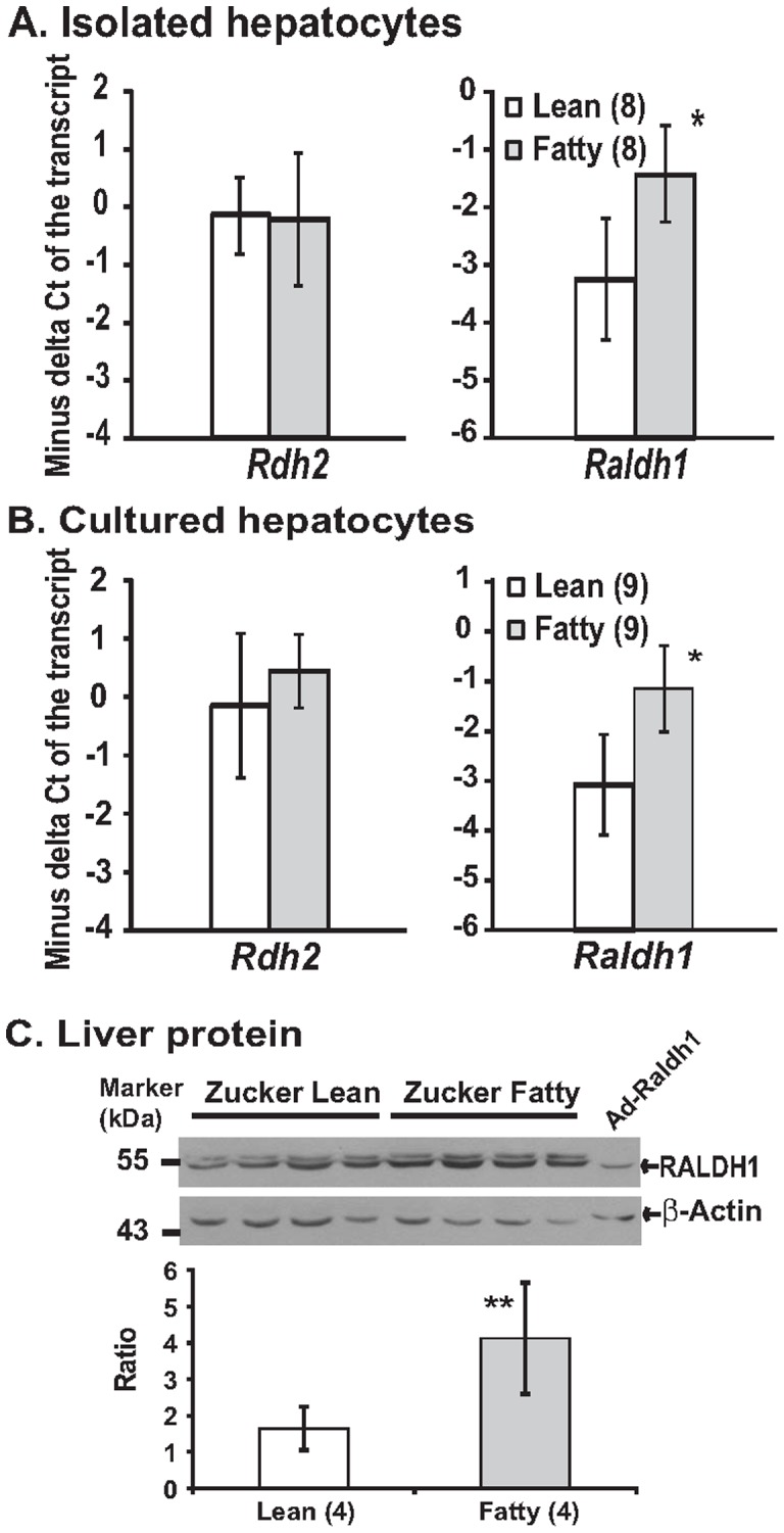
A and B. Total RNA was extracted from hepatocytes and subjected to real-time PCR analysis. Results were presented as means ± SD of the −ΔCT (the CT of 36B4– the CT of indicated transcript) from the indicated different numbers (in parenthesis) of hepatocyte isolations (* all p<0.05, for comparing the values of ZL and ZF groups of the indicated transcripts using the independent t-test). C. Whole liver tissue lyastes (100 µg/lane) of ZL (lanes 1–4) and ZF (lanes 5–8) rats, and whole cell lysate of INS-1 cells infected by Ad-Raldh1 (50 µg, lane 9) were separated in 10% SDS PAGE gels, and transferred to the PVDF membranes as described in the Material and Methods. Primary antibodies to RALDH1 (1∶1000 dilution in TBST containing 5% dry milk), and to β-Actin (1∶1000 dilution in TBST containing 5% bovine serum albumin) were recognized by goat anti-rabbit IgG conjugated to horseradish peroxidase, and visualized by chemiluminescence. The ratios of RALDH1/β-Actin were calculated after the films were scanned and analyzed. Data were presented as mean ± SD of the indicated numbers (in parenthesis) of ZL and ZF rats (** p<0.04 for comparing the ratios of ZL rats with that of ZF rats using the independent t-test).
Fig. 1C shows that the RALDH1 protein levels in the liver samples of ZF rats were higher than that of ZL rats. Since two protein bands migrating close to each other were detected in the Immuno-blot, the lysate of INS-1 cells infected by recombinant adenovirus Ad-Raldh1 expressing RALDH1 protein was used as the control to determine the correct size of it. Based on the size of the control sample, the lower band detected by the antibody should be the correct one in the liver samples. Their densities were normalized to the densities of the β-Actin in the same samples. The quantified data were presented as the ratios of RALDH1/β-Actin as shown in Fig. 1C. The ratios of ZF rats are significantly higher than that of ZL rats, demonstrating that the elevation of Raldh1 mRNA corresponds with an induction of RALDH1 protein.
Retinoids induced the expression levels of Cyp26a1 mRNA in primary rat hepatocytes
To demonstrate that retinoids catabolism can contribute to the regulation of gene expression in primary rat hepatocytes, the expression levels of Cyp26a1 mRNA (a RA responsive gene) in response to retinol (ROL) and retinal (RAL) treatments were determined using real-time PCR. Fig. 2A shows a 24-h time course of the expression levels of Cyp26a1 mRNA in Sprague-Dawley (SD) rat primary hepatocytes treated with 5 µM RA. More than 1000-fold induction of Cyp26a1 mRNA level was observed at 3 h after the RA treatment, the earliest time point checked. The Cyp26a1 mRNA level was further induced at 6 h and maintained elevated at 12 h after treatment. At 24 h after the treatment, its level dropped comparing to that at 12 h, but still remained significantly higher than that at time 0.
Figure 2. The expression levels of Cyp26a1 mRNA in response to RA treatment over a 24-h time period (A), to different dosages of ROL or RAL (B), and to an antagonist of the RAR activation (C).
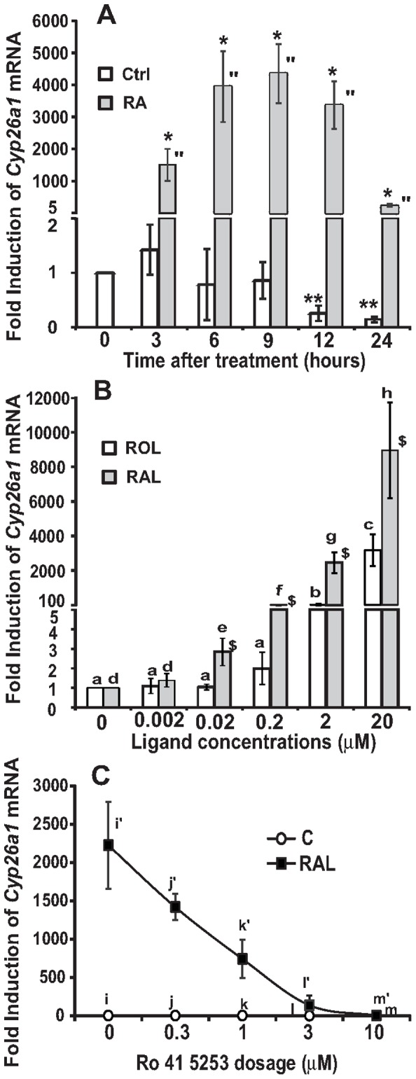
Primary hepatocytes were isolated from SD rats, and pre-treated as described in the Materials and Methods. Total RNA was isolated, and subjected to real-time PCR analysis. A. A 24-h time course of Cyp26a1 mRNA expression induced by RA (5 µM) in primary rat hepatocytes (* for comparing the vehicle control with the RA groups at the indicated time points using the independent t-test; ** and '' for comparing the different time points of vehicle control or RA group using one way ANOVA; all p<0.05). B. The expression levels of Cyp26a1 mRNA in primary hepatocytes treated with increasing concentrations of ROL or RAL for 6 hours ($ for comparing the fold induction of ROL group with that of RAL group at the indicated concentrations using the independent t-test; c > b > a; h > g > f > e > d for comparing the different dosages of ROL or RAL using one way ANOVA; all p<0.05). C. Inhibition of the RAR activation disrupted the induction of Cyp26a1 by RAL treatment. Primary hepatocytes were incubated in medium A without or with 2 μM RAL in the absence or presence of increasing concentrations of Ro 41–5253 for 6 hours (i/j > l, i'/j' > k' > l' > m' for comparing the different dosages of Ro 41–5253 in the absence or presence of RAL using one way ANOVA, all p<0.05). Results were presented as means ± SD of three independent hepatocyte isolations.
To determine the effects of ROL and RAL on the induction of Cyp26a1 expression, primary rat hepatocytes from SD rats were treated with increasing concentrations of ROL or RAL for 6 h. As shown in Fig. 2B, the levels of Cyp26a1 mRNA were induced by ROL and RAL in a dosage dependent manner. ROL and RAL started to induce Cyp26a1 expression at 2 μM and 0.02 μM, respectively. When the induction folds of Cyp26a1 expression in response to ROL and RAL treatments were compared, RAL groups had significantly higher values than ROL groups did at concentrations of 0.02 μM, 0.2 μM, 2 μM, and 20 μM. It suggests that RAL is more readily converted to RA than ROL to induce Cyp26al expression.
Inhibition of RAR activation blocked the RAL-mediated induction of Cyp26a1 expression in primary rat hepatocytes
We have shown that the activation of RAR or/and RXR can induce Cyp26a1 expression [16]. To confirm the involvement of RAR activation in the RAL-mediated induction of Cyp26a1 expression, primary SD rat hepatocytes were treated with increasing concentrations of Ro 41–5253, a specific antagonist for RAR activation [34]. As shown in Fig. 2C, RAL at 2 µM dramatically induced the Cyp26a1 expression, further confirming the result shown in Fig. 2B. This induction was dose-dependently inhibited with increasing concentrations of Ro 41–5253. When the concentration of Ro 41–5253 reached 10 μM or higher, the induction of Cyp26a1 expression by RAL was completely suppressed. This reverse association between the concentration of Ro 41–5253 and the induction of Cyp26a1 expression by RAL demonstrated that the inhibition of RAR activation disrupted the retinoid-mediated induction of Cyp26a1 expression in primary hepatocytes, indicating the important role of RAR activation in this induction.
Retinoids induced the expression levels of Cyp26a1 mRNA and CYP26A1 protein in HL1C rat hepatoma cells through the activation of RAR and RXR
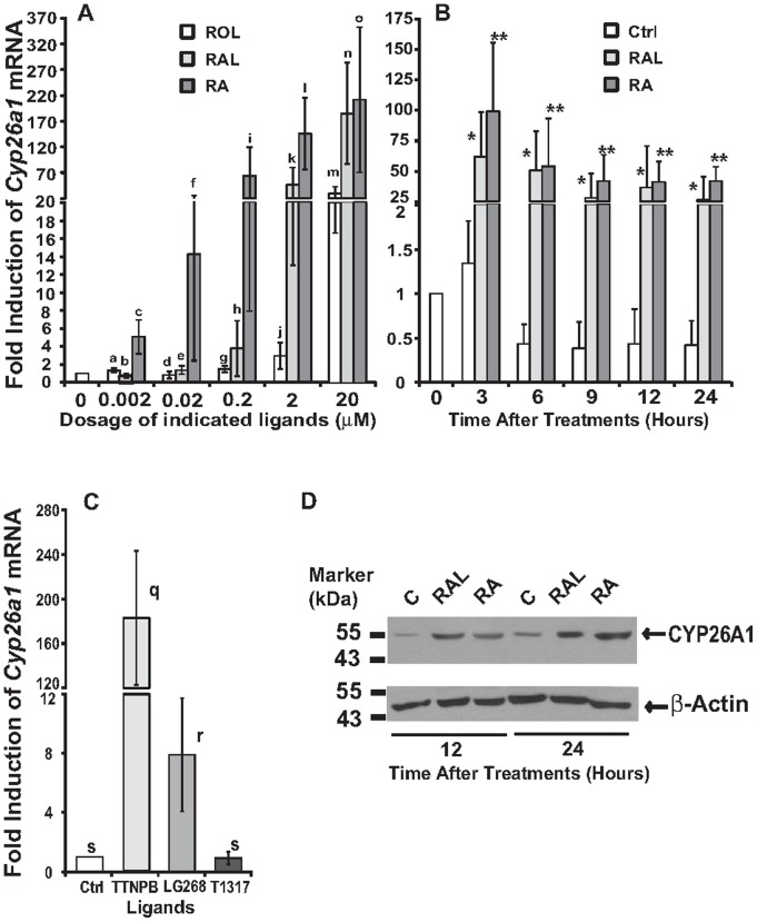
To screen for cultured cell lines which we can study the effects of retinoid metabolism on the expression of RA responsive gene, we analyzed the expression of Cyp26a1 mRNA and CYP26A1 protein in response to ROL, RAL and RA treatments in HL1C rat hepatoma cells [35]. Fig. 3A shows the expression levels of Cyp26a1 mRNA in HL1C cells treated with the indicated concentrations of retinoids. ROL, RAL and RA started to induce Cyp26a1 mRNA expression at 20 μM, 2 μM, and 0.2 μM, respectively, demonstrating the different potencies of these retinoids. At 2 μM or 20 μM, the fold induced by RAL was comparable to that by RA. The highest induction fold of Cyp26a1 mRNA mediated by RA in HL1C cells was near 300-fold which was much lower than that in primary hepatocytes (1000-fold) as shown in Fig. 2. The effects of RAL and RA on Cyp26a1 mRNA expression in HL1C cells over a 24-h time period were also examined as shown in Fig. 3B. Both RAL and RA induced Cyp26a1 mRNA expression as early as 3 h. At 6 h after the treatment, the elevated level of Cyp26a1 mRNA began to drop in comparison to its level at 3 h. At 9, 12, and 24 h time points, its level further declined, but remained significantly higher than that at time 0. The induction was still elevated at 24 h after treatment.
Figure 3. The effects of retinoids on the expression levels of Cyp26a1 mRNA and CYP26A1 protein in HL1C cells. A.
The dose-dependent induction of Cyp26a1 by ROL, RAL, and RA. HL1C cells were treated with ROL, RAL, or RA at the doses of 0, 0.002 µM, 0.02 µM, 0.2 µM, 2 µM, and 20 µM for 6 h (a/b < c, d/e < f, g/h < i, j < k/l, m < n/o, a/d/g/j < m, b/e/h < k < n, c/f < i/l/o for comparing ROL, RAL and RA at the indicated concentrations, and for comparing each ligand at the different dosages using one way ANOVA; all p<0.05). B. The expression levels of Cyp26a1 mRNA overtime (3, 6, 9, 12, and 24 h) in HL1C cells treated with 5 µM RAL or RA (* or ** for comparing RAL or RA with vehicle control groups at the indicated time points using the independent t-test; all p<0.05). C. The expression levels of Cyp26a1 mRNA in HL1C cells treated with RAR (TTNPB), RXR (LG268), and LXR (T1317) agonists. HL1C cells were treated with 1 μM TTNPB, LG268, and T1317 for 6 h (q > r>s using one way ANOVA; all p<0.05). Total RNA was extracted and subjected to real-time PCR analysis. Data were presented as mean ± SD of three independent treatments. D. Whole cell lysates (50 µg/sample) of HL1C cells treated the vehicle control, 5 µM RAL or RA for 12 (lanes 1–3) and 24 (lanes 4–6) hours were separated in 8% SDS PAGE gels, and transferred to the PVDF membranes. Primary antibodies to CYP26A1 (1∶1000 dilution in TBST containing 5% dry milk), and to β-Actin (1∶1000 dilution in TBST containing 5% bovine serum albumin) were recognized by goat anti-rabbit IgG conjugated to horseradish peroxidase, and visualized by chemiluminescence. The films were scanned and presented as described in the Material and Methods.
To examine whether the activation of RAR or RXR was sufficient for the induction of Cyp26a1 mRNA expression, HL1C cells were treated with TTNPB, LG268, and T1317, agonists for RAR, RXR and LXR activation, respectively. As shown in Fig. 3C, the induction folds of the TTNPB, LG268 and T1317 groups are 183.0±60.0, 8.0±3.8, and 1.0±0.4. These results demonstrated that the activation of RAR and RXR, but not LXR, induced the Cyp26a1 mRNA expression in HL1C cells, indicating that RA activated both RAR and RXR to induce Cyp26a1 mRNA expression. The induction fold of TTNPB group is much higher than that of LG268 group, similar to that observed in primary hepatocytes [16].
Fig. 3D shows the expression levels of CYP26A1 protein in HL1C cells treated with RAL or RA for 12 and 24 hours. The equal loading of protein samples was confirmed by the similar levels of β-Actin protein among the samples. This result demonstrated that the induction of Cyp26a1 mRNA resulted in significant elevation of CYP26A1 protein at both time points. All these results suggest that ROL and RAL may be converted to RA, which causes the RAR activation and the subsequent induction of Cyp26a1 mRNA and CYP26A1 protein levels in HL1C cells.
RA, but not RAL, induced the Srebp-1c expression through the activation of RXR, but not RAR, in INS-1 rat insulinoma cells
We have screened the cultured cell lines in which RAL does not induce Srebp-1c expression for a tool to study the conversion of RAL to RA, and shown that RA, but not RAL, induced the Srebp-1c expression dose-dependently in 833/15 INS-1 cells [16]. To further confirm that INS-1 cells are an ideal tool to study the conversion of RAL to RA, we analyzed the effects of RAL and RA on Srebp-1c expression in 834/40 INS-1 cells, another clonal INS-1 cells [36]. Fig. 4A shows that RA started to significantly induce the Srebp-1c expression at 0.1 μM while RAL did not change its expression at any concentration tested. Fig. 4B shows the induction of the Srebp-1c expression by RA in 834/40 INS-1 cells over a 24-h time period. RA, but not RAL, induced the Srebp-1c expression as early as 3 h and peaked at 6 h. At 9, 12, and 24 h, the induction folds declined in comparison to that at 3 and 6 h, but still maintained significantly higher than that at 0 h. These results and the results shown previously [16] indicate that INS-1 cells may lack the enzymatic activities that convert RAL to RA.
Figure 4. The Srebp-1c mRNA levels in INS-1 cells treated with retinoids or ligands.
A. 834/40 INS-1 cells were treated with increasing concentrations of RAL or RA for 6 h. B. 834/40 cells were treated with 2 µM of RAL or RA for indicated time. C. 833/15 cells were treated with 1 µM of specific ligands of RXR (LG268), RAR (TTNPB) or in combination. Total RNA was extracted from INS-1 cells and subjected to real-time PCR analysis. Results were presented as means ± SD of fold inductions (c > b > a for comparing the different dosages of RA-treated groups using one way ANOVA, and e > d > f for comparing control with LG 268 in the absence or presence of TTNPB using one way ANOVA; * for comparing RAL with RA groups at the indicated dosages, and ** for comparing RA with RAL or vehicle control groups at the indicated time points using independent t-test; n = 3, all P<0.05).
To determine whether the activation of RAR or RXR mediates the effects of RA on the Srebp-1c expression, INS-1 cells were treated with TTNPB (1 μM), LG268 (1 μM), or TTNPB + LG268 for 6 h. Fig. 4C shows that LG268 and LG268 + TTNPB induced the Srebp-1c expression while TTNPB alone did not induce it. Thus, RXR activation mediates the RA-induced Srebp-1c expression in INS-1 cells. All these results demonstrated that the RA-mediated activation of RXR caused the induction of Srebp-1c in INS-1 cells, which makes INS-1 cell lines an ideal tool to study the role of RA production in the regulation of lipogenic gene expression.
The over-expression of RALDH1 resulted in the RAL-mediated induction of Srebp-1c in INS-1 cells
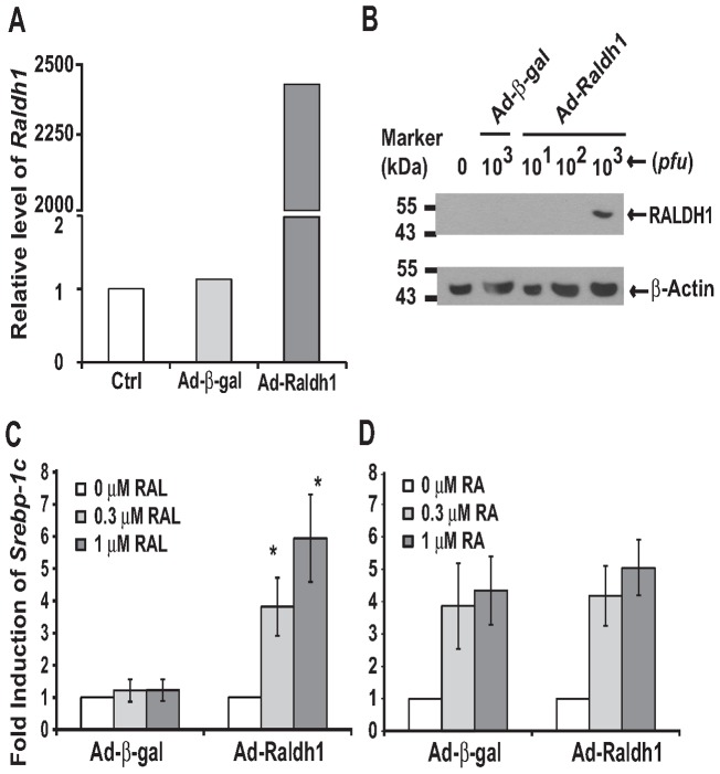
To further study the effects of the elevated Raldh1 mRNA expression in ZF hepatocytes, its full length cDNA was cloned and inserted it into pACCMV5 for the generation of recombinant adenovirus Ad-Raldh1. As shown in Fig. 5, INS-1 cells infected with Ad-Raldh1 had the over-expression of Raldh1 mRNA (Fig. 5A) and RALDH1 protein (Fig. 5B). The equal loading of the protein samples was confirmed by the similar levels of β-Actin protein among the samples. In INS-1 cells infected with Ad-Raldh1 for overnight (∼18 h), but not in those infected with Ad-β-gal, RAL started to significantly induce the Srebp-1c expression at both 0.3 μM and 1 μM (Fig. 5C). On the other hand, RA induced the Srebp-1c expression in cells infected by either Ad-β-gal or Ad-Raldh1 (Fig. 5D). In those cells with the over-expression of RALDH1, the induction folds of Srebp-1c by RAL treatment were comparable to that by RA. These results indicate that the over-expression of RALDH1 introduced the RAL-mediated induction of Srebp-1c in INS-1 cells, indicating the successful conversion of RAL to RA.
Figure 5. The over-expression of RALDH1 resulted in the RAL-mediated induction of Srebp-1c in 833/15 INS-1 cells. A.
The adenovirus-mediated Raldh1 mRNA expression. B. Immuno-blot of the over-expression of RALDH1 protein in INS-1 cells. Whole cell lysates (50 µg/sample) of the control cells (lane 1), cells infected by the indicated pfu of Ad-β-gal (lane 2) or Ad-Raldh1 (lanes 3–5) were separated in 8% SDS protein gels, and transferred to the PVDF membranes. Primary antibodies to RALDH1 (1∶1000 dilution in TBST containing 5% dry milk), and to β-Actin (1∶1000 dilution in TBST containing 5% bovine serum albumin) were recognized by goat anti-rabbit IgG conjugated to horseradish peroxidase, and visualized by chemiluminescence. The films were scanned and presented as described in the Material and Methods. C. RAL only induced Srebp-1c expression in cells over-expressing RALDH1, but not β-gal. D. RA induced Srebp-1c expression in cells over-expressing either β-gal or RALDH1. Results were presented as means ± SD of fold inductions (* for comparing the different dosages of RAL in cells infected by Ad-Raldh1 using one way ANOVA, n = 3, all p<0.05).
Discussion
In insulin resistant animals, hyperinsulinemia is associated with the elevated hepatic gluconeogenesis and lipogenesis, which should have been respectively suppressed and stimulated by insulin [37]. Our lab has shown that retinoids regulate the expression levels of genes involved in hepatic glucose and fatty acid metabolism. These regulations contributed to the modulation of the insulin-mediated expression of these genes in primary hepatocytes [14]–[16]. The implication of these observations is that the dynamic regulation of RA production may be responsible for the hepatic insulin resistance.
To confirm that the treatments of ROL and RAL can regulate gene expression probably through the production of RA, we analyzed the expression levels of Cyp26a1, an RA responsive gene [38], in primary hepatocytes (Fig. 2). Retinoids induced the expression of Cyp26a1 in a dose- and time-dependent manner. This induction can be blocked in the presence of RAR antagonist, indicating the requirement of RAR activation. This result and the fact that RAL induced the Cyp26a1 expression indicate that RA production occurs.
To find the cultured cell lines that can be used as tools for further studies of the retinoid catabolism, we have analyzed the retinoid-mediated gene expression in HL1C rat hepatoma cells (Fig. 3). ROL, RAL, and RAL also induced the expression levels of Cyp26a1 mRNA in HL1C cells in a dose- and time-dependent manner, which is similar to the results obtained in primary hepatocytes. This induction is mediated by the activation of RAR and RXR. The induction of Cyp26a1 mRNA results in the elevated levels of CYP26A1 proteins in HL1C cells. However, the elevated levels of CYP26A1 protein are not as high as that of Cyp26a1 mRNA at 12 and 24 hours (Fig. 3B and 3D), indicating the potential roles of protein translation and/or stability in the determination of the CYP26A1 protein levels in HL1C cells. The maximal induction folds by reintoids are higher in primary hepatocytes than in HL1C cells. ROL and RAL respectively induce the expression of Cyp26a1 mRNA at 2 and 0.02 µM in primary hepatocytes and at 20 and 2 µM in HL1C cells. These results indicate that primary hepatocytes seem to be more readily to convert ROL to RAL and then to RA. Alternatively, HL1C cells are more readily to convert RAL to ROL so that less RA is generated. Whether it is true or not deserves further investigation. It also indicates that HL1C cells can be used as a tool to study the metabolism of retinoids and its effects on the regulation of the expression of RA responsive genes.
It has been shown that the short chain dehydrogenase/reductase family 16C member 5 (SDR16C5), RDH2, RDH10, RALDH1-4 are expressed in the liver [33]. Since Raldh4 mRNA (encoding RALDH4) is for the production of 9-cis RA [27], we focused on Rdh2 and Raldh1. Their mRNA levels in both freshly isolated and cultured primary hepatocytes of ZL and ZF rats were compared. The mRNA level of Raldh1, but not that of Rdh2, is higher in ZF than that in ZL rat hepatocytes. In addition, the hepatic RALDH1 protein level is also higher in ZF rats than in ZL rats (Fig. 1). The elevated expression levels of Raldh1 mRNA and RALDH1 protein levels may be resposible for the altered hepatic lipogenesis in ZF rats. To test the hypothesis, we made recombinant adenovirus Ad-Raldh1 to over-express RALDH1 protein in INS-1 rat insulinoma cells. We have shown previously [16] and here (Fig. 4) that RA, but not RAL, induced Srebp-1c expression in INS-1 cells. It seems that INS-1 cells lack the enzymatic activities converting RAL to RA. This makes INS-1 cells an ideal tool to test our hypothesis that the elevated expression of RALDH1 in ZF rats causes the increase of RA production, and in turn, the induction of RA responsive gene expression. After the over-expression of Raldh1 mRNA and RALDH1 protein in INS-1 cells, RAL started to induce the Srebp-1c expression as RA did. This indicates that RAL was oxidized to RA by RALDH1 before the induction of Srebp-1c. The elevated levels of hepatic Raldh1 mRNA and RALDH1 protein in ZF rats may contribute to the regulation of hepatic lipogenesis.
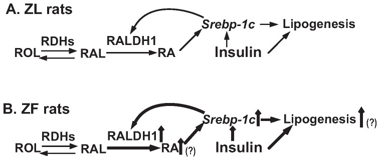
It has been shown that the mRNA level of Raldh1 is elevated in the kidney of db/db mice [39]. Both db/db mice and ZF rats develop obesity due to the mutation of leptin receptor [4]. It has been shown that ICR mice fed high cholesterol diet have the elevated expression levels of Raldh1 and Raldh2. This is attributed to the oxysterol-induced expression of SREBP-1c which directly binds to the sterol regulatory response elements (SREs) at the proximal promoter regions of Raldh1 and Raldh2, and induces their expression [32]. Since the expression of Srebp-1c is induced by insulin [40] and RA [16] in primary hepatocytes, the regulation of Raldh1 expression becomes a converge point for a feed-forward mechanism by which VA status regulates lipogenesis in the liver. It is reasonable to hypothesiz that in hepatocytes of ZL rats, RA derived from ROL synergizes with insulin to induce the expression of Srebp-1c mRNA (Fig. 6A). The mature SREBP-1c protein supports the expression of RALDH1, which promotes the production of RA to maintain the homeostasis of Srebp-1c expression in the liver of ZL rats (Fig. 6A). ZF rats are hyperphagic due to the defect of leptin receptor, which leads to the development of obesity, insulin resistance, hyperinsulinemia and hyperlipidemia [41], [42]. We have shown the ZF rat hepatocytes have elevated expression levels of Srebp-1c and Fas mRNA [43]. We think that the excessive supply of dietary VA caused by hyperphagia and hyperinsulinemia in ZF rats probably work together to trigger an elevation of RA production and an induction of Srebp-1c expression in their hepatocytes, respectively. The elevation of the mature SREBP-1c protein induces the expression of Raldh1 mRNA, which leads to more RA production, and further enhances the expression levels of Srebp-1c and its down-stream lipogenic genes. This creates a feed-forward mechanism by which the Srebp-1c expression is maintained at a higher level in the liver of ZF rats (Fig. 6B). Whether the proposed feed-forward mechanism is true or not, and what mechanism is responsible for the up-regulation of Raldh1 expression in the ZF rat liver deserve further investigation.
Figure 6. The hypothesized role of the RA production in the feed-forward induction of the expression of Srebp-1c and its downstream lipogenic genes in the liver of ZL (A) and ZF (B) rats.
A. In ZL rats, the hepatic expression of Srebp-1c is controlled by the RA production and insulin stimulation. The SREBP-1c also regulates RA production via the induction of Raldh1 expression until a homeostasis is reached. B. The hyperphagia of ZF rats due to the leptin receptor deficiency causes the over-supply of dietary VA, and hyperinsulinemia. The possible elevation of RA production in combination with insulin stimulation leads to higher expression of hepatic Srebp-1c in ZF rats, which disrupts the homeostasis. The expression of Raldh1 mRNA is further induced by SREBP-1c. Therefore, the hepatic expression of Srebp-1c in ZF rats is maintained at a higher level than that in ZL rats. The consequence is the elevated hepatic lipogenesis in ZF rats. The up arrows next to the texts indicate induction. The intensified weight of the lines indicates the induction of that part of the pathway. The question marks indicate the steps remained to be confirmed in this hypothesis.
In summary, the results shown here suggest that ROL and RAL are metabolized into RA to regulate gene expression in rat primary hepatocytes and hepatoma cells. In primary rat hepatocytes, the responsible enzymes are most likely RDH2 and RALDH1. The expression of Raldh1 mRNA is higher in primary hepatocytes from ZF rats than that from ZL rats, which leads to the elevated RALDH1 protein levels in the liver of ZF rats. Over-expression of RALDH1 introduces the RAL-mediated induction of Srebp-1c in INS-1 cells. Thus, we hypothesize that the change of RA production from the over-supply of dietary VA due to the hyperphagia of ZF rats results in higher Srebp-1c expression in ZF hepatocytes. The elevated SREBP-1c expression can further induce Raldh1 expression to create a feed-forward mechanism that could be one of the reasons responsible for the increased lipogenesis in the liver of ZF rats.
Materials and Methods
Reagents
The reagents for primary hepatocyte isolation and culture have been published previously [44]. Source of LG268 was reported previously as well [15]. All other compounds were purchased from Sigma (Saint Louis, MO) unless described otherwise. The reagents for cDNA synthesis and real-time PCR were obtained from Applied Biosystems (Foster city, CA). Medium 199, liver perfusion medium and liver digest buffer were obtained from Invitrogen (now Life Technologies, Grand Island, NY 14072). Dulbecco's Modification of Eagle Medium (DMEM), RPI-1640 medium, fetal bovine serum, antibiotics, and trypsin-EDTA solution were obtained from Fisher Scientific (Pittsburgh, PA 15275).
Animals
Male Sprague-Dawley (SD) rats were purchased from Harlan Breeders (Indianapolis, IN). Male Zucker lean (ZL) and ZF rats were bred at UTK. Rats were housed in colony cages, and fed a standard rodent diet before isolation of primary hepatocytes. All procedures were approved by the Institutional Animal Care and Use Committee at the University of Tennessee at Knoxville (Protocol numbers 1642 and 1582).
Cultures of primary rat hepatocytes, HL1C rat hepatoma cells, 293 human embryonic kidney cells, and INS-1 rat insulinoma cells
Methods for the primary rat hepatocyte preparation and analysis of RNA were described previously [44]. For the hepatocyte isolation and pretreatment, the rat was euthanized with carbon dioxide. A catheter was inserted into portal vein and connected to a peristaltic pump with liver perfusion medium and liver digestive buffer. The inferior vena cava was cut open to allow the outflow of the media at 10 ml/minute. After completion of the digestion, the liver was excised and put into a tissue culture dish containing liver digest buffer for removing the connection tissues and allowing the release of hepatocytes. The medium containing hepatocytes was filtered through a cell strainer (100 µm), and precipitated by 50× g centrifugation for 3 minutes (min). The cell pellet was washed twice with DMEM containing 5% fetal bovine serum (FBS), 100 U/ml sodium penicillin, and 100 µg/ml streptomycin sulfate after re-suspension and precipitation. After the wash, the isolated hepatocytes were plated onto 60-mm collagen type I coated dishes at 3×10⁶ cells/ per dish and incubated in 4 ml of the same medium at 37°C and 5% CO2 for 3 to 4 hours (h). The attached cells were washed once with 4 ml of PBS, and incubated in medium A (Medium 199 supplemented with 100 nM dexamethasone, 100 nM 3, 3′, 5-triiodo-l-thyronine (T3), 100 units/ml penicillin, and 100 μg/ml streptomycin sulfate) containing 1 nM insulin for overnight (14–16 h) until being used for the indicated experiments.
The HL1C cells [35] which was stably transfected with a reporter gene construct containing a fragment of Pck1 promoter sequence were kindly provided by Dr. Donald K. Scott at University of Pittsburgh. They were maintained in DMEM containing 4.5 g/L glucose, 4% FBS, 100 U/ml of penicillin and 100 μg/ml streptomycin sulfate. They were incubated in 60 mm dishes with serum free DMEM containing the indicated reagents shown in the figure legends for the indicated length of time before being subjected to analysis.
The 833/15 and 834/40 clonal insulinoma INS-1 cells [36] were cultured in medium B (RPMI 1640, 2 mM L-glutamine, 1 mM Na-pyruvate, 50 μM β-mercaptoethanol, 100 U/ml of penicillin and 100 μg/ml streptomycin sulfate) containing 10% FBS as described previously. For the treatment, INS-1 cells in 60 mm dishes were incubated in medium B containing the indicated reagents shown in the figure legends for the indicated length of time before being subjected to analysis.
The HEK 293 cells were cultured in DMEM containing 4.5 g/L glucose, 8% FBS, 100 U/ml of penicillin and 100 μg/ml streptomycin sulfate at 37°C and 5% CO2.
RNA extraction and quantitative real-time PCR
Total RNA was extracted from the treated cells on one 60 mm dish using 1 ml of RNA STAT 60 reagent (TEL-TEST, Inc, Friendswood, TX) according to the manufacture's protocol. The contaminated DNA was removed using the DNA-free TM kit (Applied Biosystems). First strand cDNA was synthesized from 2 µg of DNA-free RNA with random hexamer primers using cDNA synthesis kit (Applied Biosystems). The real-time PCR primer sequences for detecting Cyp26a1 [45], Srebp-1c [16], and Raldh1 [46] have been reported previously. The primers for detecting Rdh2 (sense 5′-CAAGTTCTTCTACCTCCCCATGA-3′, and anti-sense 5′-TCCAGTAGAAAAGGGCATCCA-3′) were designed using software Primer Express 3.0 (Applied Biosystems). Each SYBR green based real-time PCR reaction contains, in a final volume of 14 µl, cDNA from 14 ng of reverse transcribed total RNA, 2.33 pmol primers, and 7 µl of 2× SYBR Green PCR Master Mix (Applied Biosystems). Triplicate PCR reactions were carried out in 96-well plates using 7300 Real-Time PCR System. The conditions are 50°C for 2 min, 95°C for 10 min, followed by 40 cycles of 95°C for 15 seconds (s) and 60°C for 1 min. The gene expression level was normalized to that of invariable control gene, 36B4. Data are presented as either minus Δ cycle threshold C T [43] or the induction fold for which the control group is arbitrarily assigned a value of 1 using ΔΔC T method as previously described [44].
Cloning of the rat Raldh1 cDNA and generating Ad-Raldh1 recombinant adenovirus for the over-expression of RALDH1
To clone the rat Raldh1 cNDA, sense 5′-AGCCAAACCAGCAATGTCTTC-3′, and anti-sense 5′-CACTCTGCTTCTTAGGAGTTC-3′ primers were designed based on the its mRNA sequence (reference number NM_022407.3) from the National Center for Biotechnology Information. The Raldh1 cDNA containing the entire coding sequence was amplified using SD rat primary hepatocyte cDNA as a template. The amplicon was ligated into pCR®2.1 vector through TA Cloning® Kit (Invitrogen) and processed according to the manufacture's protocol. The full length cDNA with the correct sequence confirmed by DNA sequencing was inserted to pACCMV5 for making pACCMV5-Raldh1.
To generate Ad-Raldh1 recombinant adenovirus, plasmids pACCMV5-Raldh1 and JM17 were co-transfected into 293 cells using FuGENE®6 Transfection Reagent (Roche Applied Science, Indianapolis, IN). Transfected 293 cells were incubated in DMEM containing 2% FBS at 37°C and 5% CO2 until the formation of the plaques due to the lysis of the cells. The crude lyaste was collected and stored at −80°C. The presence of Raldh1 cDNA sequence in the viral DNA was confirmed by PCR.
Preparation and purification of recombinant adenoviruses
To generate the crude lysate, HEK 293 cells grew in 100 mm tissue culture plates at 80% confluence were infected by the original crude lysate containing Ad-Raldh1. The ratio of the medium to crude lysate is 10 to 1. After the lysis of the cells at around 48 h post infection, the cell culture medium (the crude lysate) was collected, and stored at −80°C until being used.
For purification of the recombinant adenovirus, NP-40 was first added into the crude lysate to reach the final concentration at 0.5%. The mixture was shaken gently at room temperature for 30 min and subjected to centrifugation at 8,000 rpm and 4°C for 15 min. The supernatant was transferred to a clean bottle, and 0.5× volume of 20% PEG8000/2.5 M NaCl was added. The preparation was shaken gently at 4°C overnight. The resulting mixture was transferred to centrifuge bottles and spun at 13,000 rpm at 4°C for 15 min. The precipitated pellet was re-suspended in a small volume of PBS (2–3 ml), and spun at 8,000 rpm and 4°C for 10 min to remove insoluble matters. Solid CsCl was added to the supernatant until its final density reached 1.34 g/ml. The mixture was spun at 90,000 rpm and 25°C for 3 h using OptimaTM MAX-XP Ultracentrifuge (Beckman Coulter, Inc.). The corresponding band containing pure viral particles was collected in a total volume less than 1 ml for desalting. The PD-10 column SephadexTM G-25 M (Amersham Pharmacia Biotech AB, Sweden) was equilibrated with 5 ml PBS. The purified virus in CsCl solution was loaded onto the column and eluted with 5 ml PBS. The flow through was collected into ten fractions. The optical densities (OD) at 260–nm of these fractions were determined using Spectronic® GENESYSTM 5 Spectrophotometer (Thermo Scientific). The fractions containing significant values of absorbance (usually at around fractions 7–9) were collected and pooled. Bovine serum albumin and glycerol were added to the pooled solution to make the stock viral solution with the final concentrations of them at 0.2% and 10%, respectively. After the filtration of the stock solution for sterilization, its OD was determined to estimate the plaque forming units (pfu). We used that 1 OD equals to 1×10¹² pfu/ml. The final purified virus stock was frozen at −80°C until being used in the indicated experiments.
Immuno-blot and quantification of the protein bands
INS-1 cells were infected with purified Ad-Raldh1 for 24 h. The cells in 60 mm dishes were washed once with 3 ml PBS and scrapped from the dish into 400 μl of whole-cell lysis buffer (1% Triton X-100, 10% glycerol, 1% IGEPAL CA-630, 50 mM Hepes, 100 mM NaF, 10 mM EDTA, 1 mM sodium molybdate, 1 mM sodium β-glycerophsphate, 5 mM sodium orthovanadate, 1.9 mg/ml aprotinin, 5 μg/ml leupeptin, 1 mM benzamide, 2.5 mM PMSF, pH 8.0). The lysates were placed on ice for at least 20 min before they were subjected to centrifugation at 13,000 rpm for 20 min. The protein concentration in the supernatant was determined with PIERCE BCA protein assay kit (Rockford, IL). Proteins (40 μg/lane) in whole cell lysates were separated on SDS-PAGE, transferred to BIO-RAD Immuno-Blot PVDF membrane (Hercules, CA) and detected with primary antibodies to RALDH1 (for Fig. 5, catalog #2052-1, Epitomics, Burlingame, CA 94010), RALDH1 (for Fig. 1, catalog #AP1465a, Abgent, San Diego, CA 92121), CYP26A1 (catalog # CYP26A11-A, Alpha Diagnostics International, TX 78244), and β-Actin (#4970 s, Cell Signaling Technology, Danvers, MA 01923) according to the protocols provided by the manufacturers. Bound primary antibodies were visualized by chemiluminescence (ECL Western Blotting Substrate, Thermo Scientific) using a 1∶2,000 dilution of goat anti-rabbit IgG (#7074P2, Cell signaling Technology) conjugated to horseradish peroxidase. Membranes were exposed to X-ray films (Phenix Research Products, Candler, NC) for protein band detection. The films were scanned using an HP Scanjet 3970 (Palo Alto, CA 94304) and stored as Tagged Image File Format (TIFF). The ImageJ Software (http://rsbweb.nih.gov/ij/) was used to determine the density of a protein band by subtracting the background in an area of the same size immediately in front of protein band. The ratio of the densities of RALDH1 and β-Actin in the same sample was calculated and used for statistical analysis.
Statistics
Data are presented as means ± S.D. The number of experiments represents the independent experiments using hepatocytes isolated from different animals or indicated cells cultured in different days. Levene's test was used to determined homogeneity of variance among groups using SPSS 19.0 statistical software and where necessary natural log transformation was performed before analysis. An independent-samples t-test was used to compare two conditions. Multiple comparisons were analyzed by one-way analysis of variance (ANOVA) using least significant different (LSD) when equal variance was assumed, and Games-Howell test was used when equal variance was not assumed. Differences were considered statistically significant at P<0.05.
Acknowledgments
The authors thank Dr. Hollie Raynor for checking the spell and grammar of this manuscript.
Funding Statement
This work was financially supported by research grant from Allen Foundation Inc (to GC), startup fund from the University of Tennessee at Knoxville (to GC), and Scientist Development Grant from American Heart Association (09SDG2140003, to GC). The authors also thank China Scholarship Council for the financial support (to Y.L. and R. L.) The funders had no role in study design, data collection and analysis, decision to publish, or preparation of the manuscript.
References
- 1. Yanovski SZ, Yanovski JA (2011) Obesity prevalence in the United States – up, down, or sideways? N Engl J Med 364: 987–989. [DOI] [PMC free article] [PubMed] [Google Scholar]
- 2. Schulze MB, Hu FB (2005) Primary prevention of diabetes: what can be done and how much can be prevented? Annu Rev of Public Health 26: 445–467. [DOI] [PubMed] [Google Scholar]
- 3. Haslam DW, James WP (2005) Obesity. Lancet 366: 1197–1209. [DOI] [PubMed] [Google Scholar]
- 4. Friedman JM (2009) Leptin at 14 y of age: an ongoing story. Am J Clin Nutr 89: 973S–979S. [DOI] [PMC free article] [PubMed] [Google Scholar]
- 5. Gaulton KJ, Willer CJ, Li Y, Scott LJ, Conneely KN, et al. (2008) Comprehensive association study of type 2 diabetes and related quantitative traits with 222 candidate genes. Diabetes 57: 3136–3144. [DOI] [PMC free article] [PubMed] [Google Scholar]
- 6. O'Rahilly S, Farooqi IS (2008) Human obesity as a heritable disorder of the central control of energy balance. Int J Obes 32: S55–S61. [DOI] [PubMed] [Google Scholar]
- 7. McGarry JD (2002) Banting lecture 2001: Dysregulation of fatty acid metabolism in the etiology of type 2 diabetes. Diabetes 51: 7–18. [DOI] [PubMed] [Google Scholar]
- 8. Shimomura I, Matsuda M, Hammer RE, Bashmakov Y, Brown MS, et al. (2000) Decreased IRS-2 and increased SREBP-1c lead to mixed insulin resistance and sensitivity in livers of lipodystrophic and ob/ob mice. Mol Cell 6: 77–86. [PubMed] [Google Scholar]
- 9. Spiegelman BM, Flier JS (2001) Obesity and the regulation of energy balance. Cell 104: 531–543. [DOI] [PubMed] [Google Scholar]
- 10. Bray GA, Nielsen SJ, Popkin BM (2004) Consumption of high-fructose corn syrup in beverages may play a role in the epidemic of obesity. Am J Clin Nutr 79: 537–543. [DOI] [PubMed] [Google Scholar]
- 11.Sporn MB, Robers AB, Goodman DS (1994) The Retinoids, Biology, Chemistry, and Medicine. New York: Raven Press.
- 12. Ross AC (2003) Retinoid production and catabolism: role of diet in regulating retinol esterification and retinoic acid oxidation. J Nutr 133: 291S–296. [DOI] [PubMed] [Google Scholar]
- 13. Napoli JL (2011) Physiological insights into all-trans-retinoic acid biosynthesis. Biochim Biophys Acta 1821: 152–167. [DOI] [PMC free article] [PubMed] [Google Scholar]
- 14. Zhang Y, Li R, Chen W, Li Y, Chen G (2011) Retinoids induced Pck1 expression and attenuated insulin-mediated suppression of its expression via activation of retinoic acid receptor in primary rat hepatocytes. Mol Cell Biochem 355: 1–8. [DOI] [PubMed] [Google Scholar]
- 15. Chen G, Zhang Y, Lu D, Li N, Ross AC (2009) Retinoids synergize with insulin to induce hepatic Gck expression. Biochem J 419: 645–653. [DOI] [PMC free article] [PubMed] [Google Scholar]
- 16. Li R, Chen W, Li Y, Zhang Y, Chen G (2011) Retinoids synergized with insulin to induce Srebp-1c expression and activated its promoter via the two liver X receptor binding sites that mediate insulin action. Biochem Biophys Res Commun 406: 268–272. [DOI] [PubMed] [Google Scholar]
- 17. Lucas PC, Forman BM, Samuels HH, Granner DK (1991) Specificity of a retinoic acid response element in the phosphoenolpyruvate carboxykinase gene promoter: consequences of both retinoic acid and thyroid hormone receptor binding. Mol Cell Biol 11: 5164–5170. [DOI] [PMC free article] [PubMed] [Google Scholar]
- 18. Lucas PC, O'Brien RM, Mitchell JA, Davis CM, Imai E, et al. (1991) A retinoic acid response element is part of a pleiotropic domain in the phosphoenolpyruvate carboxykinase gene. Proc Natl Acad Sci U S A 88: 2184–2188. [DOI] [PMC free article] [PubMed] [Google Scholar]
- 19. Scott DK, Mitchell JA, Granner DK (1996) Identification and characterization of a second retinoic acid response element in the phosphoenolpyruvate carboxykinase gene promoter. J Biol Chem 271: 6260–6264. [DOI] [PubMed] [Google Scholar]
- 20. Chen G, Liang G, Ou J, Goldstein JL, Brown MS (2004) Central role for liver X receptor in insulin-mediated activation of Srebp-1c transcription and stimulation of fatty acid synthesis in liver. Proc Natl Acad Sci U S A 101: 11245–11250. [DOI] [PMC free article] [PubMed] [Google Scholar]
- 21. Jones D (2008) End of the line for cannabinoid receptor 1 as an anti-obesity target? Nat Rev Drug Discov 7: 961–962. [DOI] [PubMed] [Google Scholar]
- 22. Mukhopadhyay B, Liu J, Osei-Hyiaman D, Godlewski G, Mukhopadhyay P, et al. (2010) Transcriptional regulation of cannabinoid receptor-1 expression in the liver by retinoic acid acting via retinoic acid receptor-γ. J Biol Chem 285: 19002–19011. [DOI] [PMC free article] [PubMed] [Google Scholar]
- 23. Zhao S, Li R, Li Y, Chen W, Zhang Y, et al. (2012) Roles of vitamin A status and retinoids in glucose and fatty acid metabolism. Biochem Cell Biol 90: 1–11. [DOI] [PubMed] [Google Scholar]
- 24. Duester G (2000) Families of retinoid dehydrogenases regulating vitamin A function. Eur J Biochem 267: 4315–4324. [DOI] [PubMed] [Google Scholar]
- 25. Wolf G (2010) Tissue-specific increases in endogenous all-trans retinoic acid: possible contributing factor in ethanol toxicity. Nutr Rev 68: 689–692. [DOI] [PubMed] [Google Scholar]
- 26. Bhat PV, Labrecque J, Boutin JM, Lacroix A, Yoshida A (1995) Cloning of a cDNA encoding rat aldehyde dehydrogenase with high activity for retinal oxidation. Gene 166: 303–306. [DOI] [PubMed] [Google Scholar]
- 27. Lin M, Zhang M, Abraham M, Smith SM, Napoli JL (2003) Mouse retinal dehydrogenase 4 (RALDH4), molecular cloning, cellular expression, and activity in 9-cis-retinoic acid biosynthesis in intact cells. J Biol Chem 278: 9856–9861. [DOI] [PubMed] [Google Scholar]
- 28. Mic FA, Molotkov A, Fan X, Cuenca AE, Duester G (2000) RALDH3, a retinaldehyde dehydrogenase that generates retinoic acid, is expressed in the ventral retina, otic vesicle and olfactory pit during mouse development. Mech Dev 97: 227–230. [DOI] [PubMed] [Google Scholar]
- 29. Wang X, Penzes P, Napoli JL (1996) Cloning of a cDNA encoding an aldehyde dehydrogenase and its expression in Escherichia coli. J Biol Chem 271: 16288–16293. [DOI] [PubMed] [Google Scholar]
- 30. Fan X, Molotkov A, Manabe SI, Donmoyer CM, Deltour L, et al. (2003) Targeted disruption of Aldh1a1 (Raldh1) provides evidence for a complex mechanism of retinoic acid synthesis in the developing retina. Mol Cell Biol 23: 4637–4648. [DOI] [PMC free article] [PubMed] [Google Scholar]
- 31. Ziouzenkova O, Orasanu G, Sharlach M, Akiyama TE, Berger JP, et al. (2007) Retinaldehyde represses adipogenesis and diet-induced obesity. Nat Med 13: 695–702. [DOI] [PMC free article] [PubMed] [Google Scholar]
- 32. Huq MM, Tsai NP, Gupta P, Wei LN (2006) Regulation of retinal dehydrogenases and retinoic acid synthesis by cholesterol metabolites. EMBO J 25: 3203–3213. [DOI] [PMC free article] [PubMed] [Google Scholar]
- 33. Theodosiou M, Laudet V, Schubert M (2010) From carrot to clinic: an overview of the retinoic acid signaling pathway. Cell Mol Life Sci 67: 1423–1445. [DOI] [PMC free article] [PubMed] [Google Scholar]
- 34. Keidel S, LeMotte P, Apfel C (1994) Different agonist- and antagonist-induced conformational changes in retinoic acid receptors analyzed by protease mapping. Mol Cell Biol 14: 287–298. [DOI] [PMC free article] [PubMed] [Google Scholar]
- 35. Forest CD, O'Brien RM, Lucas PC, Magnuson MA, Granner DK (1990) Regulation of phosphoenolpyruvate carboxykinase gene expression by insulin. Use of the stable transfection approach to locate an insulin responsive sequence. Mol Endocrinol 4: 1302–1310. [DOI] [PubMed] [Google Scholar]
- 36. Chen G, Hohmeier HE, Gasa R, Tran VV, Newgard CB (2000) Selection of insulinoma cell lines with resistance to interleukin-1beta- and gamma-interferon-induced cytotoxicity. Diabetes 49: 562–570. [DOI] [PubMed] [Google Scholar]
- 37. Brown MS, Goldstein JL (2008) Selective versus total insulin resistance: a pathogenic paradox. Cell Metab 7: 95–96. [DOI] [PubMed] [Google Scholar]
- 38. Wang Y, Zolfaghari R, Ross AC (2002) Cloning of rat cytochrome P450RAI (CYP26) cDNA and regulation of its gene expression by all-trans-retinoic acid in vivo. Arch Biochem Biophys 401: 235–243. [DOI] [PubMed] [Google Scholar]
- 39. Starkey JM, Zhao Y, Sadygov RG, Haidacher SJ, LeJeune WS, et al. (2010) Altered retinoic acid metabolism in diabetic mouse kidney identified by O isotopic labeling and 2D mass spectrometry. PLoS One 5: e11095. [DOI] [PMC free article] [PubMed] [Google Scholar]
- 40. Shimomura I, Bashmakov Y, Ikemoto S, Horton JD, Brown MS, et al. (1999) Insulin selectively increases SREBP-1c mRNA in the livers of rats with streptozotocin-induced diabetes. Proc Natl Acad Sci U S A 96: 13656–13661. [DOI] [PMC free article] [PubMed] [Google Scholar]
- 41. Aleixandre de Artiñano A, Miguel Castro M (2009) Experimental rat models to study the metabolic syndrome. Br J Nutr 102: 1246–1253. [DOI] [PubMed] [Google Scholar]
- 42. Unger RH (1997) How obesity causes diabetes in Zucker diabetic fatty rats. Trends Endocrinol Metab 8: 276–282. [DOI] [PubMed] [Google Scholar]
- 43. Zhang Y, Chen W, Li R, Li Y, Ge Y, et al. (2011) Insulin-regulated Srebp-1c and Pck1 mRNA expression in primary hepatocytes from zucker fatty but not lean rats Is affected by feeding conditions. PLoS One 6: e21342. [DOI] [PMC free article] [PubMed] [Google Scholar]
- 44. Chen G (2007) Liver lipid molecules induce PEPCK-C gene transcription and attenuate insulin action. Biochem Biophys Res Commun 361: 805–810. [DOI] [PubMed] [Google Scholar]
- 45. Wang Y, Zolfaghari R, Ross AC (2002) Cloning of rat cytochrome P450RAI (CYP26) cDNA and regulation of its gene expression by all-trans-retinoic acid in vivo. Arch Biochem Biophys 401: 235–243. [DOI] [PubMed] [Google Scholar]
- 46. Fujiwara K, Kikuchi M, Horiguchi K, Kusumoto K, Kouki T, et al. (2009) Estrogen receptor alpha regulates retinaldehyde dehydrogenase 1 expression in rat anterior pituitary cells. Endocr J 56: 963–973. [DOI] [PubMed] [Google Scholar]


