Summary
The balloon remodeling technique (BRT) was designed for endovascular treatment of wide-necked intracranial aneurysms. To date, the balloon catheters available have had a single lumen and suitable guidewires ranging from 0.010 to 0.012 inches. We describe the first case of aneurysm embolization using the BRT with the new double-lumen balloon catheter, Scepter C®, navigable on a 0.014-inch wire, and discuss the benefit of such a device.
Key words: balloon-assisted coil embolization, intracranial aneurysm, double lumen balloon, new device
Introduction
The balloon remodeling technique (BRT) used in the endovascular treatment of wide-necked complex cerebral aneurysms was first described by Moret in 1994; since then, the BRT has been widely used 1. The safety of the BRT has been proven in several publications, with a higher anatomic efficacy compared to standard coiling 2-4. In this technique, a balloon is inflated temporarily to cover the neck of the aneurysm to protect the parent artery lumen during coil deployment within the aneurysm. However, several drawbacks are associated with the guide-dependent balloons currently available, such as the HyperGlide and HyperForm balloon microcatheters (ev3, Plymouth, MN, USA). First, the microguidewires of these balloons are unstable and offer low trackability due to their small diameter. Second, the guidewire displacement in a single-lumen balloon catheter can lead to issues such as the wire getting glued within the balloon, the balloon failing to inflate, or the balloon deflating due to a clot in the lumen. To overcome this problem, an independent guidewire lumen can be used instead. We report the first case in which the use of a new double-lumen balloon catheter, Scepter C (MicroVention Inc., CA, USA), allowed treatment of a wide-necked anterior communicating artery (AcomA) aneurysm.
Technique
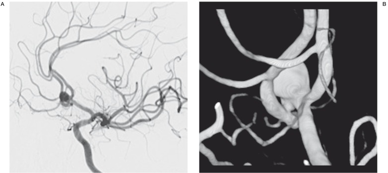
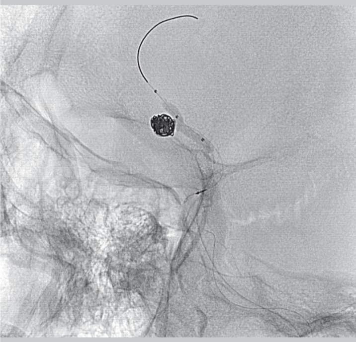
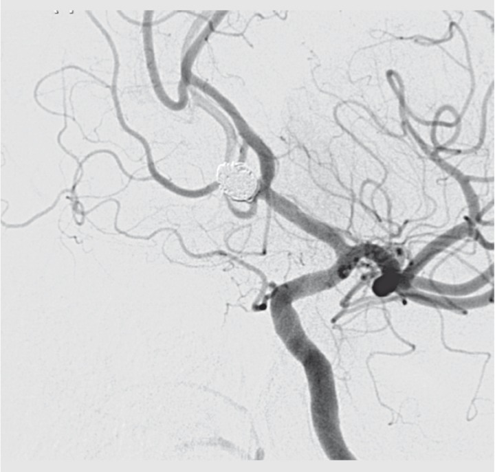
A 58-year-old man, who initially presented with a headache, was referred to our department for treatment of an unruptured AcomA wide-necked aneurysm (Figure 1). The patient had no focal neurologic deficit at the time of admission. The procedure was performed with the patient under general anesthesia and full heparinization on a biplane flat-panel angiographic unit (Integris Allura; Philips Medical Systems, Best, The Netherlands). The anticoagulation protocol during the procedure consisted of an intravenous bolus of 5000 IU heparin, with close monitoring of blood-activated clotting time (ACT) levels, which were adjusted, according to the ACT level, between 200 and seconds. The patient was given intravenous aspirin (250 mg) at the beginning of the procedure. After a full angiogram through bifemoral access, two 6F-guiding catheters (Neuropath guiding catheter; Micrus Endovascular, San Jose, CA, USA) were placed at the origin of the left internal carotid artery (ICA). The balloon microcatheter, Scepter C, was conducted through a 0.014-inch guidewire and positioned in the left A1/A2 segment, across the aneurysmal neck (Figure 2). An Echelon 10 microcatheter (ev3, Plymouth, MN, USA) was placed inside the aneurysm sac through the left A1 segment. Aneurysm coiling was performed under temporary balloon inflation until its angiographic exclusion (Figure 3). We used a 50:50 contrast/saline mixture to inflate the balloon as recommended by the manufacturer. The arterial access sheaths were removed immediately after the procedure, and the puncture sites were closed with a collagen-plug hemostatic closure device (Angio-Seal; Daig/St. Jude Medical, St. Paul, MN, USA).
Figure 1.
A) The left ICA angiogram, oblique view, shows the AcomA aneurysm with a wide neck involving both A2 segments. B) Three-dimensional image of the AcomA aneurysm, showing the left A1 segment and the left and right pericallosal arteries. The right pericallosal artery is clearly emerging from the neck of the aneurysm.
Figure 2.
The left unsubtracted ICA angiogram, oblique view, shows the inflated Scepter C balloon in the left pericallosal artery. The balloon bulges into the origin of the right A2 segment. The Traxcess 14 guidewire is positioned distally into the left A2 segment.
Figure 3.
The left ICA final angiogram, oblique view, shows total aneurysm occlusion, with patency of both A2 arteries.
Discussion
For endovascular management of wide-necked bifurcation aneurysms, various adjunctive techniques, such as the BRT or, as recently reported, Y- or X-stenting, have been proposed to permit safe endovascular occlusion 5-7. In 2003, the use of a new super-compliant balloon (HyperForm; ev3) was reported in the treatment of four complex wide-necked aneurysms in which a regular and less compliant balloon would not have conformed to the anatomy 8. More recently, another technique - the neurovascular self-expanding stent (stent-assisted coiling) - has been employed in the endovascular treatment of complex artery aneurysms 9. The major advantage of the BRT over stent-assisted techniques lies in the fact that the dual antiplatelet therapy during and following treatment recommended in stent-assisted techniques is not needed; the use of these agents may be a concern, especially in ruptured aneurysms. Stent-assisted coiling has been associated with hemorrhagic or thromboembolic complications, with permanent neurologic deficits in up to 7.4% of cases 10.
The balloons currently available for the BRT, HyperForm and HyperGlide (ev3, Plymouth, MN, USA), have a single lumen and navigate guide wires ranging from 0.010 to 0.012 inches. However, the small diameter of the microguidewires makes them more unstable with lower trackability. Moreover, guidewire displacement in a single-lumen balloon catheter can lead to issues such as the wire getting glued within the balloon, the balloon failing to inflate, or the balloon deflating due to a clot in the lumen.
The New Scepter C Occlusion Balloon Catheter is a dual coaxial lumen catheter with a nondetachable low inflation pressure compliant balloon attached to the distal end of the catheter. The catheter is designed to track over a steerable 0.014-inch guidewire. Radiopaque marker bands are located at the ends of the balloon and the distal tip of the catheter to facilitate fluoroscopic visualization. The outer surface of the catheter is coated with a hydrophilic polymer to increase lubricity. The double-lumen construction increases kink resistance for reliable balloon deflation and decreases the potential risk of balloon dysfunction by clotting in the lumen. Moreover, the large guidewire lumen allows enhanced distal navigability, due to better support. Another advantage is that this new balloon catheter is mounted on a 0.014-inch guidewire, the same size of the catheters generally used to deploy an intracranial stent. Thus, if it is necessary to deploy a stent after the BRT, it is possible—thanks to docking-type guidewires such as Traxcess (MicroVention, Tustin, CA, USA)—to exchange the balloon with the stent without the need for another catheterization of the aneurysm parent artery. In addition, the Scepter C has three radiopaque markers, compared to a single-lumen balloon, facilitating visualization (Table 1). Two cases of wide-neck aneurysm treatment using a different double-lumen balloon catheter Ascent (Micrus Endovascular, San Jose, CA, USA) were recently published 11,12. However, the balloon Scepter C used in our report is the only balloon 0.014 inches in diameter available to date with hydrophilic coating, which significantly improves trackability in tortuous vessels. Moreover, the tip of the Scepter C is twice as soft as the Ascent balloon, with, probably, a lower risk of perforation in the case of the single-catheter BRT 11,12. The high compliance property allows easy bulging into the origin of the arterial branches emerging from the aneurysm neck.
Table 1.
Comparison of technologies available.
| HyperForm / HyperGlide | Scepter C | |
|---|---|---|
| Intended use | For use in the blood vessels of the peripheral and neurovasculature where temporary occlusion is desired. These catheters offer a vessel selective technique of temporary vascular occlusion which is useful in selectively stopping or controlling blood flow for balloon-assisted embolization of intracranial aneurysm |
Same |
| Lumen configuration | Single Lumen | Dual coaxial lumen |
| Inner configuration | 0.010 | 0.0165 |
| Outer diameter | 2.2-2.8F | 2.6-2.8F |
| Balloon diameter | 3-5 mm/10-30 mm | 4 mm/10-20 mm |
| Material | Polyether block amide, polyolefin, stainless steel, PTFE, chloroprene |
Polyether block amide, polyolefin, stainless steel, PITE, polyurethane elastomeric alloy |
| No. of markers | 2 | 3 |
| Coating | Hydrophilic coating | Same |
| Guidewire compatibility | 0.010-0.012 | 0.014 or smaller |
Conclusion
We describe the first case of a large-necked intracranial aneurysm successfully treated with the BRT, using the new double-lumen balloon Scepter C. The device proved to be safe and effective for aneurysm occlusion. More experience and longer follow-up periods are required to further assess the value of this new neurovascular device.
References
- 1.Moret J, Cognard C, Weill A, et al. Reconstruction technique in the treatment of wide-neck intracranial aneurysms: long-term angiographic and clinical results. Apropos of 56 cases. J Neuroradiol. 1997;24:30–44. [PubMed] [Google Scholar]
- 2.Pierot L, Spelle L, Vitry F, et al. Immediate clinical outcome of patients harboring unruptured intracranial aneurysms treated by endovascular approach. Results of the ATENA study. Stroke. 2008;39:2497–2504. doi: 10.1161/STROKEAHA.107.512756. [DOI] [PubMed] [Google Scholar]
- 3.Cekirge HS, Yavuz K, Geyik S, et al. HyperForm balloon remodeling in the endovascular treatment of anterior cerebral, middle cerebral, and anterior communicating artery aneurysms: clinical and angiographic follow-up results in 800 consecutive patients. J Neurosurg. 2011;;114:944–953. doi: 10.3171/2010.3.JNS081131. [DOI] [PubMed] [Google Scholar]
- 4.Pierot L, Cognard C, Anxionnat R, et al. Remodeling technique for endovascular treatment of ruptured intracranial aneurysms had a higher rate of adequate postoperative occlusion than did conventional coil embolization with comparable safety. Radiology. 2010;258:546–553. doi: 10.1148/radiol.10100894. [DOI] [PubMed] [Google Scholar]
- 5.Cekirge HS, Yavuz K, Geyik S, et al. A novel “Y” stent flow diversion technique for the endovascular treatment of bifurcation aneurysms without endosaccular coiling. Am J Neuroradiol. 2011;32:1262–1268. doi: 10.3174/ajnr.A2475. [DOI] [PMC free article] [PubMed] [Google Scholar]
- 6.Saatci I, Geyik S, Yavuz K, et al. X-configured stent-assisted coiling in the endovascular treatment of complex anterior communicating artery aneurysms: a novel reconstructive technique. Am J Neuroradiol. 2011;32:E113–117. doi: 10.3174/ajnr.A2111. [DOI] [PMC free article] [PubMed] [Google Scholar]
- 7.Chow MM, Woo HH, Masaryk TJ, et al. A novel endovascular treatment of a wide-necked basilar apex aneurysm by using a Y-configuration, double-stent technique. Am J Neuroradiol. 2004;25:509–512. [PMC free article] [PubMed] [Google Scholar]
- 8.Baldi S, Mounayer C, Piotin M, et al. Balloon-assisted coil placement in wide-necked bifurcation aneurysms by use of a new, compliant balloon microcatheter. Am J Neuroradiol. 2003;24:1222–1225. [PMC free article] [PubMed] [Google Scholar]
- 9.Mocco J, Snyder KV, Albuquerque FC, et al. Treatment of intracranial aneurysms with the Enterprise stent: a multicenter registry. J Neurosurg. 2009;110:35–39. doi: 10.3171/2008.7.JNS08322. [DOI] [PubMed] [Google Scholar]
- 10.Piotin M, Blanc R, Spelle L, et al. Stent-assisted coiling of intracranial aneurysms: clinical and angiographic results in 216 consecutive aneurysms. Stroke. 2010;41:110–115. doi: 10.1161/STROKEAHA.109.558114. [DOI] [PubMed] [Google Scholar]
- 11.Clarencon F, Perot G, Biondi A, et al. The use of the Ascent(R) balloon for a two-in-one remodeling technique: feasibility and initial experience. Neurosurgery. 2011 doi: 10.1227/NEU.0b013e31822c49ad. [published ahead of print July 7 2011] post acceptance. [DOI] [PubMed] [Google Scholar]
- 12.Pukenas B, Albuquerque FC, Weigele JB, et al. Use of a new double-lumen balloon catheter for single catheter balloon-assisted coil embolization of intracranial aneurysms: technical note. Neurosurgery. 2011;69:8–12. doi: 10.1227/NEU.0b013e3182181e3a. [DOI] [PubMed] [Google Scholar]


