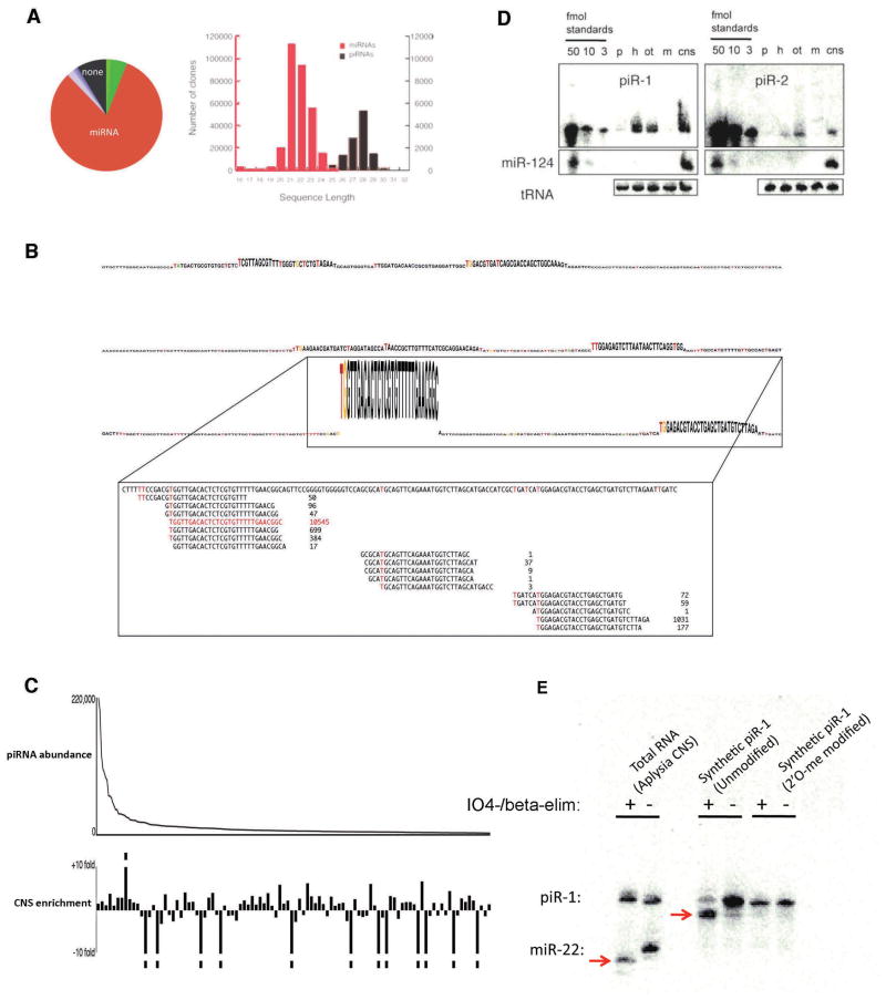
Figure 1.
A. A size histogram of cloned small RNAs from Aplysia CNS revealed two populations, and further characterization confirmed the new class of sequences (shown in black) to be piRNAs.
B. A continuous genomic region in Aplysia encoding a piRNA cluster. A representative 600 bp region within the full 21 kilobase cluster is shown here. The clone frequency of each piRNA is proportional to the height of its nucleotide bases. The clones mapping to the peak piRNA are shown in the inset and U(T) bias start sites are indicated in red.
C. The top 100 piRNAs are plotted on the x-axis in decreasing order of abundance, and their enrichment in CNS is shown as a positive deflection along the y-axis.
D. Two abundant piRNAs are probed for presence in brain (cns), ovotestis (ot), heart (h), muscle (m), and pancreas (p) by quantitative northern blot. Detection of synthetic piRNAs loaded on the far left of the blots, at a concentration of 50 fmol, 10 fmol, and 3 fmol serve as positive controls and allow quantitation. Blots are re-probed with aca-miR-124 and tRNA to control for specificity of signal and equal loading of samples.
E. Total RNA extracted from Aplysia CNS, either periodate treated with beta elimination (+) or untreated (−), was probed on northern blot for piR-1 and miR-22. piR-1 is insensitive to the treatment and is therefore modified at its 3′end. miR-22 is sensititive to the treatment (red arrow), shows an approximate 2-nt shift, and is therefore unmodified at its 3′end.

