Background: Tau phosphorylation affects synaptic transmission, but the underlying mechanism remains elusive.
Results: NMDA receptor activation leads to phosphorylation of endogenous tau, thereby regulating the interaction of tau with Fyn and postsynaptic scaffolding protein PSD95.
Conclusion: Phosphorylation of tau controls the interaction of tau with the postsynaptic PSD95-Fyn-NMDA receptor complex leading to changes in synaptic activity.
Significance: The here described physiological mechanism could go awry during the development of Alzheimer disease.
Keywords: Alzheimer Disease; Amyloid; Glutamate Receptors Ionotropic (AMPA, NMDA); Synapses; Tau
Abstract
Amyloid-β and tau protein are the two most prominent factors in the pathology of Alzheimer disease. Recent studies indicate that phosphorylated tau might affect synaptic function. We now show that endogenous tau is found at postsynaptic sites where it interacts with the PSD95-NMDA receptor complex. NMDA receptor activation leads to a selective phosphorylation of specific sites in tau, regulating the interaction of tau with Fyn and the PSD95-NMDA receptor complex. Based on our results, we propose that the physiologically occurring phosphorylation of tau could serve as a regulatory mechanism to prevent NMDA receptor overexcitation.
Introduction
Alzheimer disease (AD),4 a progressive neurodegenerative disorder, is characterized by histopathological changes in the brain comprising massive neurodegeneration and loss of neuronal connectivity (1). Brains of AD patients reveal extracellular amyloid-β (Aβ) plaques and intracellular neurofibrillary tangles mainly composed of hyperphosphorylated tau protein (2–5). Tau protein is classically known as a microtubule-interacting protein involved in regulating microtubule dynamics and stability (6–8). Data from human genetics and transgenic mouse studies strongly implicate Aβ in the etiology and pathogenesis of AD (9).
Memory deficits in AD patients, however, do not correlate well with Aβ plaque burden but, instead with synaptic marker loss (10). More importantly, learning deficits and synaptic dysfunction in transgenic animal models of AD appear before the formation of plaques, suggesting that a physiological synaptic deficit rather than neuronal loss underlies initial AD development (11–13). Several studies have suggested that Aβ could act as a homeostatic regulator of synaptic strength (14–16). These data have lent support to the notion that perturbations of Aβ levels, and their effects on synapses, might be linked directly to the learning and memory deficits in patients with AD (2, 10, 17, 18). A cellular neurophysiological correlate for learning and memory is synaptic plasticity and its two most well characterized forms, long term potentiation (LTP) and long-term depression (LTD). LTP and LTD can be induced by a transient activation of NMDA-sensitive glutamate receptors and an increase in the postsynaptic calcium concentration (19), causing an increase in synaptic transmission and spine size in LTP (20–25) or reduction in synaptic transmission and spine loss in LTD (26–32).
Interestingly, Aβ was described to localize at synaptic terminals (33), where it was found to enhance NMDA receptor-dependent transmission and to alter synaptic function (34, 35). Furthermore, Aβ can impair LTP as well as facilitate LTD (36, 37). Recent data show that lack of tau protein inhibits Aβ-induced impairment of LTP (38), suggesting that tau protein could play a role in regulating synaptic function. These data have lent fresh support to the hypothesis that the cellular action of tau and Aβ could be linked; in fact suggesting that in the AD signaling cascade Aβ could be upstream of tau.
Ten years ago it was found that Aβ fibrils were able to accelerate the formation of abnormally phosphorylated neurofibrillary tangles in a tau transgenic mouse (39) and that Aβ was inducing tau phosphorylation and toxicity in cultured septal cholinergic neurons (40). More recently it has been shown that soluble Aβ oligomers cause abnormal tau phosphorylation and loss of spines (41). Furthermore, naturally occurring Aβ dimers isolated from AD brains are sufficient to induce AD-type tau phosphorylation and neuronal death (42).
Interestingly, many kinases involved in abnormal tau phosphorylation during AD (like GSK3β and PKA) are also known to play a critical role in synaptic plasticity (43, 44), again suggesting that deregulated tau protein could be involved in perturbing synaptic function during AD. However, whether endogenous tau is localized at synapses or playing an active role in regulating synaptic transmission remains largely unknown.
To address these questions, we studied endogenous tau in organotypic hippocampal slice cultures, primary neurons and synaptosomal preparations from rat brains. We find that a small fraction of tau localizes under physiological conditions in dendrites and spines. There, tau is getting phosphorylated upon NMDA receptor activation through a signaling cascade that is also activated by preincubating neurons with Aβ. Furthermore, we show that tau interacts in a phosphorylation-dependent manner with the PSD95-Fyn-NMDA receptor complex at the postsynaptic site. Taken together, we propose that tau phosphorylation might regulate synaptic activity through changing protein interactions, a mechanism that goes awry during the development of AD.
EXPERIMENTAL PROCEDURES
Primary Cell Culture
Primary hippocampal neurons were prepared following the “Banker protocol” (45). Briefly, neurons were isolated from embryonic day 18 rats and plated on poly-d-lysine-coated glass coverslips (50 g/ml) in 24-well plates at a density of 50,000 cells/well or in 12-well plates at 100,000 cells/well. The plating medium was based on α-MEM (Invitrogen) supplemented with 10% horse serum. The medium was changed to Neurobasal medium (Invitrogen) supplemented with B27 (Invitrogen) and 0.5 mm glutamine after 2–4 h. The medium was changed after 72 h, and 5 μm cytosine arabinoside (Sigma) was added to reduce glial growth. From then on, one third of the medium was exchanged three times per week. Neurons were kept for 3 weeks and then analyzed by immunohistochemistry.
Immunostaining and Fluorescence Imaging
Cells were fixed in 4% paraformaldehyde for 15 min, permeabilized, and blocked for 30 min (0.2% Triton X-100, 0.5% FBS) and incubated with the following antibodies: chicken anti-MAP2 (Millipore; 1:2000); Tau5 (mAb against total Tau, Abcam, 1:100); PHF-1 (mAb against phosphorylated Tau, kindly provided by Peter Davis, 1:100); AT180 (mAb against phosphorylated Tau, Thermo Scientific, 1:100); AT8 (mAb against phosphorylated Tau, Thermo Scientific, 1:100); AT100 (mAb against phosphorylated Tau, Thermo Scientific, 1:100). As secondary antibodies, Alexa Fluor-labeled goat anti-mouse and goat anti-rabbit IgG antibodies were used (Invitrogen). Fluorescent images were taken with a 60× objective on a Fluoview 1000 Laser Scanning Confocal microscope (Olympus) using wavelength and filters according to the fluorophore characteristics.
Electron Microscopy
Hippocampal slices were fixed in 0.1 m cacodylate buffer (pH 7.4; 37 °C) containing 1.5% glutaraldehyde (Canemco & Marivac, Québec, Canada) and 1% paraformaldehyde (Fisher). Slices were postfixed in 1% osmium tetroxide (Canemco & Marivac) in cacodylate buffer for 1 h at room temperature. After washing in buffer, slices were dehydrated in a graded series of alcohols at room temperature, then washed twice for 10 min in 90% ethanol and embedded in LRWhite resin (Canemco & Marivac). The resin was polymerized under anaerobic conditions for 48 h at 56 °C. Ultrathin sections (∼70 nm) were cut with an ultramicrotome and placed on nickel grids coated with Formvar and carbon (MECALAB, Montréal, Québec, Canada). Sections were treated with a saturated solution of sodium metaperiodate in water for 1 min followed by 1% ovalbumin in PBS for 5 min. Sections were then incubated with AT8 primary antibody diluted in PBS containing 1% goat serum overnight. After being washed twice for 5 min in PBS containing 1% ovalbumin, followed by incubation in Colloidal gold coated with IgG (1:1) diluted in PBS for 30 min, sections were stained with uranyl acetate for 2 min and lead citrate for 5 min and analyzed with an electron microscope Philips EM CM 100.
Cell Culture Transfection
HEK-293 cells were maintained in DMEM supplemented with 10% fetal bovine serum (Invitrogen). Cells were plated in 100-mm culture dishes and transfected, using Lipofectamine 2000 reagent (Invitrogen), according to the manufacturer's instructions, with cDNAs of the 4R/0N human tau isoform cloned in pEGFP-C1 vector (Clontech): the wild type tau, tauE (Ser to Glu point mutations for Ser199, Ser202, Thr205, Ser214, Thr231, Ser235, Ser262, Ser356, Ser396, Ser400, Ser404, Ser409); tauA (Ser to Ala point mutations for Ser199, Ser202, Thr205, Ser214, Thr231, Ser235, Ser262, Ser356, Ser396, Ser400, Ser404, Ser409); tau-S199A; tau-T212A, and tau-T231A. Cells were harvested 48 h after transfection and homogenized in lysis buffer (50 mm Tris-HCl (pH 7.4), 150 mm NaCl, 5 mm EDTA, 1.5% Triton X-100, protease inhibitor (Calbiochem)).
Western Blotting
For Western blot analysis, total protein from hippocampal brain slices were obtained by boiling slices for 5 min, sonicating 2 min, and boiling 3 min in 1% SDS. A total of 15–20 μg of total protein was loaded on a 10% SDS-PAGE, separated, and transferred onto Immobilon-P transfer membranes (Millipore). The following antibodies and dilutions were used: chicken anti-MAP2 (Millipore; 1:1000); Tau5 (mAb against total Tau, Abcam, 1:2000); PHF-1 (mAb against phosphorylated Tau, kindly donated by Peter Davis, 1:2000); AT180 (mAb against phosphorylated Tau, Thermo Scientific, 1:1000); AT8 (mAb against phosphorylated Tau, Thermo Scientific, 1:1000); AT100 (mAb against phosphorylated Tau, Thermo Scientific, 1:1000); actin (monoclonal anti-β-actin, Sigma; 1:10,000); Fyn (G-4) (Santa Cruz Biotechnology, 1:2000); GluN2B (G-4) (Millipore, 1:1000). Blots were developed using ECL-Plus (GE Healthcare) or Supersignal West Femto (Pierce). Results were quantified with the ImageJ software and were normalized to the actin signal.
Aβ
Aβ 1–42 (Bachem) or scrambled Aβ (Covance) as control, was dissolved in ice-cold hexafluoroisopropyl alcohol, thoroughly vortexed, aliquoted, the hexafluoroisopropyl alcohol completely evaporated, and frozen at −80 °C until use. The Aβ was dissolved in 4.4 μl of dimethyl sulfoxide, sonicated for 30 s, and used in a 0.5 μm final concentration. To analyze the formation of oligomeric Aβ, 50 μl of slice culture medium from day 1 and day 5 was loaded on a 10–20% continuous gradient Tris-Tricine gel. Western blot analysis was performed by using anti-Aβ antibody (Millipore).
Purification of Synaptosomes
Synaptosomes were purified by discontinuous Percoll gradient procedure according to Dunkley and colleagues (46). Briefly, hippocampi and cortices from adult rats (6–12 months) were homogenized in 10 volumes of 0.32 m sucrose, buffered at pH 7.4 with Tris, using a glass/Teflon tissue grinder (clearance 0.25 mm). The homogenate was centrifuged at 1000 × g, and the supernatant was stratified on a discontinuous Percoll gradient (6, 10, and 20% v/v in Tris-buffered sucrose) and centrifuged at 33,500 × g for 5 min. The layer between 10 and 20% Percoll (synaptosomal fraction) was collected and washed by centrifugation, and the resulting pellet was used for further experimentation.
Immunoprecipitation
Dynabeads (25 μl, from Invitrogen) were washed once with PBS-Tween 2% and incubated with mAb PSD95 (Invitrogen; 1:10) in PBS-Tween 2% for 60 min at room temperature on a nutator. The beads were washed and then incubated for 1 h at 4 °C with 200 μg of protein extract in a total of 500 μl of lysis buffer containing proteinase inhibitors. The beads were washed three times with PBS-Tween buffer. The immunoprecipitated proteins were eluted by boiling for 10 min in Laemmli buffer and analyzed by Western blotting.
Slice Preparation
Hippocampal slices were prepared from p6/7 rat pups as described previously and maintained in culture for 10 days as described earlier (47, 48). For electrophysiological recording, slices were transferred to a submerged recording chamber maintained at 32 °C. For biochemistry, slices were submerged for 5 min in ACSF (119 mm NaCl, 2.5 mm KCl, 26 mm NaHCO3, 1 mm NaH2PO4, 11 mm glucose) containing NMDA (20 μm) before being transferred to ACSF without NMDA.
Electrophysiological Protocols
Extracellular field recordings from CA1 were made with an Axoclamp-2B amplifier (Axon Instruments), and data were acquired using Clampex 10 software (Axon Instruments). Borosilicate glass recording electrodes were filled with ACSF and positioned in CA1. Field EPSCs were evoked by stimulating the Schaffer collaterals at 0.2 Hz with a bipolar tungsten electrode, connected to a stimulus isolation unit (A.M.P.I.). Baseline recording lasted for 20 min.
Simultaneous whole cell recordings were obtained under visual guidance from pairs of nearby control and tau-expressing CA1 pyramidal neurons (GFP-tagged tau WT, tauA, or tauE cloned into Sindbis virus expression vector, Invitrogen). The recording chamber was perfused with ACSF containing 0.1 mm picrotoxin (Sigma), 4 mm CaCl2, 4 mm MgCl2, and 4 mm 2-chloroadenosine (Sigma), bubbled with a Carbogen mix (5% CO2,95% O2). All recordings were performed at 30 °C. Patch pipettes (3–5 megohms) were filled with internal solution containing 115 mm cesium methanesulfonate, 20 mm CsCl, 10 mm HEPES, 2.5 mm MgCl2, 4 mm Na2ATP, 0.4 mm Na3GTP, 10 mm sodium phosphocreatine, 0.6 mm EGTA, at pH 7.25 and 290 mosm. Whole cell recordings were carried out using a MultiClamp700B amplifier (Axon Instruments). Bipolar tungsten stimulation electrodes were placed over Schaffer collateral fibers 250 μm lateral to the recording site in hippocampal slice cultures. EPSC amplitudes were obtained from an average of 100 sweeps at each holding potential. All recordings were done by stimulating two independent synaptic inputs; results from each pathway were averaged and counted as n = 1. The AMPA receptor-mediated EPSC was measured as peak inward current at −60 mV, the NMDA receptor-mediated component was measured as the late component (80–85 ms after stimulus) of the outward current at +40 mV.
Statistical Analysis
Bar diagrams represent the experimental mean; the error bars represent the standard error. For statistical analysis we used the Student t test with a p value of 0.05.
RESULTS
Aβ Affects Tau Phosphorylation at Specific Sites
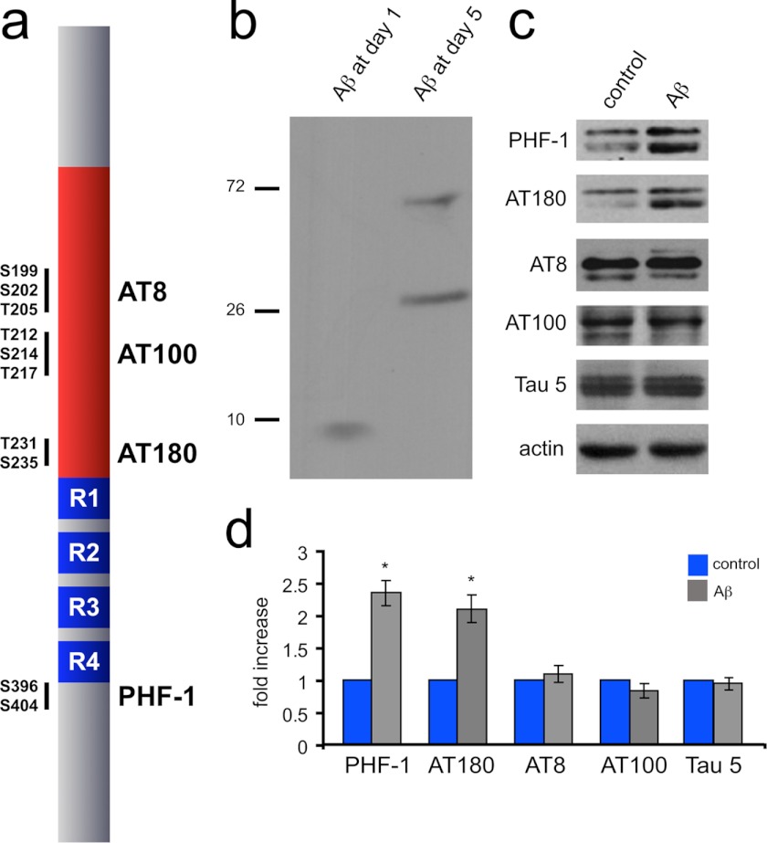
A hallmark of AD is the increased phosphorylation of tau protein at specific sites, recognized by the phosphospecific antibodies AT8, AT100, AT180, and PHF-1 (Fig. 1a). To determine whether Aβ increases tau phosphorylation in general or rather acts on specific sites, we incubated 10-day-old organotypic hippocampal slices with Aβ for 5 days. While we added monomeric Aβ to the slice cultures, Aβ formed oligomers over the incubation time of 5 days (Fig. 1b). Western blot analysis of total protein revealed basal levels of phosphorylated tau protein at all tested sites (AT8, AT100, AT180, and PHF-1, Fig. 1c). Five days incubation with Aβ increased tau phosphorylation significantly at Ser396/Ser404 (PHF-1) and Thr231/Ser235 (AT180) by more than 2-fold, whereas phosphorylation at Ser199/Ser202 (AT8) and Thr212/Thr217/Ser214 (AT100) were not changed (Fig. 1d). The total amount of tau protein did not change with Aβ incubation (indicated by the Tau 5 antibody, Fig. 1, b and c). Taken together, Aβ initially phosphorylates specific sites on tau, rather than increasing global tau phosphorylation at all sites as seen in late AD. This suggests that certain tau phosphorylation sites could depend on additional signaling pathways other than those directly elicited by Aβ.
FIGURE 1.
Aβ increases tau phosphorylation in organotypic slice cultures. a, tau protein (here 4R/0N) contains multiple phosphorylation sites, such as Ser199/202-Thr205, Thr212/217-Ser214, and Thr231-Ser235 located in the proline-rich domain (in red) or Ser396/404 located behind the repeat domain region (in blue). The corresponding phospho-specific antibodies AT8, AT100, AT180, and PHF-1 can detect the phosphorylation at these sites. b, Aβ forms oligomers after 5-day incubation with hippocampal slice cultures. 50 μl of slice culture medium was taken at days 1 and 5, and Aβ was revealed via Western blotting. Monomeric Aβ from day 1 (1 h in culture) forms oligomers after 5 days in culture. Numbers indicate size in kDa. c, Western blot analysis of 5-day Aβ-treated organotypic hippocampal slices showed an increase of tau phosphorylation at sites Ser396/404 (PHF-1) and Thr231-Ser235 (AT180), but not at sites Ser199/202-Thr205 (AT8) and Thr212/217-Ser214 (AT100). The amount of total tau protein (Tau 5) was not affected by treatment with Aβ. d, tau protein showed a significant increase in Aβ-induced phosphorylation at PHF-1 and AT180 sites (Ser396/404 and Thr231-Ser235) compared with control. *, p < 0.05, n = 4. Error bars represent the S.E.
Endogenously Phosphorylated Tau Protein Is Present in Dendrites and Synaptosomal Fractions
We detected a considerable amount of tau protein being phosphorylated at all four sites in untreated hippocampal slice cultures, indicating that tau phosphorylation at those sites might occur under physiological conditions and is not necessarily linked to AD (Fig. 1c). This is consistent with earlier reports where tau phosphorylation was observed under nonpathological conditions (e.g. Ref. 49).
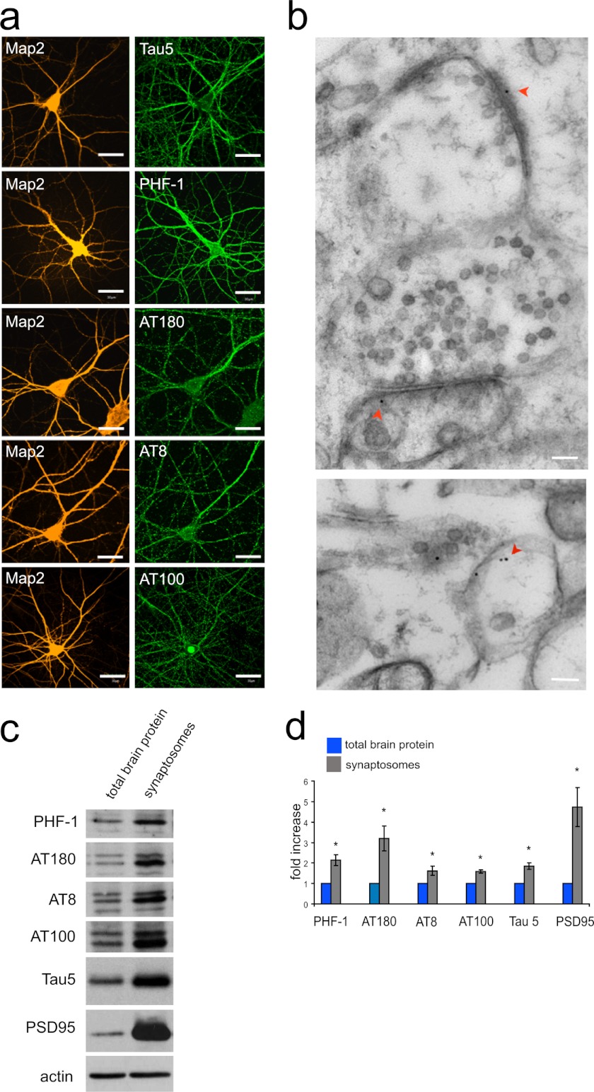
To further elucidate the presence and localization of endogenously phosphorylated tau, we immunostained phosphorylated tau protein in dissociated hippocampal neurons (3 weeks in culture, Fig. 2a). To differentiate axonal from dendritic compartments, we additionally stained for MAP2, a microtubule-associated protein found in dendrites. Staining for total tau protein by the antibody Tau5 showed a strong labeling of axons but also to some extent of dendrites (Fig. 2a, top row). Interestingly, the different phosphorylation specific tau antibodies revealed an increased localization of endogenous phospho-tau to the somatodendritic compartment for PHF-1 and a dendritic localization for AT180 and AT8 staining (Fig. 2a). Only immunostaining with AT100 did not show any preference of dendritic over axonal localization. These results suggest that tau phosphorylation occurs continuously under physiological conditions and that phosphorylated tau is mainly found in dendrites. To elucidate further whether endogenously phosphorylated tau could be found postsynaptically, we performed electron microscopy on hippocampal slice cultures. Given its strong dendritic signal in Fig. 2a, we decided to analyze phosphorylation of the tau AT8 site. We observed tau AT8 reactivity in 10 synapses (from four different preparations). Seven times phosphorylated tau localized postsynaptically (Fig. 2b, upper), twice presynaptically and once pre- and postsynaptically (Fig. 2b, lower). Given the phospho-tau localization in spines and dendrites, we wanted to further analyze tau phosphorylation at synapses with a more quantitative method. We therefore prepared hippocampal synaptosomal fractions from adult rats and analyzed their phospho-tau contents. Phosphorylated tau as well as total tau (detected by Tau5 antibody) was enriched in synaptosomes compared with total brain lysate (Fig. 2, c and d). The synapse-specific protein PSD95 (a marker for the postsynaptic density) was used as a control for the successful enrichment of synaptic proteins in the synaptosomal fraction. In summary, we find low amounts of phosphorylated tau to be present at all times in neurons; however, when phosphorylated, tau tends to be localized in dendrites and near synapses.
FIGURE 2.
Endogenously phosphorylated tau protein is present in dendrites and synapses. a, immunohistochemistry of primary neurons with tau phospho-specific antibodies (PHF-1, AT180, AT8, and AT100) revealed the presence of endogenous phosphorylated tau protein in dendrites (green). Dendrites were labeled with an anti-MAP2 antibody (orange). Total tau protein was found in dendrites and axons (Tau5). Scale bars, 30 μm. b, postsynaptic localization of tau phosphorylated at the AT8 site (red arrowhead) was revealed by immunoelectron microscopy with the AT8 antibody. Scale bars, 100 nm. c, Western blot analysis of synaptosomes for phosphorylated tau protein at sites Ser396/404 (PHF-1), Thr231-Ser235 (AT180), Ser199/202-Thr205 (AT8), and Thr212/217-Ser214 (AT100), as well as total tau (Tau5) revealed increased amounts of tau at synapses compared with total brain preparation. PSD95 protein is a control for the successful enrichment of intact synaptosomes. d, results from c were quantified. *, p < 0.05, n = 3. Error bars represent the S.E.
NMDA Receptor Activation Leads to a Transient Increase in Tau Phosphorylation
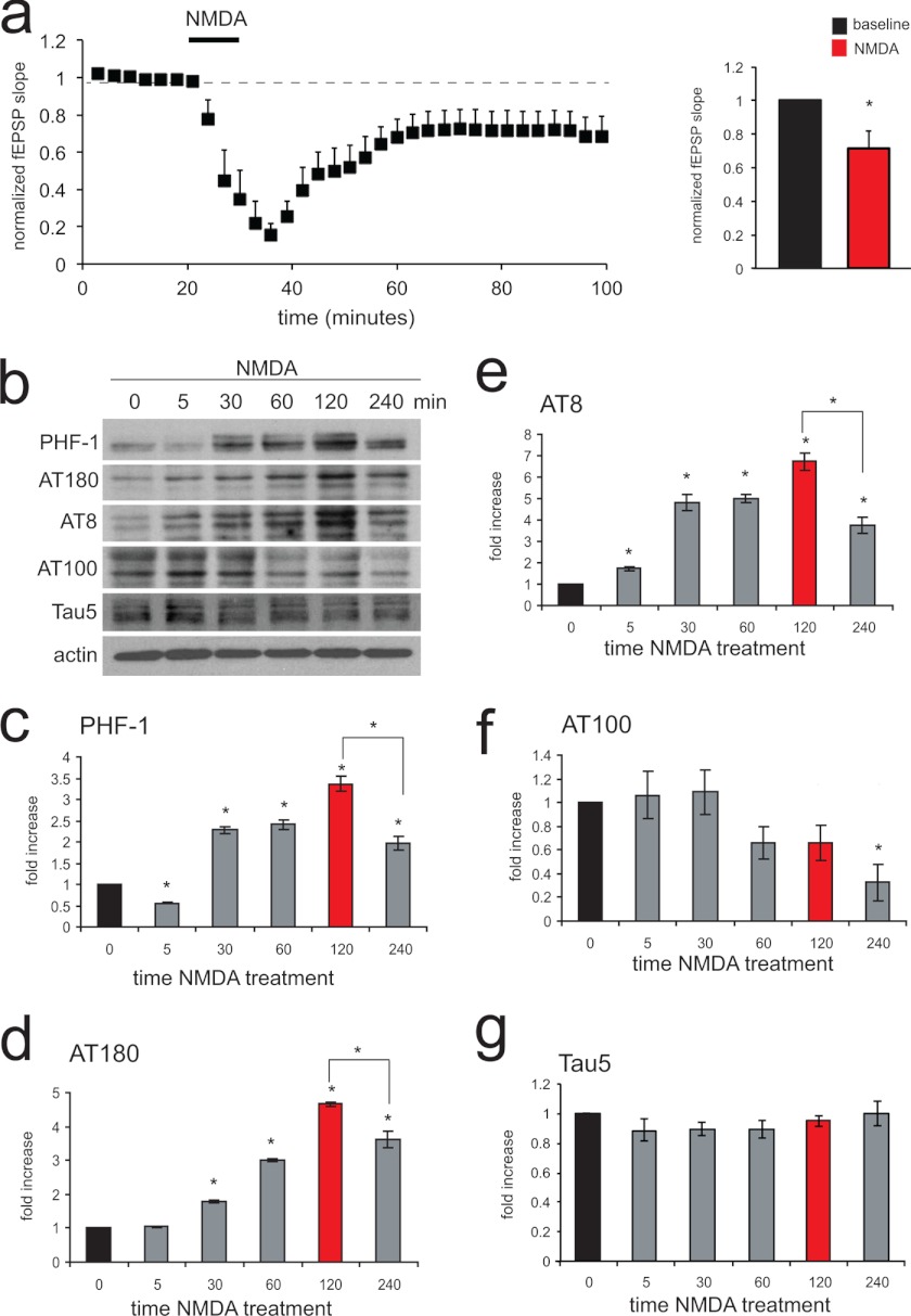
We had shown previously that Aβ engages an NMDA receptor-dependent intracellular signaling pathway leading to LTD like loss of AMPA receptors in the synapse (16). Given that phosphorylated tau was found at synapses (Fig. 2b), we wondered whether the underlying mechanism of tau phosphorylation could be mediated by NMDA receptor-dependent LTD. We took advantage of an NMDA receptor-dependent chemical induction protocol that produces a consistent and long lasting LTD (as shown by extracellular excitatory field recordings, Fig. 3a). We induced NMDA-dependent LTD in hippocampal slice cultures and analyzed tau phosphorylation at different time points. Consistent with our previous data, Western blots from total protein extracts revealed a basal level of phosphorylated tau protein at sites recognized by PHF-1, AT180, AT8, and AT100 antibodies (Fig. 3b, 0 min). After NMDA receptor activation for 5 min, phosphorylation of tau at PHF-1 (Ser396/Ser404), AT180 (Thr231/Ser235), and AT8 (Ser199/Ser202) steadily increased, reaching its maximum at 120 min (Fig. 3, c, PHF-1; d, AT180; and e, AT8). Interestingly, tau phosphorylation at those sites was reversible because the amount of tau phosphorylation was significantly decreased after 240 min compared with 120 min (Fig. 3, c–e). Conversely, tau phosphorylation at the AT100 site (Thr212/Ser217/Ser214) did not show any increase after NMDA receptor activation but a decrease (Fig. 3f). The total amount of tau protein remained constant during the NMDA incubation period (Fig. 3g). Hence, LTD-inducing NMDA receptor activation leads to an increase in tau phosphorylation at sites PHF-1, AT180, as well as AT8 and to a reduction at AT100. Again, like with Aβ, we see a selective increase of certain phosphorylation sites in tau protein rather than a nonselective general increase in phosphorylation of tau (i.e. all sites increase with NMDA except AT100). Furthermore, our data suggest that endogenous tau phosphorylation occurs at the synapse and in dendrites, contrary to the concept of having tau only being phosphorylated in the axon and then transported to the somatodendritic region.
FIGURE 3.
Phosphorylation of tau protein increases after NMDA receptor activation. a, chemically induced LTD in CA1 neurons. 10-day-old hippocampal slice cultures were incubated for 5 min in 20 μm NMDA to activate NMDA receptors and induce LTD. Right, a significant depression is seen after NMDA application (first 15 min compared with last 15 min of recording). n = 4. b, Western blot analysis revealed increased levels of tau phosphorylation after NMDA receptor activation at Ser396/404, Thr231-Ser235, and Ser199/202-Thr205 (labeled by PHF-1, AT180, and AT8) in slice cultures. The increase in tau phosphorylation is reversible (compare 120 with 240 min). No increase in phosphorylation was seen for Thr212/217-Ser214 (AT100). The amount of total tau protein (Tau 5) did not change due to NMDA treatment. c–f, quantification of NMDA receptor-dependent tau phosphorylation is shown at sites PHF-1: Ser396/404 (c), AT180: Thr231-Ser235 (d), AT8: Ser199/202-Thr205 (e), and AT100: Thr212/217-Ser214 (f). g, quantification of total tau protein (Tau 5) over the time course of NMDA treatment. *, p < 0.05, n = 4. Error bars represent the S.E.
Aβ and NMDA Receptor-dependent Tau Phosphorylation Share Common Pathways
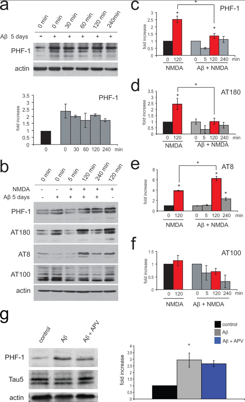
As seen in Fig. 1c, Aβ incubation for 5 days increases tau phosphorylation at PHF-1 and AT180 sites; both sites are also targeted by NMDA receptor activation (Fig. 3). However, AT8 is only increased in response to NMDA (but not Aβ), whereas AT100 does not show an increase with either treatment. We wanted to investigate further whether the combined effects of Aβ and NMDA on tau phosphorylation would activate independent (additive or exponential effect) or common intracellular signaling pathways (occlusion effect). To test this, we wanted to analyze tau phosphorylation in response to NMDA receptor activation in hippocampal slice cultures with or without prior incubation of Aβ for 5 days. To rule out changes in tau phosphorylation due to incubating slices in ACSF and to show that Aβ treatment leads to a long lasting increase in specific tau phosphorylation, we first incubated Aβ-treated slices in ACSF and quantified the changes of phosphorylation at the PHF-1 site over time. As expected, Aβ-treated slices show an increase in PHF-1 phosphorylation, which does not change over time with incubation in ACSF alone (Fig. 4a). To test whether Aβ occludes tau phosphorylation via NMDA receptor signaling (which means “preactivate” the same signaling pathway and therefore reduce the effect of NMDA), we compared normalized NMDA receptor-induced tau phosphorylation with and without prior Aβ exposure. Consistent with our previous findings, 5 days of Aβ incubation leads to an increased tau phosphorylation at the PHF-1 and AT180 sites (Fig. 4b, PHF-1 and AT180; compare the first and second lanes). Furthermore, as seen before, NMDA receptor activation without Aβ induced phosphorylation at PHF-1, AT180, and AT8 sites, again without affecting the AT100 site (compare first with last lanes, Fig. 4b). However, as seen in Fig. 4c, the NMDA receptor-dependent increase of PHF-1 tau phosphorylation is reduced in slices pretreated with Aβ compared with mock-treated slices, indicating that for the phosphorylation of the PHF-1 site, Aβ and NMDA receptor signaling share a common intracellular pathway. The same result was obtained for AT180 (Fig. 4d). As mentioned before, Aβ alone increased tau phosphorylation at PHF-1 and AT180 sites but had no effect on AT8 and AT100 phosphorylation (see Fig. 1d). We hence did not expect an occlusion of NMDA receptor-mediated phosphorylation at these sites after pretreatment with Aβ. Consistent with this idea, we still observe a large transient increase at AT8 phosphorylation after prior incubation with Aβ (Fig. 4e). The AT100 site, however, did not show any increase in tau phosphorylation, neither with Aβ alone, nor with Aβ followed by NMDA receptor activation (Fig. 4f). Finally, we wanted to test whether Aβ induces tau phosphorylation through a direct activation of NMDA receptors. We therefore incubated slice cultures with Aβ in the presence of the NMDA receptor antagonist APV and tested for tau phosphorylation at the PHF-1 site. As seen in Fig. 4g, Aβ induces tau phosphorylation even in the presence of APV, indicating that Aβ does not act through the NMDA receptor.
FIGURE 4.
Aβ partially occludes NMDA receptor-dependent tau phosphorylation. a, in hippocampal slices, Aβ-induced elevated tau phosphorylation at Ser396/404 remained constant over time in the absence of additional NMDA; n = 4. b, Western blot analysis of NMDA receptor-induced tau phosphorylation with (lanes 2–5) or without (lanes 1 and 6) prior Aβ incubation is shown (PHF-1:Ser396/404; AT180:Thr231-Ser235; AT8:Ser199/202-Thr205; and AT100:Thr212/217-Ser214). c, quantification of tau phosphorylation at PHF-1 with and without prior Aβ incubation reveals that Aβ partially occludes NMDA receptor-dependent phosphorylation at this site. d, the same appears with tau phosphorylation at AT180. e, phosphorylation at AT8, however, showed a significant increase in phosphorylation in samples treated with Aβ and NMDA. f, phosphorylation of tau protein at AT100 did not show significant changes. g, blocking the NMDA receptor with APV (50 μm) does not block the Aβ-induced increase of tau phosphorylation at the PHF-1 site. *, p < 0.05, n = 4. Error bars represent the S.E.
Taken together, we find that LTD-inducing NMDA receptor activation leads to an increase in tau phosphorylation at specific sites (PHF-1, AT180, and AT8), which can overlap with Aβ-induced phosphorylation sites (PHF-1 and AT180). However, Aβ does not induce tau phosphorylation through the activation of NMDA receptors.
Phosphomimetic Tau Facilitates LTD Induction
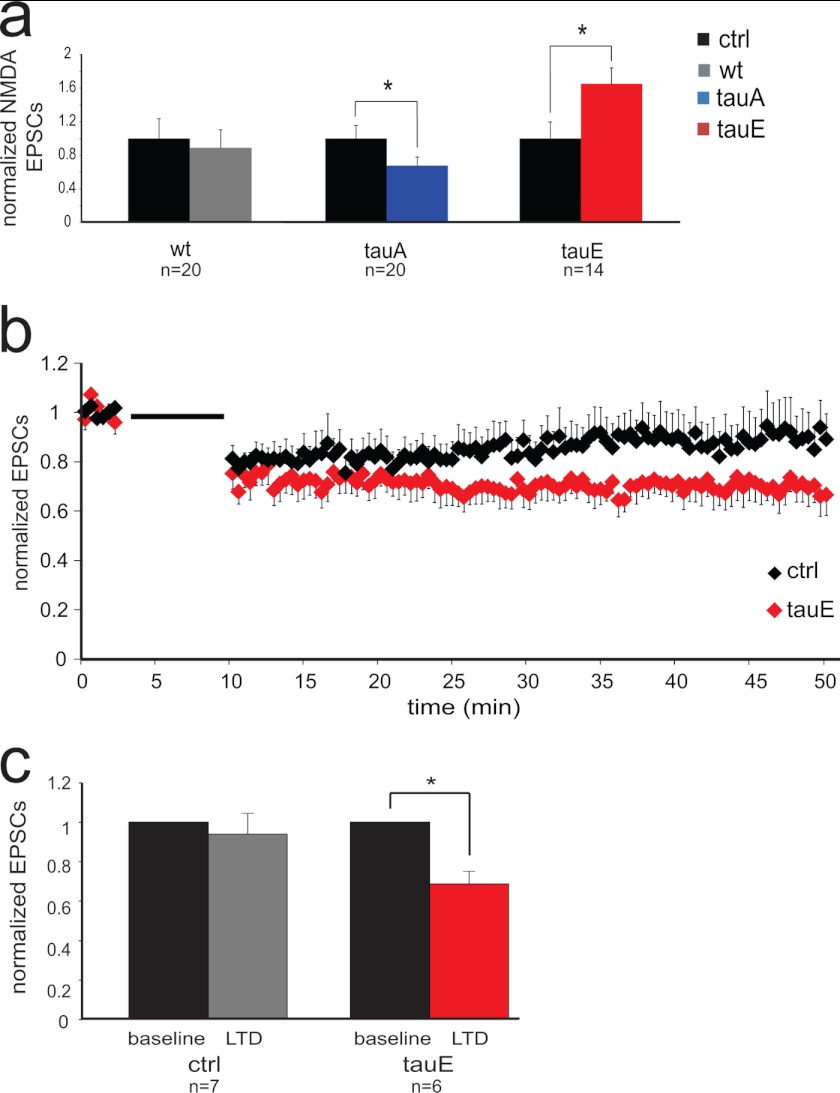
So far, our findings indicate that phosphorylated tau is enriched in dendrites and synaptosomes (Fig. 2) and that phosphorylation of tau protein is increased by Aβ as well as by NMDA receptor activation (Fig. 4). Given these results, we speculated that phosphorylated tau could play a role in NMDA receptor-dependent signaling. To address this question further, we made use of tau constructs carrying either phosphomimetic mutations (tauE, which means point-mutated serines/threonines to glutamates, including the PHF-1, AT8, and AT180 sites) or phospho-blocking mutations (tauA, which means point-mutated serine/threonine to alanine, including the PHF-1, AT8, and AT180 sites). We therefore either overexpressed tau wild type, tauA, or tauE in CA1 neurons of hippocampal slice cultures. Analyzing evoked NMDA receptor-dependent currents, by comparing control cells with tau-expressing cells, we could show that tau alone had no effect on postsynaptic NMDA receptor responses. However, the overexpression of tauA reduced NMDA receptor currents, whereas tauE overexpression increased NMDA receptor currents significantly (Fig. 5a). Contrary to changes in NMDA receptor currents, we did not detect changes in AMPA receptor-dependent transmission with tauA or tauE overexpression (data not shown). Given that NMDA receptor responses are at the core of synaptic plasticity, we wondered whether a facilitation of NMDA receptor responses would lead to a facilitated induction of LTD in cells overexpressing tauE. To test this hypothesis, we chose a weak LTD induction protocol for hippocampal slice cultures (400 stimuli, 1 Hz, CA1 pyramidal cells clamped at −20 mV in 9–10-day-old cultures). We failed to induce a robust LTD in control neurons; however, we were able to obtain a stable LTD in cells expressing tauE (Fig. 5, b and c). Thus, short term overexpression of phosphomimetic tau increases NMDA receptor-dependent synaptic transmission and facilitates induction of LTD. Taken together, we show that Aβ as well as NMDA receptor activation trigger an increase in tau phosphorylation in spines. In return phosphomimetic tau is regulating NMDA receptor-dependent transmission and LTD.
FIGURE 5.
Phosphomimetic tau enhances evoked NMDA receptor EPSCs. Whole cell recordings from CA1 hippocampal neurons in slice cultures are shown. a, mutated phospho-blocked tau (tauA) significantly reduces electrically evoked NMDA receptor EPSCs, whereas the phosphomimetic version (tauE) significantly enhances them. b, a weak LTD induction protocol results in robust LTD expression in neurons expressing tauE, but not in control neurons. c, results from b were quantified, comparing currents recorded during base line with those from the last 3 min (47–50 min). *, p < 0.05. Error bars represent the S.E. n refers to the number of recorded cells.
NMDA Receptor-dependent Tau Phosphorylation Causes Endogenous Tau to Leave the Postsynaptic Complex
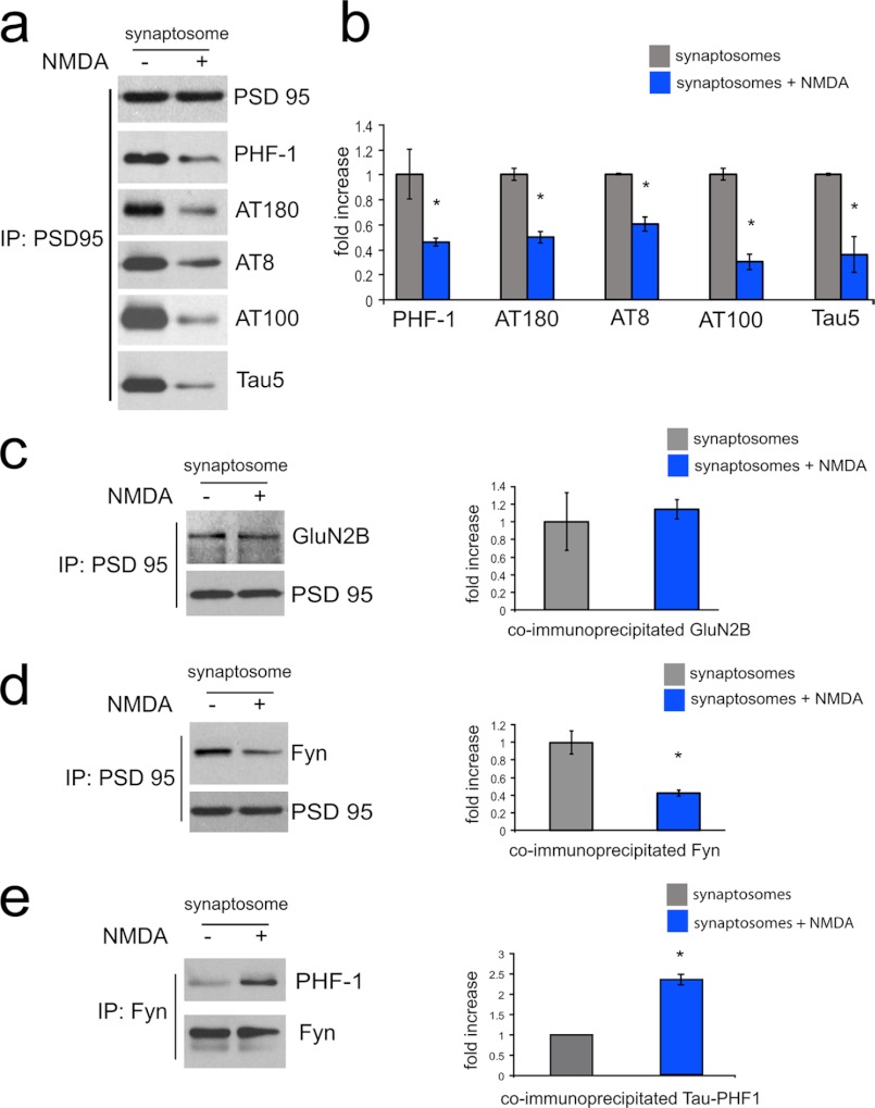
Given these effects of phosphomimetic tau on synaptic NMDA receptor-dependent transmission, we speculated whether endogenous phosphorylated tau could be part of the postsynaptic complex, i.e. tau interacts directly with proteins of the postsynaptic density. The PSD95 protein is a member of the MAGUK protein family, serving as scaffolding proteins in the postsynaptic density, linking ion channel complexes with intracellular signaling cascades. Recent publications suggested a direct interaction between tau and PSD95 (50). We therefore immunoprecipitated PSD95 from synaptosomes and tested for co-immunoprecipitated tau protein. As seen in Fig. 6, a and b, PSD95 co-immunoprecipitated tau (indicated by the Tau5 antibody) as well as phosphorylated tau (PHF-1, AT180, AT8, and AT100). Given this interaction, we wondered whether NMDA receptor activation would change the interaction between tau protein and PSD95. To test this, we treated synaptosomes with NMDA. Surprisingly, we observed a reduction in the interaction of tau with PSD95, suggesting that the activation of NMDA receptors leads to a loss of tau from the postsynaptic density complex (Fig. 6, a and b). Combining our data with those of the literature, one can suggest a multiprotein complex composed of PSD95, NMDA receptors (51), tau (Fig. 6a), and the tyrosine kinase Fyn (52, 53). We wanted to investigate further the fate of this multiprotein complex after NMDA receptor stimulation. Again, we used NMDA-treated synaptosomes to immunoprecipitate PSD95 and analyze for the co-immunoprecipitated NMDA receptor subunit GluN2B (old nomenclature: NR2B) and Fyn. NMDA receptor activation did not change the interaction between GluN2B and PSD95 (Fig. 6c); however, the interaction with Fyn was significantly reduced (Fig. 6d). Interestingly, when analyzing the interaction between tau and Fyn, we found that NMDA receptor activation increased the interaction between phospho-tau and Fyn (Fig. 6e).
FIGURE 6.
Reduced interaction between tau protein and the PSD protein complex after activation of NMDA receptors. a, immunoprecipitation (IP) of PSD95 and co-immunoprecipitated tau from synaptosomal preparations. After NMDA receptor activation, the amount of co-immunoprecipitated phosphorylated tau protein was reduced compared with untreated control, leading to a general loss of tau protein (Tau5 antibody) interaction with PSD95. b, quantification of co-immunoprecipitated phosphorylated tau protein for the sites Ser396/404 (PHF-1), Thr231-Ser235 (AT180), Ser199/202-Thr205 (AT8), Thr212/217-Ser214 (AT100), and total tau (Tau5). c, immunoprecipitation of PSD95 and co-immunoprecipitated GluN2B from the same synaptosomal preparations showing no change upon NMDA receptor activation. d, immunoprecipitation of PSD95 and co-immunoprecipitated Fyn from the same synaptosomal preparations showing a significant loss of Fyn interaction with PSD95 upon NMDA receptor activation. e, immunoprecipitation of Fyn and co-immunoprecipitated tau from the same synaptosomal preparation showing an increase in Fyn and phospho-tau interaction (here PHF-1) upon NMDA receptor activation. *, p < 0.05, n = 3. Error bars represent the S.E.
Given these results it appears that NMDA receptor activation leads to tau phosphorylation, leading to an increase of tau interaction with Fyn, but a decrease of tau interaction with PSD95. One could therefore speculate, that tau transiently increases the Fyn content at the synapse followed by a dissociation of tau/Fyn from the PSD95-NMDA receptor complex.
PSD95-Tau Interaction Is Regulated by Phosphorylation of Tau at Specific Sites
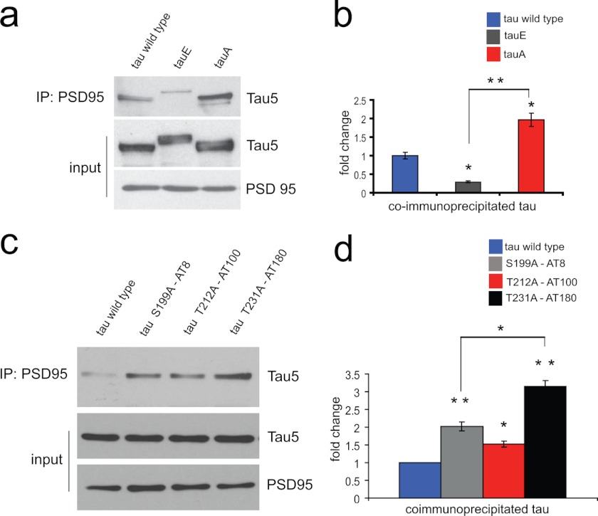
So far, our data suggest that tau protein interacts with the PSD95-GluN2B-Fyn complex and that activation of NMDA receptors regulates this interaction. Although it is very tempting to speculate that NMDA receptor-dependent tau phosphorylation is at the core of this regulation, we lack evidence for this scenario. To address this hypothesis, we wanted to analyze further the effects of tau phosphorylation on its interaction with PSD95. We therefore expressed tau, tauE, or tauA together with PSD95 in HEK cells, immunoprecipitated PSD95, and probed for associated tau protein in Western blots (Fig. 7a). The expression levels of the different tau constructs were monitored with Tau5 antibody. Compared with wild type tau protein, the interaction of tauE with PSD95 is significantly reduced, whereas that of tauA is significantly increased (Fig. 7b). These data strongly support a regulation of PSD95-tau interaction through tau phosphorylation. However, whether different phosphorylated sites in tau have varying effects on the interaction with PSD95 remains unclear. We therefore introduced phospho-blocking serine/threonine to alanine point mutations for three relatively adjacent sites: AT8, AT100, and AT180 (see Fig. 1a), representing sites that are phosphorylated in response to Aβ as well as NMDA (AT180), only NMDA (AT8), or neither (AT100). Again, we tested these tau point mutants for the interaction with PSD95 in HEK cells. All tested phospho-blocked tau constructs increased their interaction with PSD95 as revealed by Western blotting (Fig. 7c). However, although the increase with PSD95 for the AT100 mutant was very moderate, it was more pronounced for the AT8 mutant and strongest for the AT180 mutant. These data corroborate that different phosphorylation sites in tau not only represent different intracellular signaling pathways, but also have different effects on the interaction of tau with other proteins, like PSD95 in postsynaptic densities.
FIGURE 7.
Phosphorylation of tau protein regulates its interaction with PSD95 protein. HEK cells transfected with PSD95 and various tau constructs. a, immunoprecipitated PSD95 revealing the direct interaction between tau and PSD95 (left lane). The tau-PSD95 interaction decreases with phosphomimetic tauE (middle lane) and increases with the phospho-blocked mutant tauA (right lane). b, quantification of a; n = 4. c, phospho-blocking point mutations of tau protein at sites Ser199 (tau S199A-AT8), Thr212 (tau T212A-AT100), and Thr231 (tau T231A-AT180) promoting the interaction between PSD95 and tau protein compared with wild type tau. d, quantification of c, revealing that different point mutations in tau have different effects on its interaction with PSD95. *, p < 0.05, n = 4. Error bars represent the S.E.
DISCUSSION
Aβ and tau protein are the two most prominent factors in the etiology of AD. Recently, several studies suggested that at least some effects of Aβ are executed via the action of tau (38, 54). Although widely agreed upon, that hyperphosphorylated tau can act as a pathologically active form of tau in AD, the underlying mechanisms remain largely elusive. The overall idea of this project was to analyze the effects of endogenously occurring tau phosphorylation, to obtain insight on potential initial mechanisms engaged during AD. We now show that endogenous tau is found at postsynaptic sites and that NMDA receptor activation induces tau phosphorylation. The acute overexpression of phosphomimetic tau leads to an increase in NMDA receptor transmission and thereby to a facilitation of LTD induction. Furthermore, we show that tau interacts with the PSD95-NMDA receptor complex and that selective phosphorylation of tau decreases its affinity to PSD95, but increases interaction with Fyn.
Localization of Phosphorylated Tau in Dendrites
Tau protein was originally described as being overall restricted to axons (55); however, recent results from several groups could show that overexpressed tau can localize in somatodendritic compartments like spines (41, 50, 56). Our results suggest that endogenous tau is also localized in dendritic compartments where it is getting phosphorylated through the action of NMDA receptor activation. Furthermore, the NMDA receptor-mediated increase of tau phosphorylation in synaptosomes argues for the presence of unphosphorylated as well as phosphorylated tau in spines. Interestingly, tau phosphorylation through NMDA receptor activation appears to be reversible (Fig. 3), i.e. tau is likely to oscillate between a phosphorylated and unphosphorylated form at synapses.
These results strongly suggest that in dendritic compartments, phosphorylation of tau fulfills primarily a physiological function, contributing only when deregulated to tauopathies and AD.
NMDA Receptor-dependent Tau Phosphorylation and Synaptic Plasticity
Our results show that tau phosphorylation is increased after LTD-like NMDA receptor activation (Fig. 3). GSK3β as one of the most prominent tau kinases was recently described as a major effector for NMDA receptor-dependent LTD induction (43), an observation that would place tau directly downstream of the LTD signaling pathway. GSK3β targets, among others, the PHF-1, AT8, and AT180 sites in tau (57, 58), sites we identified as bona fide phosphorylation targets after NMDA receptor-dependent LTD induction (Fig. 3). Contrary to these sites, we did not observe AT100 phosphorylation after activation of NMDA receptors (Fig. 3). Interestingly, AT100 does not appear as a GSK3β target site, but was described being phosphorylated in response to the activation of the kinases p70 S6, DYRK1A, and Akt, with the latter two being directly involved in synaptic function (59–61). DYRK1A, originally analyzed for its gene being localized in the Down syndrome critical region, is involved in regulating bidirectional synaptic plasticity, thereby affecting LTP as well as LTD (62), whereas Akt is a key kinase for the induction of LTP (43). It has to be emphasized that in the context of synaptic plasticity, the role of Akt in neurons is to phosphorylate and thereby inhibit GSK3β, allowing the expression of LTP and blocking subsequent LTD induction (most likely a mechanism involved in memory consolidation, (43)). Our result, that NMDA receptor-dependent LTD does not increase tau phosphorylation at the AT100 site (Fig. 3), is in line with this observation, hinting toward a signaling pathway-dependent differential phosphorylation of tau.
We observed tau getting phosphorylated after LTD induction and being part of synaptosomes, suggesting that tau and tau kinases could be localized at the postsynaptic density (Figs. 2 and 3). Indeed, tau is able to interact directly with PSD95 (Fig. 6), a major postsynaptic scaffolding protein, which in turn interacts with NMDA receptors and Src kinases like Fyn (51). Tau, however, is also able to interact with the tyrosine kinase Fyn (52). Interestingly, we see an increase in the interaction of tau and Fyn when tau is phosphorylated as a result of NMDA receptor activation (Fig. 6e). This result is supported by recent studies showing that the interaction between Fyn and tau is regulated through tau phosphorylation at the AT8 site, increasing tau-Fyn interaction by 42-fold after tau is phosphorylated or bearing a phosphomimetic mutation at the AT8 site (63). In our experiments, tau phosphorylation at AT8 is strongly up-regulated through the activation of NMDA receptors (Fig. 3), suggesting that NMDA receptor activation could massively enhance the interaction between tau and Fyn. A prominent role of Fyn at the synapse is to phosphorylate and thereby increase NMDA receptor-dependent transmission (64). It is of particular interest that this change of NMDA receptor phosphorylation through Fyn is regulating synaptic plasticity like LTD and LTP (64). Hence, allowing Fyn to localize to the postsynaptic signaling complex will affect NMDA receptor activity and as a consequence synaptic plasticity. We show that short term expression of phosphomimetic tau leads to an increase in NMDA receptor-dependent synaptic transmission and a facilitation of LTD induction (Fig. 5). Given that a prolonged expression of phospho-tau should induce LTD-like mechanisms leading to AMPA receptor endocytosis, this result appears to be in line with a recent report showing that the prolonged expression of phosphomimetic tau in dissociated neurons can lead to a reduction in AMPA receptor-dependent synaptic transmission (NMDA receptor-dependent transmission was not tested (56)).
Given our result that tau-PSD95 interaction is weakened upon tau phosphorylation (see Fig. 7) but that tau phosphorylation strengthens the tau-Fyn interaction, we propose the following model: the Fyn kinase signaling complex at the synapse contains, among others, PSD95 interacting with NMDA receptor, Fyn, and tau. The activation of NMDA receptors leads to phosphorylation of different sites in tau, which modulates the interaction of tau with its partners: the tau-Fyn interaction is strengthened (see Fig. 6e), whereas the tau-PSD95 interaction is weakened (see Figs. 6 and 7). These changes could lead to a transient increase in synaptic Fyn and hence to a transient increase in NMDA receptor activation before tau-Fyn leaves the PSD95-NMDA receptor complex.
It is furthermore noteworthy that the interaction of tau with different proteins appears to be regulated via different phosphorylation sites: whereas AT8 seems to have a strong effect on tau-Fyn interaction (63), the AT180 site seems to be more important for tau-PSD95 interaction (Fig. 7). Future research has to clarify whether phosphorylation of tau at different sites is a common mechanism for fine-tuning its interaction with different partners.
Implications for Alzheimer Disease
The above discussed phosphorylation of tau as a consequence of NMDA receptor activation suggests that tau phosphorylation at multiple sites (“hyperphosphorylation”) could be part of a physiological intracellular signaling cascade, regulating synaptic NMDA receptor-dependent transmission. We already hypothesized in the past that an early stage of AD could involve Aβ inducing an NMDA receptor-dependent LTD-like pathway (16). Again, we see a similar picture with tau: phosphorylation of tau can be induced by NMDA receptor stimulation as well as by incubation with Aβ (Figs. 3 and 4). It has to be emphasized, however, that acute NMDA receptor-dependent tau phosphorylation was reversible (Fig. 3) whereas the increase in tau phosphorylation after 5 days of Aβ exposure was not (Fig. 4a).
Interestingly, whereas Aβ in our hands does increase tau phosphorylation at the PHF-1 and the AT180, site it has no direct effect on the AT8 or the AT100 site (Figs. 1 and 4). This result could be explained by the fact that in AD, the tau phosphorylation at the AT8 and AT100 sites generally appears at a later stage (65); i.e. we do not mimic the late stages of AD by incubating slice cultures with Aβ for only 5 days but only the early phase where an impaired synaptic transmission rather than neuronal loss would occur (12).
Taken together, we hypothesize that tau phosphorylation is part of a regulatory mechanism to control NMDA receptor activity. During AD, Aβ might perturb this regulatory pathway by increasing specific tau phosphorylation sites, leading to an increase of NMDA receptor activation and in consequence to an increase of NMDA receptor-dependent calcium influx, ultimately causing changes in synaptic plasticity and eventually neurotoxicity as described before (35, 66–68).
Acknowledgments
We thank Drs. Richard Robitaille, Jean-Claude Lacaille, and Vincent Castellucci for critical comments and support.
This work was supported by grants from the Canadian Institutes of Health Research and the Alzheimer Society of Canada.
- AD
- Alzheimer disease
- Aβ
- amyloid-β
- EPSC
- excitatory postsynaptic current
- GSK3β
- glycogen synthase kinase 3β
- LTD
- long term depression
- LTP
- long term potentiation
- PSD95
- postsynaptic density 95
- Tricine
- N-[2-hydroxy-1,1-bis(hydroxymethyl)ethyl]glycine.
REFERENCES
- 1. Samuel W., Masliah E., Hill L. R., Butters N., Terry R. (1994) Hippocampal connectivity and Alzheimer's dementia: effects of synapse loss and tangle frequency in a two-component model. Neurology 44, 2081–2088 [DOI] [PubMed] [Google Scholar]
- 2. Lue L. F., Kuo Y. M., Roher A. E., Brachova L., Shen Y., Sue L., Beach T., Kurth J. H., Rydel R. E., Rogers J. (1999) Soluble amyloid β peptide concentration as a predictor of synaptic change in Alzheimer's disease. Am. J. Pathol. 155, 853–862 [DOI] [PMC free article] [PubMed] [Google Scholar]
- 3. Ingelsson M., Fukumoto H., Newell K. L., Growdon J. H., Hedley-Whyte E. T., Frosch M. P., Albert M. S., Hyman B. T., Irizarry M. C. (2004) Early Aβ accumulation and progressive synaptic loss, gliosis, and tangle formation in AD brain. Neurology 62, 925–931 [DOI] [PubMed] [Google Scholar]
- 4. Grundke-Iqbal I., Iqbal K., Tung Y. C., Quinlan M., Wisniewski H. M., Binder L. I. (1986) Abnormal phosphorylation of the microtubule-associated protein tau (tau) in Alzheimer cytoskeletal pathology. Proc. Natl. Acad. Sci. U.S.A. 83, 4913–4917 [DOI] [PMC free article] [PubMed] [Google Scholar]
- 5. Galván M., David J. P., Delacourte A., Luna J., Mena R. (2001) Sequence of neurofibrillary changes in aging and Alzheimer's disease: a confocal study with phospho-tau antibody, AD2. J. Alzheimers Dis. 3, 417–425 [DOI] [PubMed] [Google Scholar]
- 6. Timm T., Matenia D., Li X. Y., Griesshaber B., Mandelkow E. M. (2006) Signaling from MARK to tau: regulation, cytoskeletal cross-talk, and pathological phosphorylation. Neuro-degenerative Diseases 3, 207–217 [DOI] [PubMed] [Google Scholar]
- 7. Yu J. Z., Rasenick M. M. (2006) Tau associates with actin in differentiating PC12 cells. FASEB J. 20, 1452–1461 [DOI] [PubMed] [Google Scholar]
- 8. Bullmann T., de Silva R., Holzer M., Mori H., Arendt T. (2007) Expression of embryonic tau protein isoforms persist during adult neurogenesis in the hippocampus. Hippocampus 17, 98–102 [DOI] [PubMed] [Google Scholar]
- 9. Selkoe D. J., Schenk D. (2003) Alzheimer's disease: molecular understanding predicts amyloid-based therapeutics. Annu. Rev. Pharmacol. Toxicol. 43, 545–584 [DOI] [PubMed] [Google Scholar]
- 10. Terry R. D., Masliah E., Salmon D. P., Butters N., DeTeresa R., Hill R., Hansen L. A., Katzman R. (1991) Physical basis of cognitive alterations in Alzheimer's disease: synapse loss is the major correlate of cognitive impairment. Ann. Neurol. 30, 572–580 [DOI] [PubMed] [Google Scholar]
- 11. Oddo S., Caccamo A., Shepherd J. D., Murphy M. P., Golde T. E., Kayed R., Metherate R., Mattson M. P., Akbari Y., LaFerla F. M. (2003) Triple-transgenic model of Alzheimer's disease with plaques and tangles: intracellular Aβ and synaptic dysfunction. Neuron 39, 409–421 [DOI] [PubMed] [Google Scholar]
- 12. Hsia A. Y., Masliah E., McConlogue L., Yu G. Q., Tatsuno G., Hu K., Kholodenko D., Malenka R. C., Nicoll R. A., Mucke L. (1999) Plaque-independent disruption of neural circuits in Alzheimer's disease mouse models. Proc. Natl. Acad. Sci. U.S.A. 96, 3228–3233 [DOI] [PMC free article] [PubMed] [Google Scholar]
- 13. Mucke L., Masliah E., Yu G. Q., Mallory M., Rockenstein E. M., Tatsuno G., Hu K., Kholodenko D., Johnson-Wood K., McConlogue L. (2000) High-level neuronal expression of Aβ1–42 in wild-type human amyloid protein precursor transgenic mice: synaptotoxicity without plaque formation. J. Neurosci. 20, 4050–4058 [DOI] [PMC free article] [PubMed] [Google Scholar]
- 14. Chang E. H., Savage M. J., Flood D. G., Thomas J. M., Levy R. B., Mahadomrongkul V., Shirao T., Aoki C., Huerta P. T. (2006) AMPA receptor downscaling at the onset of Alzheimer's disease pathology in double knockin mice. Proc. Natl. Acad. Sci. U.S.A. 103, 3410–3415 [DOI] [PMC free article] [PubMed] [Google Scholar]
- 15. Kamenetz F., Tomita T., Hsieh H., Seabrook G., Borchelt D., Iwatsubo T., Sisodia S., Malinow R. (2003) APP processing and synaptic function. Neuron 37, 925–937 [DOI] [PubMed] [Google Scholar]
- 16. Hsieh H., Boehm J., Sato C., Iwatsubo T., Tomita T., Sisodia S., Malinow R. (2006) AMPAR removal underlies Aβ-induced synaptic depression and dendritic spine loss. Neuron 52, 831–843 [DOI] [PMC free article] [PubMed] [Google Scholar]
- 17. Carter T. L., Rissman R. A., Mishizen-Eberz A. J., Wolfe B. B., Hamilton R. L., Gandy S., Armstrong D. M. (2004) Differential preservation of AMPA receptor subunits in the hippocampi of Alzheimer's disease patients according to Braak stage. Exp. Neurol. 187, 299–309 [DOI] [PubMed] [Google Scholar]
- 18. Masliah E., Mallory M., Alford M., DeTeresa R., Hansen L. A., McKeel D. W., Jr., Morris J. C. (2001) Altered expression of synaptic proteins occurs early during progression of Alzheimer's disease. Neurology 56, 127–129 [DOI] [PubMed] [Google Scholar]
- 19. Chittajallu R., Alford S., Collingridge G. L. (1998) Ca2+ and synaptic plasticity. Cell Calcium 24, 377–385 [DOI] [PubMed] [Google Scholar]
- 20. Sheng M., Kim M. J. (2002) Postsynaptic signaling and plasticity mechanisms. Science 298, 776–780 [DOI] [PubMed] [Google Scholar]
- 21. Bredt D. S., Nicoll R. A. (2003) AMPA receptor trafficking at excitatory synapses. Neuron 40, 361–379 [DOI] [PubMed] [Google Scholar]
- 22. Collingridge G. L., Isaac J. T., Wang Y. T. (2004) Receptor trafficking and synaptic plasticity. Nat. Rev. Neurosci. 5, 952–962 [DOI] [PubMed] [Google Scholar]
- 23. Bliss T. V., Lomo T. (1973) Long-lasting potentiation of synaptic transmission in the dentate area of the anaesthetized rabbit following stimulation of the perforant path. J. Physiol. 232, 331–356 [DOI] [PMC free article] [PubMed] [Google Scholar]
- 24. Malinow R., Malenka R. C. (2002) AMPA receptor trafficking and synaptic plasticity. Annu. Rev. Neurosci. 25, 103–126 [DOI] [PubMed] [Google Scholar]
- 25. Matsuzaki M., Honkura N., Ellis-Davies G. C., Kasai H. (2004) Structural basis of long-term potentiation in single dendritic spines. Nature 429, 761–766 [DOI] [PMC free article] [PubMed] [Google Scholar]
- 26. Lee S. H., Liu L., Wang Y. T., Sheng M. (2002) Clathrin adaptor AP2 and NSF interact with overlapping sites of GluR2 and play distinct roles in AMPA receptor trafficking and hippocampal LTD. Neuron 36, 661–674 [DOI] [PubMed] [Google Scholar]
- 27. Man H. Y., Lin J. W., Ju W. H., Ahmadian G., Liu L., Becker L. E., Sheng M., Wang Y. T. (2000) Regulation of AMPA receptor-mediated synaptic transmission by clathrin-dependent receptor internalization. Neuron 25, 649–662 [DOI] [PubMed] [Google Scholar]
- 28. Lüscher C., Xia H., Beattie E. C., Carroll R. C., von Zastrow M., Malenka R. C., Nicoll R. A. (1999) Role of AMPA receptor cycling in synaptic transmission and plasticity. Neuron 24, 649–658 [DOI] [PubMed] [Google Scholar]
- 29. Nägerl U. V., Eberhorn N., Cambridge S. B., Bonhoeffer T. (2004) Bidirectional activity-dependent morphological plasticity in hippocampal neurons. Neuron 44, 759–767 [DOI] [PubMed] [Google Scholar]
- 30. Zhou Q., Homma K. J., Poo M. M. (2004) Shrinkage of dendritic spines associated with long-term depression of hippocampal synapses. Neuron 44, 749–757 [DOI] [PubMed] [Google Scholar]
- 31. Snyder E. M., Philpot B. D., Huber K. M., Dong X., Fallon J. R., Bear M. F. (2001) Internalization of ionotropic glutamate receptors in response to mGluR activation. Nat. Neurosci. 4, 1079–1085 [DOI] [PubMed] [Google Scholar]
- 32. Xiao M. Y., Zhou Q., Nicoll R. A. (2001) Metabotropic glutamate receptor activation causes a rapid redistribution of AMPA receptors. Neuropharmacology 41, 664–671 [DOI] [PubMed] [Google Scholar]
- 33. Pham E., Crews L., Ubhi K., Hansen L., Adame A., Cartier A., Salmon D., Galasko D., Michael S., Savas J. N., Yates J. R., Glabe C., Masliah E. (2010) Progressive accumulation of amyloid-β oligomers in Alzheimer's disease and in amyloid precursor protein transgenic mice is accompanied by selective alterations in synaptic scaffold proteins. FEBS J. 277, 3051–3067 [DOI] [PMC free article] [PubMed] [Google Scholar]
- 34. Hoe H. S., Fu Z., Makarova A., Lee J. Y., Lu C., Feng L., Pajoohesh-Ganji A., Matsuoka Y., Hyman B. T., Ehlers M. D., Vicini S., Pak D. T., Rebeck G. W. (2009) The effects of amyloid precursor protein on postsynaptic composition and activity. J. Biol. Chem. 284, 8495–8506 [DOI] [PMC free article] [PubMed] [Google Scholar]
- 35. Selkoe D. J. (2002) Alzheimer's disease is a synaptic failure. Science 298, 789–791 [DOI] [PubMed] [Google Scholar]
- 36. Walsh D. M., Klyubin I., Fadeeva J. V., Cullen W. K., Anwyl R., Wolfe M. S., Rowan M. J., Selkoe D. J. (2002) Naturally secreted oligomers of amyloid β protein potently inhibit hippocampal long-term potentiation in vivo. Nature 416, 535–539 [DOI] [PubMed] [Google Scholar]
- 37. Cheng L., Yin W. J., Zhang J. F., Qi J. S. (2009) Amyloid β protein fragments 25–35 and 31–35 potentiate long-term depression in hippocampal CA1 region of rats in vivo. Synapse 63, 206–214 [DOI] [PubMed] [Google Scholar]
- 38. Shipton O. A., Leitz J. R., Dworzak J., Acton C. E., Tunbridge E. M., Denk F., Dawson H. N., Vitek M. P., Wade-Martins R., Paulsen O., Vargas-Caballero M. (2011) Tau protein is required for amyloid β-induced impairment of hippocampal long-term potentiation. J. Neurosci. 31, 1688–1692 [DOI] [PMC free article] [PubMed] [Google Scholar]
- 39. Götz J., Chen F., van Dorpe J., Nitsch R. M. (2001) Formation of neurofibrillary tangles in P301l tau transgenic mice induced by Aβ 42 fibrils. Science 293, 1491–1495 [DOI] [PubMed] [Google Scholar]
- 40. Zheng W. H., Bastianetto S., Mennicken F., Ma W., Kar S. (2002) Amyloid β peptide induces tau phosphorylation and loss of cholinergic neurons in rat primary septal cultures. Neuroscience 115, 201–211 [DOI] [PubMed] [Google Scholar]
- 41. Zempel H., Thies E., Mandelkow E., Mandelkow E. M. (2010) Aβ oligomers cause localized Ca2+ elevation, missorting of endogenous tau into dendrites, tau phosphorylation, and destruction of microtubules and spines. J. Neurosci. 30, 11938–11950 [DOI] [PMC free article] [PubMed] [Google Scholar]
- 42. Jin M., Shepardson N., Yang T., Chen G., Walsh D., Selkoe D. J. (2011) Soluble amyloid β-protein dimers isolated from Alzheimer cortex directly induce tau hyperphosphorylation and neuritic degeneration. Proc. Natl. Acad. Sci. U.S.A. 108, 5819–5824 [DOI] [PMC free article] [PubMed] [Google Scholar]
- 43. Peineau S., Taghibiglou C., Bradley C., Wong T. P., Liu L., Lu J., Lo E., Wu D., Saule E., Bouschet T., Matthews P., Isaac J. T., Bortolotto Z. A., Wang Y. T., Collingridge G. L. (2007) LTP inhibits LTD in the hippocampus via regulation of GSK3β. Neuron 53, 703–717 [DOI] [PubMed] [Google Scholar]
- 44. Grey K. B., Burrell B. D. (2010) Co-induction of LTP and LTD and its regulation by protein kinases and phosphatases. J. Neurophysiol. 103, 2737–2746 [DOI] [PMC free article] [PubMed] [Google Scholar]
- 45. Kaech S., Banker G. (2006) Culturing hippocampal neurons. Nat. Protoc. 1, 2406–2415 [DOI] [PubMed] [Google Scholar]
- 46. Dunkley P. R., Jarvie P. E., Robinson P. J. (2008) A rapid Percoll gradient procedure for preparation of synaptosomes. Nat. Protoc. 3, 1718–1728 [DOI] [PubMed] [Google Scholar]
- 47. Hayashi Y., Shi S. H., Esteban J. A., Piccini A., Poncer J. C., Malinow R. (2000) Driving AMPA receptors into synapses by LTP and CaMKII: requirement for GluR1 and PDZ domain interaction. Science 287, 2262–2267 [DOI] [PubMed] [Google Scholar]
- 48. Shi S., Hayashi Y., Esteban J. A., Malinow R. (2001) Subunit-specific rules governing AMPA receptor trafficking to synapses in hippocampal pyramidal neurons. Cell 105, 331–343 [DOI] [PubMed] [Google Scholar]
- 49. Matsuo E. S., Shin R. W., Billingsley M. L., Van deVoorde A., O'Connor M., Trojanowski J. Q., Lee V. M. (1994) Biopsy-derived adult human brain tau is phosphorylated at many of the same sites as Alzheimer's disease-paired helical filament tau. Neuron 13, 989–1002 [DOI] [PubMed] [Google Scholar]
- 50. Ittner L. M., Ke Y. D., Delerue F., Bi M., Gladbach A., van Eersel J., Wölfing H., Chieng B. C., Christie M. J., Napier I. A., Eckert A., Staufenbiel M., Hardeman E., Götz J. (2010) Dendritic function of tau mediates amyloid-β toxicity in Alzheimer's disease mouse models. Cell 142, 387–397 [DOI] [PubMed] [Google Scholar]
- 51. Sheng M., Hoogenraad C. C. (2007) The postsynaptic architecture of excitatory synapses: a more quantitative view. Annu. Rev. Biochem. 76, 823–847 [DOI] [PubMed] [Google Scholar]
- 52. Tezuka T., Umemori H., Akiyama T., Nakanishi S., Yamamoto T. (1999) PSD-95 promotes Fyn-mediated tyrosine phosphorylation of the N-methyl-d-aspartate receptor subunit NR2A. Proc. Natl. Acad. Sci. U.S.A. 96, 435–440 [DOI] [PMC free article] [PubMed] [Google Scholar]
- 53. Lee G., Newman S. T., Gard D. L., Band H., Panchamoorthy G. (1998) Tau interacts with Src-family non-receptor tyrosine kinases. J. Cell Sci. 111, 3167–3177 [DOI] [PubMed] [Google Scholar]
- 54. Roberson E. D., Halabisky B., Yoo J. W., Yao J., Chin J., Yan F., Wu T., Hamto P., Devidze N., Yu G. Q., Palop J. J., Noebels J. L., Mucke L. (2011) Amyloid-β/Fyn-induced synaptic, network, and cognitive impairments depend on tau levels in multiple mouse models of Alzheimer's disease. J. Neurosci. 31, 700–711 [DOI] [PMC free article] [PubMed] [Google Scholar]
- 55. Kosik K. S., Finch E. A. (1987) MAP2 and tau segregate into dendritic and axonal domains after the elaboration of morphologically distinct neurites: an immunocytochemical study of cultured rat cerebrum. J. Neurosci. 7, 3142–3153 [DOI] [PMC free article] [PubMed] [Google Scholar]
- 56. Hoover B. R., Reed M. N., Su J., Penrod R. D., Kotilinek L. A., Grant M. K., Pitstick R., Carlson G. A., Lanier L. M., Yuan L. L., Ashe K. H., Liao D. (2010) Tau mislocalization to dendritic spines mediates synaptic dysfunction independently of neurodegeneration. Neuron 68, 1067–1081 [DOI] [PMC free article] [PubMed] [Google Scholar]
- 57. Lovestone S., Hartley C. L., Pearce J., Anderton B. H. (1996) Phosphorylation of tau by glycogen synthase kinase-3β in intact mammalian cells: the effects on the organization and stability of microtubules. Neuroscience 73, 1145–1157 [DOI] [PubMed] [Google Scholar]
- 58. Ishiguro K., Omori A., Takamatsu M., Sato K., Arioka M., Uchida T., Imahori K. (1992) Phosphorylation sites on tau by tau protein kinase I, a bovine derived kinase generating an epitope of paired helical filaments. Neurosci. Lett. 148, 202–206 [DOI] [PubMed] [Google Scholar]
- 59. Ryoo S. R., Jeong H. K., Radnaabazar C., Yoo J. J., Cho H. J., Lee H. W., Kim I. S., Cheon Y. H., Ahn Y. S., Chung S. H., Song W. J. (2007) DYRK1A-mediated hyperphosphorylation of tau: a functional link between Down syndrome and Alzheimer disease. J. Biol. Chem. 282, 34850–34857 [DOI] [PubMed] [Google Scholar]
- 60. Pei J. J., An W. L., Zhou X. W., Nishimura T., Norberg J., Benedikz E., Götz J., Winblad B. (2006) P70 S6 kinase mediates tau phosphorylation and synthesis. FEBS Lett. 580, 107–114 [DOI] [PubMed] [Google Scholar]
- 61. Ksiezak-Reding H., Pyo H. K., Feinstein B., Pasinetti G. M. (2003) Akt/PKB kinase phosphorylates separately Thr212 and Ser214 of tau protein in vitro. Biochim. Biophys. Acta 1639, 159–168 [DOI] [PubMed] [Google Scholar]
- 62. Ahn K. J., Jeong H. K., Choi H. S., Ryoo S. R., Kim Y. J., Goo J. S., Choi S. Y., Han J. S., Ha I., Song W. J. (2006) DYRK1A BAC transgenic mice show altered synaptic plasticity with learning and memory defects. Neurobiol. Dis. 22, 463–472 [DOI] [PubMed] [Google Scholar]
- 63. Bhaskar K., Yen S. H., Lee G. (2005) Disease-related modifications in tau affect the interaction between Fyn and Tau. J. Biol. Chem. 280, 35119–35125 [DOI] [PubMed] [Google Scholar]
- 64. Salter M. W., Kalia L. V. (2004) Src kinases: a hub for NMDA receptor regulation. Nat. Rev. Neurosci. 5, 317–328 [DOI] [PubMed] [Google Scholar]
- 65. Luna-Muñoz J., Chávez-Macías L., García-Sierra F., Mena R. (2007) Earliest stages of tau conformational changes are related to the appearance of a sequence of specific phospho-dependent tau epitopes in Alzheimer's disease. J. Alzheimers Dis. 12, 365–375 [DOI] [PubMed] [Google Scholar]
- 66. Venkitaramani D. V., Chin J., Netzer W. J., Gouras G. K., Lesne S., Malinow R., Lombroso P. J. (2007) β-Amyloid modulation of synaptic transmission and plasticity. J. Neurosci. 27, 11832–11837 [DOI] [PMC free article] [PubMed] [Google Scholar]
- 67. Mattson M. P. (2010) ER calcium and Alzheimer's disease: in a state of flux. Sci. Signal. 3, pe10. [DOI] [PMC free article] [PubMed] [Google Scholar]
- 68. Demuro A., Parker I., Stutzmann G. E. (2010) Calcium signaling and amyloid toxicity in Alzheimer disease. J. Biol. Chem. 285, 12463–12468 [DOI] [PMC free article] [PubMed] [Google Scholar]


