
Figure 6.
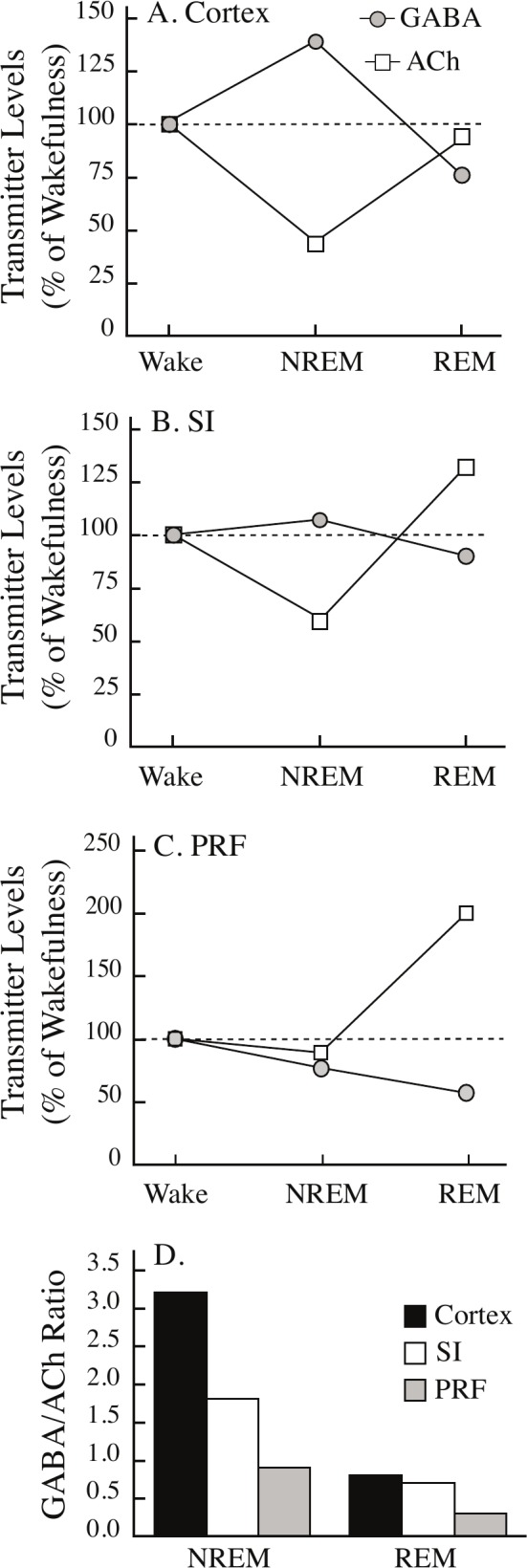
The ratio of GABA-to-ACh differs across brain regions and sleep states. Extracellular levels of GABA and acetylcholine (ACh) in theA, cerebral cortex, B, substantia innominata (SI) region of the basal forebrain, and C, pontine reticular formation (PRF) show differential changes during rapid eye movement sleep (REM) and non-REM sleep (NREM) relative to wakefulness (Wake). D. The ratio of GABA-to-ACh is plotted by sleep state and brain region. The GABA-to-ACh ratio was set to 1.0 during wakefulness. The Table 1 legend provides citations for all measures of GABA and ACh.