



Abstract
Among the ectopic breasts, an ectopic breast tissue on vulva is an extremely rare case, especially in adult period. To our knowledge only 38 cases of ectopic breast tissue on vulva are documented in the world literature, out of which only 10 cases of unilateral ectopic breast tissue on vulva has been reported. Because of its rarity here we report a case of unilateral ectopic breast tissue on vulva in an adult female.
Keywords: Ectopic breast tissue, Vulva, Adult female
Introduction
Ectopic breast tissue is a residual tissue that persists during the embryonic development along ectodermal primitive milk streaks. Incomplete involution anywhere along the bilateral primitive embryonic milk streak can result in accessory or ectopic breast tissue [1]. It may occur anywhere along the bilateral primitive embryonic milk line, extending from the axilla to the groin [2]. Thoracic, abdominal, inguinal and vulvar ectopic breasts whether isolated, multiple or in conjunction with axillary breast tissue are much less frequent [3]. Clinically it is commonly confused with other swelling like lipoma. Diagnosis is possible only after histopathological examination of tissue.
Case History
Forty-five years old female patient referred with history of swelling at vulva on left side since 5 years. Initially it was size of peanut but very gradually increased over the period of 5 years and attended the present size of lemon. Swelling was not associated with pain or any discharge but patient was worried because swelling has gradually increased in size. Now patient had discomfort while walking due to friction of swelling with left thigh for which she sought medical advice.
Local examination revealed single ovoid, pedunculated swelling on left labia majora which measured about 4 cm × 4 cm. (Fig. 1) Swelling was not warm and non tender. It was firm rubbery in consistency. No abnormalities of vulval skin were noted. Swelling was not fixed to skin or underlying structures. There was no inguinal lymphadenopathy. Excisional biopsy was performed. Histopathological examination revealed features of normal breast tissue. (Fig. 2)
Fig. 1.
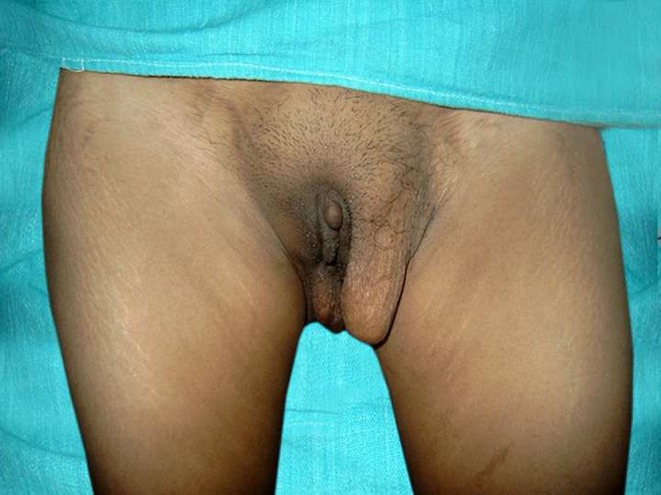
Photograph showing ectopic mammary gland on left labia majora in standing position
Fig. 2.

Photograph showing microscopic features of ectopic breast tissue composed of glands and fibrocollagenous and vascular scanty stroma. Glands are arranged in lobules and are lined by double layered epithelium i.e. inner epithelium and outer myoepithelium. (H .and E. ×100)
Discussion
Ectopic breast tissue occurs along the milk lines, extending bilaterally from the mid-axillae through the normal breasts and then inferiorly to the medial groins. In women, the inferior extensions of the milk lines transverse the vulva bilaterally. The embryological anlage of the milk line is the milk ridge, which later undergoes complete atrophy except for short segments that remain in the pectoral region to give rise to normal breasts. Ectopic or supernumerary breasts develop from portions of the milk ridges that fail to atrophy. Clinically, ectopic breasts are encountered in 1% to 6% of women [3].
Various theories about causation of supernumerary breast have been put forward. Schultze (1892) described a pair of band-like ectodermal thickenings in mammalian embryos running diagonally along the ventrolateral body wall from the axillary to the pubic region which he called “Milk-lines.”In the course of normal development these lines undergo fragmentation to form a series of ectodermal accumulations which he called “Anlagen” or rudiments of future mammary glands . Incomplete suppression of redundant Anlagen leads to the appearance of supernumerary breasts, which may be located anywhere along the milk lines on one or both sides [4].
Deaver & McFarland (1918) demonstrated a strong hereditary factor in the causation of the supernumerary breasts. According to them, unusual sites of ectopic breast tissue (neck, face, arms, ears, legs and buttocks) may be attributed to misplacement of embryonic mammary Anlagen or to mammary differentiation of apocrine sweat glands [4].
According to Darwin’s theory, the anomaly may be regarded as an atavistic or reversionary manifestation, in which, for some obscure reason, a remote ancestral characteristic unexpectedly makes its appearance. However, this theory does not adequately explain the occurrence of breasts in those unusual sites such as neck, face, arms, ears, legs and buttocks as reported by Deaver and McFarland [4].
The clinical presentation of ectopic breasts is highly variable. In some cases the glandular tissue of an ectopic breast is partly or entirely replaced by fat and may be diagnosed clinically as a lipoma, especially if the nipple and areola are unapparent [3]. Vulval breasts, although congenital in origin, do not as a rule attract attention until they become enlarged or active at the time of puberty or in association with pregnancy and lactation. At times the aberrant glands may remain quiescent and unnoticed throughout puberty and repeated pregnancies as occurred in our case [4].
Vulval breasts can undergo many physiological and pathologic changes that occur in the normally situated breast [5]. They respond to hormonal influences at puberty, in the menstrual cycle, during pregnancy and lactation. Complications common to normal breasts including neoplastic changes (both benign and malignant) may also occur [4]. Diagnosis of ectopic breast tissue on vulva is possible only after histopathological examination of tissue. FNAC may be helpful in preoperative diagnosis of ectopic breast tissue [6].
Vulval breasts serve no useful function in the human; on the contrary they act as the potent source of diseases. Therefore whenever discovered they should be promptly removed [4]. Also clinician should be aware that ectopic breast tissue can occur in this location so whenever swelling is detected at vulva ectopic breast tissue should be considered as one of the differential diagnosis.
References
- 1.Dordevic M, Jovanovic B, Mitrovic S, Dordevic G. Ectopic mammary tissue in vulva. Vojnosanit Pregl. 2008;65(5):407–409. doi: 10.2298/VSP0805407D. [DOI] [PubMed] [Google Scholar]
- 2.Aksoy HM, Aksoy B, Portakal S, Ozdemir A. An uncommon location for accessory breast tissue: inner thigh. Eur J Plast Surg. 2008;31:339–340. doi: 10.1007/s00238-008-0286-3. [DOI] [Google Scholar]
- 3.Rosen PP. Abnormalities of mammary growth and development. Rosen’s breast pathology. Philadelphia: Lippincott William’s & Wilkins; 2009. pp. 28–31. [Google Scholar]
- 4.Tow SH, Shanmugaratnam K. Supernumerary mammary gland in the vulva. Br Med J. 1962;2(5314):1234–1236. doi: 10.1136/bmj.2.5314.1234. [DOI] [PMC free article] [PubMed] [Google Scholar]
- 5.Rosai J. Female reproductive system. Rosai and Ackerman’s Surgical Pathology, Vol-2. St. Louis: Mosby Elsevier; 2005. p. 1483. [Google Scholar]
- 6.Kapila K, al-Rabah NA, Junaid TA. Ectopic breast tissue on the vulva diagnosed by fine needle aspiration. Acta Cytol. 1998;42(6):1480–1481. doi: 10.1159/000332190. [DOI] [PubMed] [Google Scholar]