Abstract
This prospective cohort study was conducted to find the role of tumor neovascularization in skin melanoma measured by preoperative Doppler ultrasound flowmetry in determining the 15-year outcome. Setting: Department of Surgery, University of Wales College of Medicine, Cardiff, UK. Seventy-one primary melanomas in 67 patients were studied with a 10 MHz Doppler ultrasound flowmeter. The flow signals were recorded on an audiotape. The peak systolic frequency, mean systolic frequency, and minimum diastolic frequency were measured on a spectrum analyzer. The follow-up (median 144 months) information is complete till December 2005 on 63 patients. Blood flow signals were detected in 41 lesions; these were labeled Doppler flow positive. No flow was detected in 22 lesions, labeled Doppler flow negative. Among the Doppler flow positive group, 39% patients have died with metastatic melanoma, whereas none of the patients with a Doppler-negative lesion have died or developed any recurrence. Higher peak systolic frequency (above 2,500 MHz.) was associated with a hazard ratio for death due to melanoma of (HAZARD RATE = 5.99). Higher risk of death, locoregional, and systemic recurrences were associated with higher peak systolic frequency. Doppler flowmetry performed preoperatively is a noninvasive, quick, and simple method to assess tumor blood flow which may help in predicting long-term survival and planning neoadjuvant therapies aimed at inhibiting angiogenesis or targeting tumor vasculature.
Keywords: Tumor blood flow, Neovascularization, Malignant melanoma, Recurrence, Doppler ultrasound flowmetry, Cohort study, Survival
Introduction
The vertical tumor thickness in cutaneous melanoma, first described by Alexander Breslow, correlates with long-term recurrence in most patients [1]. In 1989, we reported for the first time an association between tumor recurrence and vascularity assessed by blood flow measurement and histological vascular quantitation [2].
The adverse outcome of thick melanomas may be related to the degree of neovascularization. In the present work, we studied the association between the parameters of tumor blood flow and the long-term outcome, to see if the association demonstrated at 2 years persisted over a much longer period.
Patients and Methods
We have investigated the tumor vascularity in 71 primary melanomas in 67 patients (4 patients had 2 melanomas each), by the Doppler ultrasound flowmetry between March 1984 and December 1986. All new consecutive patients coming to Melanoma Clinic of the Department of Surgery, University Hospital of Wales, Cardiff, the UK, were selected after an informed consent. Of these, data on 63 patients were used for the prognostic assessment.
Four exclusions were as follows: One patient died of previously metastatic melanoma and one case had stage II melanoma at presentation (regional lymph node metastasis), and two patients moved out and were lost to follow-up. Four patients presented with two lesions each; in these four cases, the information on thicker lesions has been used for analysis (the thinner of the two lesions were three in situ melanomas and one superficial spreading melanoma of 0.68 mm thickness). The follow-up information is complete until December 2005.
Method of Recording Tumor Blood Flow
A thin pencil ultrasonic continuous wave transducer probe of 7 mm diameter was applied over the skin melanoma with a drop of ultrasonic coupling gel prior to any biopsy or surgery. The method has been described previously in detail [3]. The statistical analysis and variable coding are described in detail in Appendix.
Results
The mean age of patients was 56.20 (standard deviation 14.29; range 29–84) years. The median tumor thickness was 0.9 mm (interquartile range 0.3, 2.14 mm) and median peak systolic frequency was 2159.5 Hz (interquartile range 1476.5, 3440). Blood flow was detected in 41 lesions, and these were labeled “Doppler flow positive.” No blood flow was present in 22 lesions, and these were called “Doppler flow negative”. Among the Doppler-positive lesions, 13 lesions were ulcerated, whereas only one Doppler-negative lesion was ulcerated. The proportion of ulcerated lesions was statistically different in Doppler-positive and Doppler-negative groups (p = 0.023). Among the Doppler-positive group, 20 lesions were ≤1.5 mm in thickness and 21 were thicker than 1.5 mm. Among the Doppler-negative group, all except one (21/22) lesions were thinner than 0.75 mm (Table 1). In order to analyze the data according to Clark’s level, the data were categorized in category 1 (including Clark’s level I, II, and III) and category 2 (Clark’s level IV and V). Table 2 provides information on tumor thickness categories and result of Doppler flowmetry.
Table 1.
Association of considered covariates with Doppler status
| Variables | Doppler (Positive) (n = 41) | Doppler (Negative) (n = 22) | p-value |
|---|---|---|---|
| Age (years) | |||
| <=50 (n = 20) | 11 | 9 | |
| >50 (n = 43) | 30 | 13 | 0.271 |
| Ulcer | |||
| No (n = 49) | 28 | 21 | |
| Yes (n = 14) | 13 | 1 | 0.023 |
| Thickness (mm) | |||
| <=1.5 (n = 41) | 20 | 21 | |
| >1.5 (n = 22) | 21 | 1 | 0.000 |
| Clark’s level category | |||
| 1 (I/II/III) (n = 38) | 17 | 21 | |
| 2 (IV/V) (n = 25) | 24 | 1 | 0.000 |
Table 2.
Tumor thickness and doppler flowmetry
| Tumor thickness | Doppler flow positive | Doppler flow negative |
|---|---|---|
| Less than 0.75 mm | 6 | 21 |
| 0.751 to 1.5 mm | 14 | 0 |
| 1.51 to 4.0 mm | 11 | 1 |
| More than 4.0 mm | 10 | 0 |
| Total | 41 | 22 |
Doppler Status and Survival
The Doppler ultrasound flowmetry detected blood flow signals in 41 lesions. Among the Doppler flow-positive group, 39% of lesions were associated with death due to melanoma. In the remaining 22 lesions, no flow was detected. None of the 22 cases with a Doppler-negative lesion have died of melanoma or developed any local, lymph nodal, or systemic recurrence (Table 3) in 15 years. One patient with Doppler-negative lesion died on the 10th postoperative day due to coronary artery thrombosis (autopsy confirmed). Five more patients with a Doppler-negative tumor have died without any recurrence due to old age-related diseases.
Table 3.
Survival status of patients in relation to results of doppler flowmetry
| Status | Doppler positive | Doppler negative |
|---|---|---|
| Died with recurrence | 16 | 0 |
| Died without recurrence | 4 | 6 |
| Alive without recurrence | 18 | 16 |
| Alive with LNR | 3 | 0 |
| Total | 41 | 22 |
| Risk of death with recurrence | 0.39 | 0 |
| Risk difference | 0.39; 95%C.I. 0.24 to 0.53 | Fisher’s 2 tailed p = 0.0005 |
LNR regional lymph node recurrence
Results in Intermediate Thickness Lesions (0.751–4.0 mm Thick)
The results in this group are presented in Table 4. All patients with Clark’s level I tumor (8 cases) were flow negative. All except one case (24 cases) with Clark’s level IV and V lesions were flow positive. In 12 of 17, Clark’s level III and 5 of 13 lesions with Clark’s level II melanoma blood flow was detected by the Doppler flowmetry. Thirteen of 14 ulcerated lesions exhibited blood flow.
Table 4.
Survival in patients with 0.751 mm to 4.0 mm thick melanomas
| Status | Doppler positive | Doppler negative |
|---|---|---|
| Died with recurrence | 6 | 0 |
| Died without recurrence | 3 | 1 |
| Alive without recurrence | 13 | 0 |
| Alive with LNR | 3 | 0 |
| Total | 25 | 1 |
| Risk of death with recurrence | 0.24 | 0 |
| Risk difference | 0.24; 95%C.I. 0.07 to 0.40 | Fisher’s 2 tailed p = 1. |
LNR regional lymph node recurrence
The peak systolic, mean systolic, and minimum diastolic frequencies were strongly correlated to each other. The correlation coefficient between the peak systolic and mean systolic frequencies was 0.83 and between the peak and diastolic frequencies was 0.78. The correlation coefficient between the mean systolic and minimum diastolic frequencies was 0.66. This correlation between potential predictor variables suggests collinearity. Hence, only peak systolic frequency has been used for further analysis.
The current outcome status is as follows: alive with no recurrence, 32 cases (50%); dead with local or systemic recurrence, 16 cases (25%); and dead with other causes without recurrence, 12 (19%). Nine patients developed a recurrence in the regional nodal basin, and all had a flow-positive lesion. Remaining 54 cases had no lymph node recurrence. Of these, 3 are alive after lymph node recurrence (treated by regional node dissection). Four patients developed local recurrence and all 4 had a Doppler-positive melanoma. Remaining 59 cases remain free from local recurrence; of these 22 had a Doppler-negative lesion and 37 had a Doppler flow-positive lesion. Fifteen patients developed systemic recurrence, and all these patients had presented with a Doppler flow-positive lesion. Remaining 48 cases were free from systemic recurrence and of these 22 had a flow-negative lesion and 26 cases had flow-positive lesion.
Survival Analysis
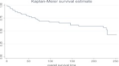
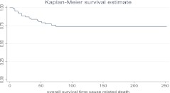
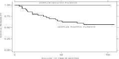
The median duration of follow-up of the patients was 144 months (range 0.4–252 months). A total of 28 (44.4%) patients died. Of 63 patients, only 16 (25.4%) died with recurrence. The 5-year overall survival of the patients was 76% (95% CI 64.1–84.9%) and 5-year cause-specific survival (melanoma-related death) was 77.4% (95% CI 64.9–86.0%). Figures 1 and 2 show the pattern of overall survival (all-cause deaths) and cause-specific deaths (melanoma-related), respectively, for the whole cohort. Figure 3 demonstrates the Kaplan-Meier survival probability curves for Doppler-positive and Doppler-negative cases.
Fig. 1.
Depicting Kaplan Meier survival probability curve for whole cohort for overall survival time in months
Fig. 2.
Showing Kaplan Meier survival probability curve for survival time for melanoma related death in months
Fig. 3.
Showing Kaplan Meier survival probability curve for doppler positive and doppler negative cases
Under univariate analysis of death due to melanoma, the variables such as ulcer, peak, tumor thickness category, and Clark’s level were found significant. The results under univariate Cox model are presented in Table 5. The patients who had the ulcer were 14 times more likely to die (HAZARD RATE 14.86; 95% CI 5.05–43.76) than those without an ulcer. The risk of death due to melanoma was 13 times more in case of thickness more than 1.5 mm (HAZARD RATE 13.3; 95% CI 5.05–43.76) as compared to cases with thin melanoma less than 1.5 mm thick.
Table 5.
Hazard ratio of death on Cox regression analysis
| Variables | Unadjusted hazard ratio UHR (95%CI) | Adjusted hazard ratio AHR (95%CI)* |
|---|---|---|
| Age (years) | ||
| <=50 (n = 20) | 1.00 | |
| >50 (n = 43) | 2.37 (0.68 to 8.34) | |
| Ulcer | ||
| No (n = 49) | 1.00 | |
| Yes (n = 14) | 14.86 (5.05 to 43.76) | |
| Clark’s level | ||
| 1 (I/II/III) (n = 38) | ||
| 2 (IV/V) (n = 25) | 6.64 (2.13 to 20.66) | |
| Thickness (mm) | ||
| <=1.5 (n = 41) | 1.00 | 1.00 |
| >1.5 (n = 22) | 13.30 (3.76 to 47.06) | 5.80 (1.56 to 21.54) |
| Peak systolic frequency(MHz) | ||
| <=2,500 (n = 21) | 1.00 | 1.00 |
| >2,500 (n = 15) | 5.88 (1.86 to 18.57) | 5.99(1.55 to 23.16) |
*Results are adjusted in relation to age
Survival Analysis and Doppler Flowmetry
On univariate analysis, patients having higher peak systolic frequency (above 2,500 MHz) were more likely to die of melanoma than those with lower peak systolic frequency (HAZARD RATE 5.88; 95% CI 1.86–18.57; SE 3.45, z = 3.02; p = 0.003).
There was no melanoma-related death among Doppler-negative patients.
The following variables were included in the final stepwise Cox regression: peak systolic frequency category, thickness, and Clark’s level. Age category (>50 years and ≤50 years) was adjusted in the final model. The proportional hazard assumption was checked and was found to be satisfied. In the final multivariable Cox analysis, only thickness and peak systolic frequency category remained as significant factors. The patients with thickness more than 1.5 mm were five times more likely to have died in comparison to the patients with less than 1.5 mm thickness after adjusting other important covariates (HAZARD RATE 5.80; 95% CI 1.56–21.54). Similarly, the risk of death was five times more among the patients who had higher peak systolic frequency (above 2,500 MHz) after controlling other potential covariates (HAZARD RATE 5.99; 95% CI 1.55–23.16). Among the Doppler-positive cases, only these two factors (thickness category above 1.5 mm and peak systolic frequency category above 2,500 MHz) remained significant in multivariable Cox proportional hazard regression analysis for death due to melanoma.
Discussion
The vertical tumor thickness is established as the most important predictor of outcome in patients with malignant melanoma of skin. Why should thickness play such an important role in influencing the biological behavior of melanoma? Is it just an expression of increasing tumor volume or a surrogate for the increasing biologic aggressiveness with release of progressively increasing amounts of molecules causing tumor invasion and metastasis?
Several workers have shown a prognostic value of tumor vascularity in skin melanoma. In one study, angiogenin, vascular endothelial growth factor (VEGF), fibroblast growth factor (b-FGF), and interleukin-8 (IL-8) were measured in sera of 125 melanoma patients. Serum levels of angiogenin, VEGF, b-FGF, and IL-8 were significantly increased in patients with melanoma compared to healthy controls. Elevated serum levels of VEGF, b-FGF, and IL-8 were strongly associated with poor survival [4].
The tumor thickness was strongly correlated with peak systolic and mean systolic frequencies. Such strong correlation between the thickness and the frequency measurements indicates that with progressive tumor growth the blood flow velocity increases pari passu with thickness. The neovascular network with rapid blood flow offers a path for abundant supply of nutriments for rapid proliferation and opens up avenues for hematogenous dissemination. A high peak systolic frequency indicates high velocity of blood flow through the tumor.
Doppler flowmetry allows preoperative assessment of the biological aggressiveness and long-term outcome, whereas the vertical thickness information is available only after the tumor excision. In tumors with marked vascularity, it may help to administer preoperative chemotherapy or chemoradiation in a manner akin to neoadjuvant therapy in breast cancer. In this regard, newer agents such as tyrosine kinase inhibitors, angiostatin, or newer antimitotic drugs are worthy of trial. After two to three courses of chemotherapy, the response of the melanoma may be assessed and the same drugs may be used following excision. Serial assessment of the tumor vascularity may predict the response to therapy as profuse blood flow through the tumor tissue may enhance the delivery of drugs in the tumor. The overall response of surgery alone in thick tumors (>4 mm thick) is so poor that the proposed approach of neoadjuvant therapy is worthy of investigation.
Survival of all the patients with Doppler-negative tumors for more than 15 years with no death due to melanoma recurrence is a very significant biological finding. Such excellent outcome has been reported for the “horizontal growth phase melanomas” by Clark’s group [5, 6]. It is possible that the absence of “detectable blood flow” is a feature of horizontal growth phase lesions. Doppler flowmetry allows demarcation between two categories of lesions (horizontal growth phase and vertical growth phase lesions) preoperatively. The similar association between tumor vasculature and survival has been reported by other authors [7, 8]. Lassau et al. [8] in the largest study on color Doppler sonography (CDS) assessed the tumor vascularity in 111 skin melanomas before surgical resection and demonstrated intratumor vessels in 43 lesions. The neovascularization visualized with CDS was significantly linked to relapse. Similar to our observation, they found a strong correlation between the vascularization and Breslow tumor thickness. They reported that Breslow thickness and vascularity confer equivalent prognostic information.
We propose that a “tumor vascularity database” should be established in countries with a high prevalence of melanoma to prospectively record the tumor vascularity assessed by Laser and ultrasonic Doppler flowmetry in a large cohort of patients to evaluate its biological significance. The flow signals should be subjected to more detailed signal analysis, namely, time-averaged mean frequency, normalized area under the spectrum, and relative flow index, for a detailed insight in tumor vascularity.
Conclusion
Doppler flowmetry predicts long-term survival. It may help in planning neoadjuvant therapies aimed at inhibiting angiogenesis or targeting tumor vasculature.
We suggest that tumor vascularity should be included in the preoperative assessment of skin melanoma.
Appendix
Variable Coding
- Outcome Variables
- Survival status was coded as 0 for alive patients and 1 for patients who died of melanoma. Patients who died without metastasis were coded as 0. Those alive at the end of follow-up period are coded as 0, regardless of presence or absence of recurrence. These cases are treated as “censored” for survival analysis. The survival status and presence or absence of the recurrence was ascertained by going through the hospital records of the patients and where necessary by information through the family general practitioner. The local recurrence was defined as a hard nodule at or near the scar (within a distance of 5 cm) confirmed by aspiration cytology or biopsy. Nodules located beyond 5 cm in the direction of lymph nodal basin were termed “in transit lymphatic metastasis.” The systemic recurrence was defined as enlargement of liver, distant lymph nodes, multiple skin deposits, deposits seen on ultrasound, chest X-ray, or CT scan of brain, chest or abdomen. The onset of local or systemic recurrence was defined as the period elapsing between the date of surgery and the date when recurrence was first noticed in the follow-up clinic.
- The survival period in months was recorded from the date of operation to the date of death or date when the patient was last seen. The survival period was used as a continuous variable.
- Predictor Variables
- Doppler flow: This was coded as Doppler flow positive (if flow signals were present) or Doppler flow negative (if flow signals were absent).
- Frequency measurements: Peak systolic frequency (P), mean systolic frequency (M), and minimum diastolic frequency (D) in hertz were used as continuous variables. The peak systolic frequency was categorized into two broad groups: up to 2,500 MHz or less and more than 2,500 MHz.
- Other considered independent covariates were age (≤50 or >50 years), ulcer (no = 0 or yes = 1); tumor thickness (≤1.5 or >1.5 mm), Clark’s level of invasion (categorized into two levels—Clark’s level I, II, and III = 1; Clark’s level IV and V = 2).
Statistical Analysis
The association of each categorical covariate with Doppler status was tested using Fisher’s exact test. Pearson’s correlation coefficients were obtained among peak systolic, mean systolic, and diastolic frequencies to assess collinearity.
The survival probability was calculated by the Kaplan-Meier method. Log rank test was performed to compare the survival probability in relation to each categorical covariate. Univariate Cox proportional hazard analysis was carried out to provide hazard ratio of death with each covariate. The variables found significant under univariate analysis along with clinically significant covariates were included in the stepwise multivariable Cox model. The inclusion probability of a variable was 0.05, whereas the exclusion probability of the variable was 0.10 under stepwise regression. The proportionality assumption was checked using Schoenfeld residuals. The statistical analyses were performed on STATA 9.0 (Texas, the USA).
Footnotes
Leslie E. Hughes is deceased
Contributor Information
Anurag Srivastava, Phone: +91-11-9868397729, FAX: +91-11-26588663, Email: dr.anuragsrivastava@gmail.com.
Robert E. Mansel, Email: ManselRE@cardiff.ac.uk
David J. T. Webster, Email: djtwebster@doctors.org.uk
Alok Dwivedi, Email: dwivedak@ucmail.uc.edu.
References
- 1.Breslow A. Thickness cross sectional area and depth of invasion in the prognosis of cutaneous melanoma. Ann Surg. 1970;172:902–908. doi: 10.1097/00000658-197011000-00017. [DOI] [PMC free article] [PubMed] [Google Scholar]
- 2.Srivastava A, Hughes LE, Woodcock JP, Laidler P. Vascularity in cutaneous melanoma detected by Doppler sonography and histology: correlation with tumor behaviour. Br J Cancer. 1989;59:89–91. doi: 10.1038/bjc.1989.17. [DOI] [PMC free article] [PubMed] [Google Scholar]
- 3.Srivastava A, Laidler P, Hughes LE, Woodcock JP, Shedden EJ. Neovasclarization in human cutaneos melanoma: a quantitative morphological and Doppler ultrasound study. Eur J Cancer Clin Oncol. 1986;22:1205–1209. doi: 10.1016/0277-5379(86)90322-6. [DOI] [PubMed] [Google Scholar]
- 4.Ugurel S, Rappl G, Tilgen W, Reinhold U. Increased serum concentration of angiogenic factors in malignant melanoma patients correlates with tumor progression and survival. J Clin Oncol. 2001;19(2):577–583. doi: 10.1200/JCO.2001.19.2.577. [DOI] [PubMed] [Google Scholar]
- 5.Clark WH, Jr, Elder DE, Guerry D, Epstein MN, Greene MH, Horn MV. A study of tumor progression. The precursor lesions of superficial spreading and nodular melanoma. Human Pathol. 1984;15:1147–1165. doi: 10.1016/S0046-8177(84)80310-X. [DOI] [PubMed] [Google Scholar]
- 6.Barnhill RL, Levy MA. Regressing thin cutaneous malignant melanomas (< or =1.0 mm) are associated with angiogenesis. Am J Pathol. 1993;143(1):99–104. [PMC free article] [PubMed] [Google Scholar]
- 7.Vlaykova T, Laurila P, Muhonen T, Hahka-Kemppinen M, Jekunen A, Alitalo K, et al. Prognostic value of tumor vascularity in metastatic melanoma and association of blood vessel density with vascular endothelial growth factor expression. Melanoma Res. 1999;9(1):59–68. doi: 10.1097/00008390-199902000-00008. [DOI] [PubMed] [Google Scholar]
- 8.Lassau N, Lamuraglia M, Serge Koscielny S, Spatz A, Roche A, Leclere J, Avril MF. Prognostic value of angiogenesis evaluated with high-frequency and colour Doppler sonography for preoperative assessment of primary cutaneous melanomas: correlation with recurrence after a 5 year follow-up period. Cancer Imag. 2006;6(1):24–29. doi: 10.1102/1470-7330.2006.0009. [DOI] [PMC free article] [PubMed] [Google Scholar]


