Abstract
Background:
The loss of glenohumeral internal rotation range of motion in overhead athletes has been well documented in the literature. Several different methods of assessing this measurement have been described, making comparison between the results of studies difficult.
Hypothesis:
Significant differences in the amount of internal rotation range of motion exist when using different methods of stabilization.
Study Design:
Descriptive laboratory study.
Methods:
Three techniques were used bilaterally in random fashion to measure glenohumeral internal rotation range of motion: stabilization of the humeral head, stabilization of the scapula, and visual inspection without stabilization. An initial study on 20 asymptomatic participants was performed to determine the intrarater and interrater reliability for each measurement technique. Once complete, measurements were performed on 39 asymptomatic professional baseball players to determine if a difference existed in measurement techniques and if there was a significant side-to-side difference. A 2-way repeated-measures analysis of variance was used.
Results:
While interrater reliability was fair between all 3 methods, scapular stabilization provided the best intrarater reliability. A statistically significant difference was observed between all 3 methods (P < .001). Internal rotation was significantly less in the dominant shoulder than in the nondominant shoulder (P < .001).
Conclusion:
Differences in internal rotation range of motion measurements exist when using different methods. The scapula stabilization method displayed the highest intrarater reproducibility and should be considered when evaluating internal rotation passive range of motion of the glenohumeral joint.
Clinical Relevance:
A standardized method of measuring internal rotation range of motion is required to accurately compare physical examinations of patients. The authors recommend the use of the scapula stabilization method to assess internal rotation range of motion by allowing normal glenohumeral arthrokinematics while stabilizing the scapulothoracic articulation.
Keywords: goniometry, overhead athlete, shoulder
The assessment of physiologic mobility of the glenohumeral joint in overhead athletes has received significant attention in recent literature.¶ In particular, internal rotation (IR) range of motion (ROM) of the glenohumeral joint and tightness of the posterior glenohumeral joint capsule have been the focus of many discussions.2,3The dominant shoulder of overhead athletes exhibits significantly greater external rotation and a decrease in IR in comparison to the nondominant shoulder.1,3,4,9,10,12,19
Several theories exist regarding the cause of this unique motion characteristic of the overhead athlete. These include posterior capsular tightness,5,7,8 osseous adaptation,3,9,16 and muscular tightness.3,19 Controversy exists regarding the exact mechanism of loss of IR. However, most authors agree that a significant loss of IR may lead to several pathologies. Warner et al17 noted a significant decrease in glenohumeral joint IR in patients with subacromial impingement. Wilk and Andrews18 reported a reverse capsular pattern in patients with subacromial impingement syndrome in whom IR was most limited, followed by a loss of abduction and then external rotation. Burkhart et al5 stated that an asymptomatic shoulder that exhibits a moderate degree of IR loss was more susceptible to developing pathologies such as “dead arm” syndrome and superior labral anterior and posterior (SLAP) lesions. Furthermore, these authors described an acronym for the clinical observation of decreased IR, termed glenohumeral IR deficit (GIRD). Recently, Wilk et al (unpublished data, 2008) reported a correlation between GIRD and shoulder injuries in professional baseball pitchers followed over a 3-year period. Furthermore, they reported that pitchers with GIRD exhibited a 2.4-times greater risk of shoulder injuries than pitchers without GIRD. These findings support the importance of accurately assessing IR in the overhead athlete.
Although the importance of assessing glenohumeral joint IR ROM has been well established, controversy exists regarding the most accurate method to measure this motion. Several techniques and corresponding values of IR ROM have been reported, including active ROM assessed in regard to the vertebral level that can be reached behind the back1 and passive ROM measured at 90° of shoulder abduction. Numerous authors have emphasized the importance of using scapulothoracic joint stabilization to restrict scapular movement.7,10,14 Unfortunately, in many instances, the illustrations and/or descriptions were not clearly stated by the investigators. Consequently, tremendous variability exists in published mean ROM values, from 83° in asymptomatic pitchers by Brown et al,4 to 62° in professional pitchers by Wilk and Andrews,18 to 36° in throwers by Meister et al.14 Thus, the lack of a uniform method of measuring IR makes it difficult to compare passive ROM data from one study to another, or to compare patient clinical findings to those of published studies.
There exist 3 methods of assessing IR passive ROM of the glenohumeral joint at 90° of shoulder abduction. These methods include providing stabilization of the scapula, stabilization of the humeral head, or visual inspection only (with no stabilization). Clinically, it has been our observation that each method renders significantly different IR passive ROM values compared to the other methods, and thus the 3 are not comparable.
The purpose of this study was to determine the reliability and compare the results of 3 different methods of assessing glenohumeral joint IR passive ROM in asymptomatic overhead athletes. We hypothesized that significant differences exist in the amount of IR ROM between these 3 clinical techniques.
Materials and Methods
Participants
Two groups of asymptomatic overhead athletes volunteered for this study. The first group consisted of 20 males (mean age, 27 ± 6 years; mean height, 170 ± 7 cm; mean weight, 72 ± 15 kg) in whom each method of measurement was conducted in the nondominant shoulder. The second group consisted of 39 professional baseball players (mean age, 27 ± 4.2 years; mean height, 190.5 ± 5 cm; mean weight, 93.4 ± 10.4 kg) who were analyzed during spring training physicals. Of these 39 participants, 32 were pitchers, 6 catchers, and 1 an outfielder. Twenty-nine were right-hand dominant and 10 were left-hand dominant. All participants were asymptomatic of upper extremity injuries at the time of the study. Informed consent was obtained prior to testing. The research protocol was approved by the institutional review board of the American Sports Medicine Institute.
Procedure
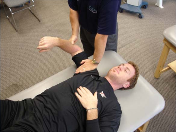
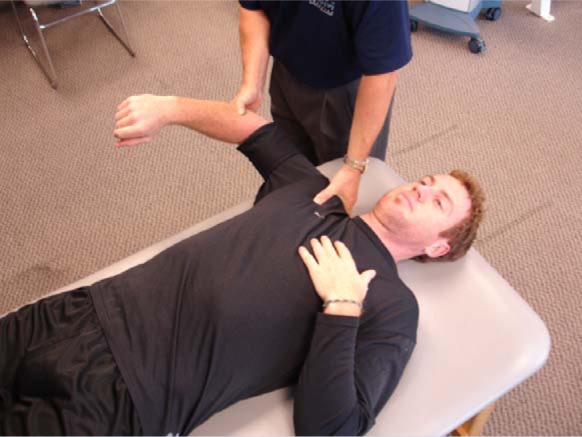
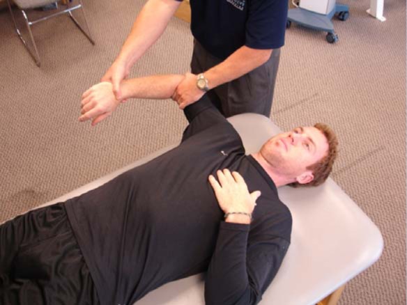
Testing was performed with the individuals positioned supine with the shoulder at 90° of abduction and 10° of horizontal adduction (scapular plane), with 90° of elbow flexion. The shoulder was positioned in the scapular plane rather than the coronal plane to minimize any pretension of capsular or muscle soft tissue. Glenohumeral IR ROM was measured using 3 different techniques. In the first technique, stabilization of the humeral head was performed by placing the palm of the hand over the clavicle, coracoid process, and humeral head (Figure 1). In the second method, stabilization of the scapula was done by grasping the coracoid process and the spine of the scapula posteriorly (Figure 2). In the third method, stabilization was not performed. Instead, the arm was passively internally rotated until the humeral head or scapula was observed to begin to elevate based on visual inspection (Figure 3).
Figure 1.

Glenohumeral internal rotation passive range of motion measurement using stabilization to the humeral head by placing the palm of the hand over the clavicle, coracoid process, and humeral head. The patient is positioned supine with the shoulder at 90° of abduction and approximately 10° of horizontal adduction (scapular plane).
Figure 2.

Glenohumeral internal rotation passive range of motion measurement using stabilization of the scapula by grasping the coracoid process and the spine of the scapula posteriorly. The patient is positioned supine with the shoulder at 90° of abduction and approximately 10° of horizontal adduction (scapular plane).
Figure 3.

Glenohumeral internal rotation passive range of motion measurement without stabilization. The arm is passively internally rotated until the humeral head or scapula is observed to begin to elevate based on visual inspection. The patient is positioned supine with the shoulder at 90° of abduction and approximately 10° of horizontal adduction (scapular plane).
In order to determine the reliability of each method, 3 teams consisting of 1 physical therapist and 1 athletic trainer performed IR ROM positioning and measuring, respectively, on each of the 20 participants from the first group within 5 minutes of each other. Five trials were performed on 5 separate days.
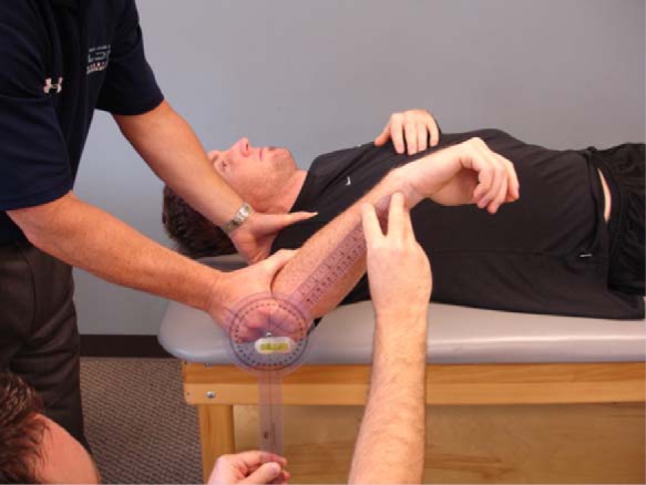
To determine if differences existed between each method, 2 examiners were consistently used in the second group of 39 individuals, 1 to position the shoulder and the other to read the measurements. Measurements were made with a standard goniometer with a special bubble level attachment. The center of rotation of the goniometer was placed over the tip of the olecranon while 1 arm was positioned along the length of the ulna, aligned with the ulnar styloid process. The other arm was positioned inferiorly perpendicular to the ground, using the bubble level to assure proper alignment (Figure 4). One measurement was taken using each method in a randomized fashion. The order of arm dominance tested was also randomized. The examiner positioning the shoulder was blinded to the results of the measurements.
Figure 4.

Range of motion measurements using a standard goniometer with a bubble attachment. The bubble is used to assure that the axis of the goniometer is perpendicular to the ground during measurement. Note that the bubble is aligned within the center of the goniometer.
Statistics
Reliability was assessed using intraclass correlation coefficients and a P level of < .05 was considered significant. Two-way repeated measures analyses of variance were used to compare the differences between methods and between the dominant and nondominant shoulder. Using the Mauchly test of sphericity, adjustment was made for any variance in the data and, therefore, corrected the F value and associated probabilities from the repeated-measures analysis of variance. Paired t tests were conducted for post hoc pair-wise comparisons. A P value <.05 was considered to be significant. All statistical analyses were performed using SPSS 10.0.5 (SPSS Inc, Chicago, Illinois).
Results
The assessment of the reliability of each method showed that, while all 3 techniques produced similar interrater reliability, the scapular stabilization method had the highest intrarater reproducibility (Table 1).
Table 1.
Reliability of 3 methods of measuring internal rotation.a
| Internal Rotationb (mean) | Intrarater ICC | Interrater ICC | |
|---|---|---|---|
| No stabilization | 58° | 0.48 | 0.47 |
| Scapular stabilization | 46° | 0.62 | 0.43 |
| Humeral head stabilization | 40° | 0.51 | 0.45 |
ICC, intraclass correlation coefficient.
A statistically significant difference was observed between each method of stabilization (P < .001).
The mean results of each method of IR measurement for the dominant and nondominant shoulder from the second group of 39 baseball players are shown in Table 2. A statistically significant decrease in IR ROM was observed on the dominant shoulder compared to the nondominant shoulder for each method of measurement (P < .001, F = 54.2). A statistically significant difference was also found between the 3 methods (P < .001, F = 598). There was also a statistically significant interaction (P = .10, F = 5.71) between shoulder and the type of test performed. A post hoc pair-wise t test showed statistically significant correlation between test types (Table 3). The visual inspection method (no stabilization) allowed for the greatest amount of IR ROM, while the humeral head stabilization method allowed for the least amount of IR ROM. The altered ROM observed between each method of measurement was consistent bilaterally.
Table 2.
Internal rotation range of motion.a
| Dominant Shoulder (mean ± SD) | Nondominant Shoulder (mean ± SD) | Comparison Dominant – Nondominant P Value | |
|---|---|---|---|
| No stabilization | 52.3° ± 8.4° | 65.2° ± 8.4° | < .001 |
| Scapular stabilization | 43.9° ± 8.1° | 53.5° ± 9.1° | < .001 |
| Humeral head stabilization | 35.8° ± 8.7° | 45.3° ± 8.4° | < .001 |
A statistically significant difference was also observed between each method of stabilization for the dominant and nondominant shoulders (P < .001). SD, standard deviation.
Table 3.
Correlation between test types.a
| Post Hoc Paired t Test | ||
|---|---|---|
| Correlation | Significance (P Value) | |
| VI_Dom & SS_Dom | .792 | <.001 |
| VI_Dom & H_Dom | .729 | <.001 |
| SS_Dom & H_Dom | .940 | <.001 |
| VI_ND & SS_ND | .835 | <.001 |
| VI_ND & H_ND | .840 | <.001 |
| SS_ND & H_ND | .889 | <.001 |
SS, scapula stabilized; VI, visual inspection; H, humeral head stabilization; Dom, dominant; ND, nondominant.
Discussion
The most significant finding with this study was the significant differences (P < .001) found when comparing the 3 methods of assessing shoulder IR passive ROM. The humeral stabilization method produced the least amount of passive ROM on both extremities: 36° on the dominant shoulder and 45° on the nondominant side. In contrast, the scapular stabilization method demonstrated 44° of IR on the dominant side and 54° of IR on the nondominant side. The visual inspection method rendered the greatest amount of IR on both sides, 52° and 65°, respectively.
The results of this study illustrate significant differences in IR ROM in the throwing shoulder compared to the nonthrowing shoulder. This was observed with all 3 methods. These findings are consistent with those previously reported in the literature.1,3,4,9,10,12,19 Most authors have noted bilateral differences of 7° to 9°, with the dominant throwing shoulder exhibiting less IR ROM and greater external rotation.9,14,19 Although others have noted greater differences bilaterally, Brown et al4 reported a mean bilateral difference of 15°, whereas Ellenbecker et al10 noted an 11° difference.
In this study, we demonstrated a bilateral difference that ranged from 8° to 13° depending on the method of assessment. The method of visual inspection (no stabilization) measured the greatest difference of 13°, in comparison with the humeral head stabilization, which produced the smallest difference of 8°. Importantly, significant differences existed between the dominant and nondominant shoulders during all 3 methods.
We believe that the 3 methods of measurement have a significant effect on the validity of measurement of pure glenohumeral joint IR ROM. The visual inspection method provides minimal stabilization to the scapula, thus allowing the scapula to move (producing an anterior tilt and protraction) and resulting in greater shoulder IR ROM. Thus, the increase in motion is not a product of pure glenohumeral motion, but rather a combination of scapulothoracic motion and glenohumeral motion.
In contrast, the humeral head stabilization method permitted the least amount of IR. This technique may restrict the normal arthrokinematics of the glenohumeral joint. When stabilization or pressure is applied to the anterior humeral head during IR passive ROM, the normal anterior translation of the humeral head is restricted,11 possibly minimizing IR motion. This technique may also generate tension on the glenohumeral joint capsule via direct contact with the articulating surfaces, which may restrict normal glenohumeral motion. The amount of pressure on the humeral head significantly affects the amount of IR; for instance, greater posteriorly directed pressure results in less IR.
The last method of assessing IR was the method that stabilized the scapula.15 In this technique, the examiner applied light pressure anteriorly to the coracoid process with the thumb, and applied pressure on the spine of the scapula with the fingers. During this technique, the examiner attempts to palpate and stabilize any scapular motion, while allowing for normal glenohumeral motion. During passive IR, the scapula undergoes an anterior tilt. The goal of this technique is to passively move the humerus until scapular motion occurs, then measure the degree of IR before the compensatory scapulothoracic joint motion contributes to the overall motion. This technique may be the most clinically relevant. Some clinicians have recommended stabilizing the scapula with pressure on the anterior acromion. Although useful, that method is difficult to perform without also altering glenohumeral movement. In some individuals, because of the anatomy of the joint and the size of the anterior deltoid, palpation of the anterior acromion is difficult due to soft tissue bulk.
The loss of IR ROM observed in overhead athletes has received significant attention over the past several years. Theories regarding the cause of this loss of motion vary, although tightness of the posterior capsule has been cited by several sources6-8 as a potential mechanism, based mainly on clinical observation with limited scientific research. A recent study by Borsa et al,3 that examined the anterior and posterior laxity of the glenohumeral joint in asymptomatic professional baseball players, noted that posterior capsular tightness did not exist and all the players exhibited greater posterior translation than anterior translation. They reported no correlation between capsular laxity and loss of IR ROM. The theories regarding posterior capsular tightness and loss of IR ROM may be due, in part, to the technique used to assess IR ROM. To accurately assess posterior capsular tightness, the clinician should perform a posterolaterally directed translatory force with the arm at 90° of abduction, approximately 30° of horizontal adduction, and neutral rotation.
The important finding of this study is the significant differences found between the assessment techniques for performing IR passive ROM. This is obvious when one reviews the literature to determine the acceptable or normal amount of IR ROM in the overhead athlete. Variability continues to exist due to the lack of consistency in the assessment technique. Wilk et al19 and Crockett et al9 reported a mean of 62° of IR on the throwing shoulder using the visual inspection (no stabilization) method. Brown et al4 reported an average of 83° of IR, but did not mention if stabilization was used. Ellenbecker et al10 noted an average of 45° of IR in male tennis players, compared to a mean of 52° in females. Stabilization of the scapulothoracic joint was provided through control of the coracoid process and acromion. Meister et al14 stabilized the scapula in Little League throwers (ages 8-16 years), although the exact method was not discussed. They reported an average IR of 36°. More recently, Wilk et al (unpublished data, 2008) reported an average of 46° of IR in a study using the scapular stabilization technique.
These data emphasize the importance of consistency and standardization in assessing IR in the overhead throwing athlete. We recommend that the clinician and researcher adopt a standardized technique to measure glenohumeral IR passive ROM. This will allow the clinician to compare patient findings and researchers to compare data accurately. Furthermore, the referring physician and treating rehabilitation specialist should perform the same standardized technique to ensure consistency and accuracy with patient care. We recommend utilizing the technique that stabilizes the scapula. This technique allows the normal arthrokinematics of the glenohumeral joint to occur, while not allowing excessive scapulothoracic joint motion to contribute to the total IR arc of motion. It also allows the examiner to detect when the scapulothoracic joint motion begins to contribute to IR. This technique allows for a greater appreciation of the end-feel assessment. The method that stabilizes the humeral head is too restrictive and does not permit normal glenohumeral joint arthrokinematics. As a result of these findings, we are currently using the scapular stabilization technique clinically.
The reliability of the 3 methods was somewhat low, ranging from 0.43 to 0.62 with intraclass correlation coefficients. When examining the intraclass reliability, the scapular stabilization technique was shown to exhibit the highest reliability (0.62) and the lowest was visual inspection (0.48). Further research is needed to examine techniques to enhance both the intrarater and interrater reliability of these 3 methods. The low reliability values found in this study are a concern.
Although this study attempts to determine the reliability of IR measurements, a gold standard has yet to be determined. Results of this study are applicable to asymptomatic overhead athletes.
Footnotes
References
- 1. Bigliani LU, Codd TP, Connor PM, Levine WN, Littlefield MA, Hershon SJ. Shoulder motion and laxity in the professional baseball player. Am J Sports Med. 1997;25:609-613 [DOI] [PubMed] [Google Scholar]
- 2. Borsa PA, Dover GC, Wilk KE, Reinold MM. Glenohumeral range of motion and stiffness in professional baseball pitchers. Med Sci Sports Exerc. 2006;38:21-26 [DOI] [PubMed] [Google Scholar]
- 3. Borsa PA, Wilk KE, Jacobson JA, et al. Correlation of range of motion and glenohumeral translation in professional baseball pitchers. Am J Sports Med. 2005;33:1392-1399 [DOI] [PubMed] [Google Scholar]
- 4. Brown LP, Niehues SL, Harrah A, Yavorsky P, Hirshman HP. Upper extremity range of motion and isokinetic strength of the internal and external shoulder rotators in major league baseball players. Am J Sports Med. 1988;16:577-585 [DOI] [PubMed] [Google Scholar]
- 5. Burkhart SS, Morgan CD, Kibler WB. Shoulder injuries in overhead athletes: the “dead arm” revisited. Clin Sports Med. 2000;19:125-158 [DOI] [PubMed] [Google Scholar]
- 6. Burkhart SS, Morgan CD, Kibler WB. The disabled throwing shoulder: spectrum of pathology. Part I: pathoanatomy and biomechanics. Arthroscopy. 2003;19:404-420 [DOI] [PubMed] [Google Scholar]
- 7. Burkhart SS, Morgan CD, Kibler WB. The disabled throwing shoulder: spectrum of pathology. Part III: the SICK scapula, scapular dyskinesis, the kinetic chain, and rehabilitation. Arthroscopy. 2003;19:641-661 [DOI] [PubMed] [Google Scholar]
- 8. Burkhart SS, Morgan CD, Kibler WB. The disabled throwing shoulder: spectrum of pathology. Part II: evaluation and treatment of SLAP lesions in throwers. Arthroscopy. 2003;19:531-539 [DOI] [PubMed] [Google Scholar]
- 9. Crockett HC, Gross LB, Wilk KE, et al. Osseous adaptation and range of motion at the glenohumeral joint in professional baseball pitchers. Am J Sports Med. 2002;30:20-26 [DOI] [PubMed] [Google Scholar]
- 10. Ellenbecker TS, Roetert EP, Bailie DS, Davies GJ, Brown SW. Glenohumeral joint total rotation range of motion in elite tennis players and baseball pitchers. Med Sci Sports Exerc. 2002;34:2052-2056 [DOI] [PubMed] [Google Scholar]
- 11. Howell SM, Galinat BJ, Renzi AJ, Marone PJ. Normal and abnormal mechanics of the glenohumeral joint in the horizontal plane. J Bone Joint Surg Am. 1988;70:227-232 [PubMed] [Google Scholar]
- 12. Johnson L. Patterns of shoulder flexibility among college baseball players. J Athl Train. 1992;27:44-49 [PMC free article] [PubMed] [Google Scholar]
- 13. Meister K. Injuries to the shoulder in the throwing athlete: part one, biomechanics/pathophysiology/classification of injury. Am J Sports Med. 2000;28:265-275 [DOI] [PubMed] [Google Scholar]
- 14. Meister K, Day T, Horodyski M, Kaminski TW, Wasik MP, Tillman S. Rotational motion changes in the glenohumeral joint of the adolescent/Little League baseball player. Am J Sports Med. 2005;33:693-698 [DOI] [PubMed] [Google Scholar]
- 15. Norkin CC, White DJ. Measurement of Joint Motion: A Guide to Goniometry. 3rd ed. Philadelphia: F.A. Davis Company; 2003 [Google Scholar]
- 16. Reagan KM, Meister K, Horodyski MB, Werner DW, Carruthers C, Wilk K. Humeral retroversion and its relationship to glenohumeral rotation in the shoulder of college baseball players. Am J Sports Med. 2002;30:354-360 [DOI] [PubMed] [Google Scholar]
- 17. Warner JP, Micheli LJ, Arslanian LE, Kennedy J, Kennedy R. Patterns of flexibility, laxity, and strength in normal shoulders and shoulders with instability and impingement. Am J Sports Med. 1990;18:366-375 [DOI] [PubMed] [Google Scholar]
- 18. Wilk KE, Andrews JR. Rehabilitation following arthroscopic subacromial decompression. Orthopedics. 1993;16:349-358 [DOI] [PubMed] [Google Scholar]
- 19. Wilk KE, Meister K, Andrews JR. Current concepts in the rehabilitation of the overhead throwing athlete. Am J Sports Med. 2002;30:136-151 [DOI] [PubMed] [Google Scholar]


