Abstract
The overhead throwing motion is a coordinated effort of muscle units from the entire body, culminating with explosive motion of the upper extremity. The throwing motion occurs at a rapid pace, making analysis difficult in real time. Electromyographic studies and high-speed video recordings have provided invaluable details regarding the involved musculature, the sequence of muscle involvement, and associated kinematic variables. The goal of the present article is to provide an overview of the kinetic chain—that is, a detailed description of the muscular coordination during each phase of pitching—and to describe specific types of pitches. An enhanced understanding of the components of the kinetic chain and the phases of the throwing motion can provide important information for rehabilitation, performance enhancement, and injury prevention.
Keywords: pitching, throwing shoulder, kinetic chain, pitching motion, shoulder injuries
The overhand pitching motion consists of a sequence of body movements that start when the pitcher lifts the lead foot, progresses to a linked motion in the hips and trunk, and culminates with a ballistic motion of the upper extremity to propel the ball toward home plate. The effective synchronous use of selective muscle groups maximizes the efficiency of the kinetic chain (Table 1). The lower extremity and trunk generate and transfer energy to the upper extremity. Coordinated lower extremity muscles (quadriceps, hamstrings, hip internal and external rotators) provide a stable base for the trunk (core musculature) to rotate and flex. The extremely rapid rate of this motion makes assessment difficult. The time elapsed between front foot contact and ball release is only 0.145 seconds,30 followed by an additional half second for the ball to reach home plate.7 Maximum humeral internal rotation velocity during throwing may reach 7500 to 7700 degrees per second.8,21 Extreme degrees of external rotation of the shoulder, coupled with forward linear trunk motion, allow a greater distance for the accelerating force to be applied to the ball, generating top velocity.26 An intricate relationship between the dynamic stabilizers (rotator cuff, pectoralis major, and latissimus dorsi) and static stabilizers is required to simultaneously supply the range of motion, force, and stability of the glenohumeral joint. This integrated effort relies on the trapezius, rhomboids, levator scapulae, and serratus anterior muscles for stabilization, positioning, and synchronous scapular motion. The scapula acts synchronously with the rotator cuff to maintain the glenohumeral center of rotation within a physiologic range during the pitching motion.13,18,20
Table 1.
Significant temporal events of pitching.
| Key Events in the Throwing Motion | Percentage of Time From Stride Foot Contact (0%) to Ball Release (100%) |
|---|---|
| Maximum pelvis rotational angular velocity21,29 | 25-39 |
| Max upper torso rotation angular velocity during cocking phase21,29 | 51-52 |
| Elbow begins extending just prior to max external rotation21 | 81 |
| Transition from late cocking to acceleration29 | 81 |
| Maximum shoulder external rotation21,29 | 81 |
| Shoulder begins internal rotation at beginning of acceleration21 | 81 |
| Max elbow extension velocity, during acceleration21,29 | 90-95 |
| Maximum forward trunk tilt angular velocity21,29 | 93-96 |
| Maximum shoulder internal rotation angular velocity, 3 to 4 milliseconds after ball release7,21,29 | 102-104 |
Because the throwing motion occurs almost exclusively above 90° of abduction, the inferior glenohumeral ligament and capsule act as the primary static anterior restraint. The deltoid elevates the humerus while the rotator cuff adjusts the position of the humeral head on the glenoid.16 The pectoralis major and latissimus dorsi power the shoulder forward.16
A pitcher’s velocity, consistency, and durability may be linked to kinematic and kinetic factors as well as the temporal association of segmental body motions. Optimization of these parameters allows for efficient and consistent transfer of energy from proximal to distal components. Understanding the variables that optimize function may prevent injury by reducing the forces imparted to the shoulder and elbow joints. Breakdown of the kinetic chain will reduce its efficiency, making top velocity more difficult (Table 2).
Table 2.
Common points of breakdown of the kinetic chain.
| Points of Breakdown in Kinetic Chain | Result |
|---|---|
| Premature forward motion during windup/stride | Arm lags behind body |
| Stride foot lands in closed position | Decreased pelvic/trunk rotation; must throw across body, decrease force generation in kinetic chain |
| Stride foot lands in open position | Premature pelvic rotation, uncouples kinetic chain; arm lags behind body; shoulder must generate increased forces to maintain velocity |
| Pitcher throws in upright position (diminished forward trunk tilt) | Acceleration forces imparted to ball of shorter distances; less velocity generated |
| Increased lead knee flexion at ball release | Less forces generated by torso flexing/rotating over front side |
| Diminished external rotation in late cocking | Acceleration forces act on ball over shorter distance |
| Scapular dyskinesis | Diminished external rotation in late cocking (decreased scapular retraction); subacromial impingement |
The Kinetic Chain’s Involvement in the Pitching Motion
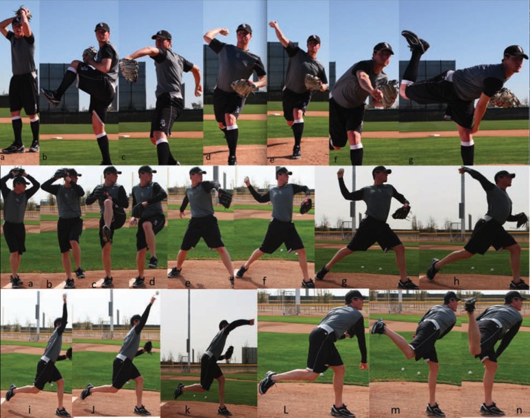
The pitching motion consists of 6 phases (Figure 1): windup, early cocking/stride, late cocking, acceleration, deceleration, and follow-through.10,12,17,22 These phases are intricately coupled, resulting in efficient generation and transfer of energy from the body into the arm and, ultimately, the hand and ball. Each segment starts as the adjacent proximal segment reaches top speed, culminating with top speed of the most distal segment.27 The windup and stride position the lower extremity and trunk for the most effective performance of the kinetic chain. The legs and trunk serve as the main force generators of the kinetic chain.1 The scapula is key in facilitating this energy transfer distally to the hand.10,12,18 Scapular dysfunction prohibits optimum energy transfer.18 In previous work, Kibler and Chandler calculated that a 20% decrease in kinetic energy delivered from the hip and trunk to the arm requires a 34% increase in the rotational velocity of the shoulder to impart the same amount of force to the hand.19
Figure 1.
A, components of the throwing motion as viewed from the front (home plate): windup (a, b), early cocking/stride (c), late cocking (d), acceleration (e), deceleration/follow-through (f, g); B, the throwing motion viewed from the side: windup (a-c), early cocking/stride (d, e), late cocking (f, g), acceleration (h-j), deceleration (k), follow-through (l-n).
The complex interaction of the lower extremities and core musculature in the kinetic chain reduces the kinetic contributions of the shoulder joint.31 Thus, the pitching motion should not be thought of as an upper extremity action, rather an integrated motion of the entire body that culminates with rapid motion of the upper extremity. Improvement of velocity can result from optimization of the kinetic chain, which likely also reduces the kinetic contributions of the shoulder to produce top velocity.3,19,21,31,35 Reduced kinetic stresses on the shoulder may prevent injury, leading to greater durability and health of the throwing shoulder.
High-speed 3-dimensional video analysis has furthered our understanding of the kinetic chain’s role in the overhead throwing motion. Variables such as lead knee flexion, forward trunk tilt, peak elbow extension, maximum shoulder external rotation, and maximum pelvis angular velocity have all been correlated with increased pitching velocity.21,31,35 Body height, as well humeral and radial length, has also been associated with increased velocity.21 Werner et al found that velocity was affected most by the pitcher’s body weight, the time from stride foot contact to maximum shoulder external rotation, knee flexion at stride foot contact, elbow angle at stride foot contact, maximum shoulder external rotation, maximum upper trunk rotation speed, peak elbow extension angular velocity, knee flexion at ball release, and forward trunk tilt at ball release.35
The Phases of the Pitching Motion
Windup
The windup and stride position the body to optimally generate the forces and power required to achieve top velocity. The windup begins with the initial movement of the contralateral lower extremity, and it culminates with elevation of lead leg to its highest point and with separation of the throwing hand from the glove.22
The pitcher keeps his center of gravity over his back leg (Figure 2) to allow generation of maximum momentum once forward motion is initiated. If the pitcher’s body and momentum fall forward prematurely, the kinetic chain will be disrupted and greater shoulder force will be required to propel the ball at top velocity.2,12
Figure 2.
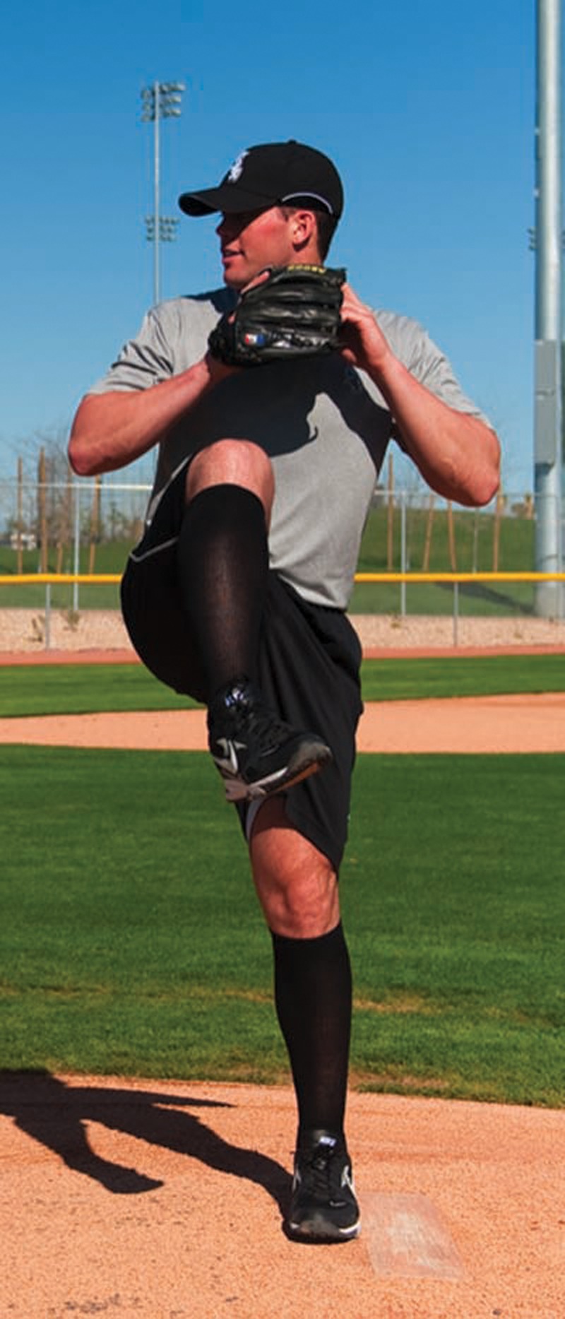
During windup and stride, the pitcher keeps his center of gravity back (over stance leg) for as long as possible to allow maximum generation and transfer of momentum and force to the upper extremity and ball.
Early Cocking/Stride
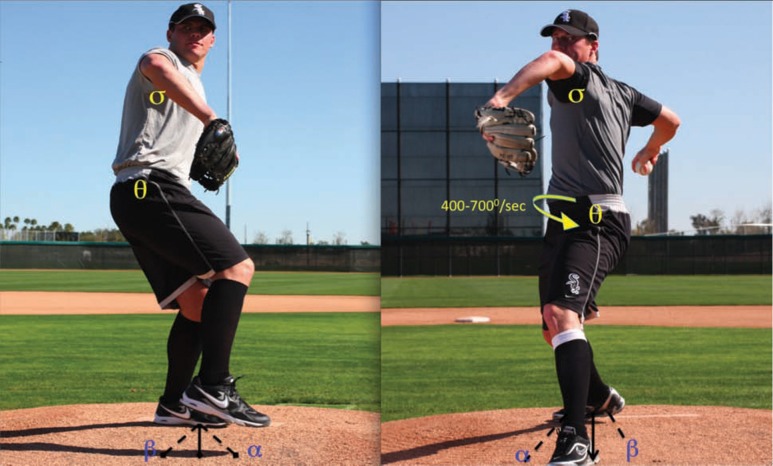
The early cocking/stride phase begins once the lead leg reaches its maximum height and the ball is removed from the glove (Table 3), and it ends when the lead foot contacts the pitching mound. The stride functions to increase the distance over which linear and angular trunk motions occur, allowing for increased energy production for transfer to the upper extremity.5 The stance knee and hip extend and lead to the initiation of pelvic rotation and forward tilt, followed by upper torso rotation. The pelvis achieves maximum rotational velocities of 400 to 700 degrees per second during this phase.12 The abdominal obliques eccentrically contract to prevent excess lumbar hyperextension during upper torso rotation and flexion. The stance leg gluteus maximus fires to maintain slight dominant-sided extension and provide pelvis and trunk stabilization during coiling.33
Table 3.
Early cocking/stride phase of pitching motion.
| Begins as lead leg reaches max height.5,8,10 |
| Stride increases the distance over which angular/linear acceleration occurs. |
| Pelvic tilt and rotation begin.21,31,32 |
| Deltoid is active early in phase to aid in abduction of shoulder.14,17 |
| Suprasinatus, infraspinatus, and teres minor are active late, to initiate shoulder external rotation.14 |
| Ends at stride (lead) foot contact.5,8,10 |
The supraspinatus, infraspinatus, and teres minor externally rotate the shoulder and position of the humeral head on the glenoid.4,22 The serratus anterior and the scapular retractors (middle trapezius, rhomboid, and levator scapulae) position the glenoid in upward rotation and retraction, providing a stable base on which the humerus can rotate.4 The lead foot should land in line with the stance foot, pointing toward home plate or in a few degrees of internal rotation (Figure 3).12 If the foot lands too far closed (stride foot in front of stance foot), pelvis rotation may be limited causing the pitcher to throw across his body.
Figure 3.
During early cocking/stride, the pelvis externally rotates (θ) earlier and to a greater extent than the torso (σ). It reaches 400 to 700 degrees per second. The lead foot should land in line with the stance foot and home plate. If it lands in a closed position (α), the pitcher must throw across his body; if the lead foot lands in an open position (β), it will cause uncoupling of kinetic chain owing to premature overrotation of the pelvis.
Late Cocking
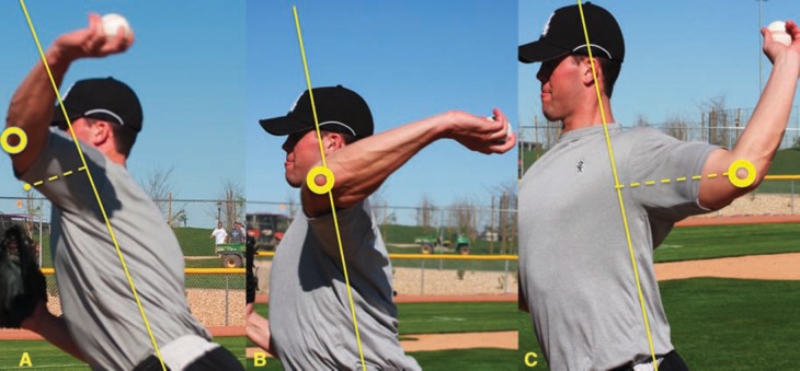
The late cocking phase occurs between lead foot contact and the point of maximal external rotation of the throwing shoulder (Table 4). The scapula is brought into a position of retraction; the elbow flexes; and the humerus undergoes abduction and external rotation. The pelvis reaches its maximum rotation, and the upper torso continues to rotate and tilt forward and laterally. The lead knee begins to extend, forming a solid base for trunk flexion. As the torso rotates, the anterior deltoid and pectoralis major contract to bring the throwing extremity into horizontal adduction (Figure 4), to achieve the 15° to 20° for the launch position of late cocking.5,12 Maximum shoulder internal rotation torque occurs just before maximum shoulder external rotation.11
Table 4.
Late cocking phase of the pitching motion.
| Occurs between foot contact and max external rotation.5,8,10 |
| Scapula retracts and tilts upward via actions of the rhomboids, levator, trapezius.14,24 |
| Humeral abduction and external rotation—mainly owing to infraspinatus/teres minor (electromyographic data).14,16,17 |
| Supraspinatus functions primarily in glenohumeral compression, humeral head depression.4,8,12,23,28 |
| Pelvis reaches max rotation.21,32 |
| Increased torso rotational and angular velocities.21,31,32 |
| Phase ends as subscapularis, pectoralis major, and latissimus dorsi eccentrically contract to terminate external rotation.5,10,16,17 |
Figure 4.
Horizontal adduction (A) and horizontal abduction are used to describe the position of the arm in the sagittal plane of motion with reference to the center of the torso (horizontal neutral; B). Horizontal abduction (arm behind torso; C) occurs early in the throwing motion, whereas horizontal adduction (arm in front of torso) occurs from the end of late cocking and beyond.
Near the end of arm cocking (64% of time from foot contact until ball release), maximum valgus torque is experienced at the elbow.31 The flexor and pronator muscles of the forearm generate a counter varus torque (64 N⋅m).8 Maximum elbow flexion is limited by eccentric contraction of the triceps, followed by concentric contraction as the elbow extends at the termination of late cocking and throughout acceleration.12
Adaptive increases in elevation and upward rotation of the scapula are important, ensuring sufficient subacromial space to accommodate the 80° to 100° of humeral abduction in the throwing position without impingement.5,6,10,22,24 The teres minor and the infraspinatus are active, delivering extreme amounts of external rotation.4,14 The supraspinatus is the least active of the rotator cuff muscles during this phase. The rotator cuff muscles provide a compressive force of 550 to 770 N8,12,22 during late cocking, resisting shoulder distraction caused by the torque of the rapidly rotating upper torso.8 The biceps muscle reaches peak activity as it flexes the elbow, limits anterior translation, and provides a compression force on the humeral head.15 As the shoulder approaches maximum external rotation, the subscapularis, pectoralis major, and latissimus dorsi are eccentrically contracting, applying a stabilizing anterior force to the glenohumeral joint, and halting external rotation.4,14 These muscles, along with the serratus anterior, prevent further scapular retraction and initiate protraction signaling the end of the late cocking phase.22 At termination of the late cocking phase, the arm is positioned in 95° of elbow flexion, 165° to 175° of external rotation, 90° to 95° degrees of abduction, and 10° to 20° degrees horizontal adduction (Figure 5).4,5,8,10,12,22 Increased amounts of shoulder external rotation help to allow the accelerating forces to act over the longest distance,25 allowing greater prestretch and elastic energy transfer to the ball during acceleration.21,31,35
Figure 5.
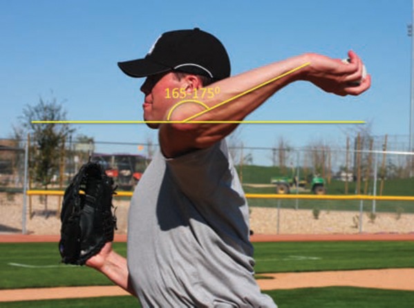
Extreme amounts of external rotation in the range of 165° to 175° are achieved by the throwing extremity during the late cocking phase.
Acceleration
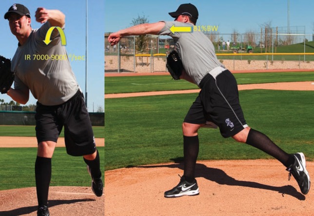
The acceleration phase is defined as the time between maximum external rotation of the shoulder and ball release (Table 5). The trunk continues to rotate and tilt, initiating the transfer of potential energy through the upper extremity. The scapula protracts to maintain a stable base as the humerus undergoes horizontal adduction and violent internal rotation. This rapid motion delivers the arm from as much as 175° of external rotation to 100° of internal rotation (at ball release) in only 42 to 58 milliseconds.25 A decreased time to maximum shoulder internal rotation and increased trunk tilt at ball release have been associated with an increase in ball velocity.31 The subscapularis reaches maximum activity during this phase along with the pectoralis major and latissimus dorsi, producing the violent internal rotation of the humerus reaching forces as high as 185% of its maximum muscle test strength.14 Internal rotation velocities as high as 7000 to 9000 degrees per second have been reported.5,25 The serratus anterior reaches maximum activity during acceleration as it promotes scapular protraction, providing a stable glenoid for humeral rotation.14,16,22,26 During acceleration, the elbow initially flexes from 90° to 120°, then rapidly extends to near 25° just before ball release.25,31 Elbow extension results from a combination of the centrifugal force generated by the rotating torso and the concentric contraction of the triceps; it is immediately followed by shoulder internal rotation.12,16 The biceps brachii supplies elbow flexion torque, reaching a maximum value of 61 N⋅m just before ball release.8 Maximum elbow extension angular velocity occurs just before ball release and may reach mean angular velocity of 2251 degrees per second.21,35 Ball release is aided by wrist flexion to neutral in the 20 milliseconds preceding release and radioulnar pronation to 90° in the 10 milliseconds prior to release.34
Table 5.
Acceleration phase of the pitching motion.a
| Occurs from max external rotation until ball release.5,8,10 |
| Scapula protracts and antetilts—increased activity of serratus anterior (electromyographic data).14,16,24 |
| Shoulder muscle forces shift from eccentric (during LC) to concentric anteriorly.25 |
| Shoulder muscle forces shift from concentric (LC) to eccentric posteriorly.25 |
| Subscapularis reaches it maximum activity, generating humeral internal rotation.14 |
| Max shoulder internal location velocity occurs at or milliseconds after ball release.21,31 |
| Max elbow extension angular velocity occurs at ball release.21,31 |
LC, late cocking.
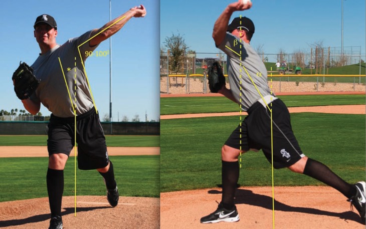
The nondominant rectus abdominus, abdominal obliques, and lumbar paraspinous muscles all show significant activity over their dominant-side counterparts during acceleration to accentuate pelvic and trunk rotation and tilt.33 Concentric rectus femoris contraction contributes to lead leg hip flexion and knee extension, providing a stable front side to help create increased angular momentum of the trunk. Increased forward trunk tilt allows the pitching extremity to accelerate through a greater distance, allowing more force to be transferred to the ball.3,21,31,35 Forward trunk tilt reaches a mean of 32° to 55° and a maximum angular velocity of 300 to 450 degrees per second at ball release (Figure 6).12,21,31,35 Less maximum lead knee flexion angular velocity, increased knee extension (mean 58°),35 and knee extension angular velocity at the instant of ball release are associated with increased velocity (Figure 7).21
Figure 6.
Trunk tilt during acceleration: Trunk forward flexion (α) has been associated with an increase in velocity generation. Also note that during the acceleration phase, the arm is maintained in 90° to 100° of abduction and in horizontal adduction (β), which allows increased linear distance for acceleration forces to be imparted to the baseball. The trunk also assumes a position of lateral tilt (*) in the direction away from the throwing extremity.
Figure 7.
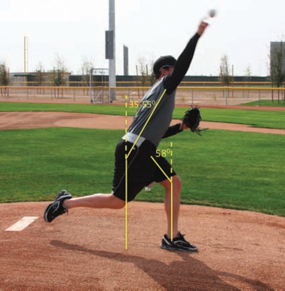
Increased angles of knee extension and forward trunk flexion at ball release have been associated with increased velocities of the pitched ball. The increased knee extension provides a solid base for the trunk to rotate and flex upon, whereas the additional forward flexion allows for an increased distance for the accelerating forces to act upon the ball before release.
Deceleration
The deceleration phase occurs between ball release and maximum humeral internal rotation and elbow extension (Table 6). The phase ends with completion of humeral rotation to 0°, shoulder abduction to 100°, and an increase in horizontal arm adduction to 35°.22 This is the most violent phase of the throwing cycle, resulting in the greatest amount of joint loading encountered during throwing. Excessive posterior (400 N) and inferior shear forces (300 N) occur, as do elevated compressive forces (> 1000 N) and adduction torques.5,8,16,17,22 The posterior shoulder soft tissue structures (teres minor, infraspinatus, and posterior deltoid) dissipate these enormous forces during the acceleration phase as the arm continues to adduct and internally rotate (Figure 8).4,14 These massive eccentric contractile requirements of the posterior shoulder musculature are likely responsible for the posterior capsular and soft tissue reaction commonly seen in throwers and for the glenohumeral internal rotation deficit seen in pitchers.2 After ball release, the upper extremity is outstretched to home plate; the elbow8,25 is flexed 25°; and the arm is abducted an average of 93° and set in 6° of horizontal adduction.8
Table 6.
Deceleration phase of pitching motion.
| Most violent phase of pitching motion. |
| Excessive distraction, posterior/inferior shear forces on glenohumeral joint.8,34 |
| Explosive eccentric loading of rotator cuff to resist distraction.4,14,17,34 |
| Marked eccentric biceps activity.14 |
| Trapezius, rhomboids, and serratus anterior are highly active to decelerate the shoulder girdle and stabilize the scapula.14,16 |
Figure 8.
During deceleration, the posterior shoulder musculature must dissipate the forces generated to propel the ball forward. Slowing the upper extremity, which reaches internal rotation (IR) velocities of 7000 to 9000 degrees per second, generates distraction forces of as high as 81% body weight (BW).34
During deceleration, there is marked biceps and brachialis activity decelerating the rapidly extending elbow and pronating forearm.5,16 The trapezius, rhomboids, and serratus anterior also assist in the deceleration of the shoulder girdle and in the stabilization of the scapula.12 The teres minor is highly active during this phase, resisting anterior humeral head translation, horizontal adduction, and internal rotation.12,13
Follow-through
As follow-through proceeds, the body continues to move forward with the arm until motion has ceased. Horizontal adduction increases to 60°, and muscle firing decreases in general.22 The follow-through phase culminates with the pitcher in a fielding position.8 The decreased joint loading and minimal forces during this phase render it an unlikely culprit for injury.
Differences in Pitchers Characteristics
Competition level
Pitchers at the various competition levels show varying muscle recruitment patterns and use of the kinetic chain to differentially generate top velocities. Professional pitchers predominantly use the subscapularis and latissimus dorsi for acceleration, whereas amateurs use more of the rotator cuff muscles with an active pectoralis minor and a relatively quiescent latissimus dorsi.14 Amateur pitchers display substantial biceps activity during late cocking and deceleration, compared to minimal biceps activity.14 Professional pitchers often lose maximum shoulder external rotation and trunk forward tilt at ball release but maintain pelvis, upper trunk, shoulder, and elbow angular velocities: an adaptation that compensates for position variables and maintains baseline velocity.9
Types of Pitches
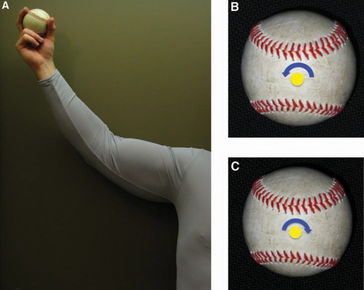
A successful pitcher alters pitch velocity and movement characteristics to keep batters off balance and deter their anticipation of a particular pitch type. Commonly used pitches are the slider, the changeup, and the curveball. The slider is meant to mimic the appearance of a fastball in arm speed and motion with slightly decreased ball velocity and the addition of horizontal plane ball movement (“break”) to fool the hitter. The arm motion and grip remain the same as the fastball, but the pitcher supinates the forearm until ball release during late acceleration, generating rotation of the ball around a central axis (Figure 9), which generates horizontal plane motion from right to left for a right-handed pitcher and vice versa for a left-handed thrower. The fingers are located in the 3 and 9 o’clock position at ball release rather than on top of the ball, as in a fastball (Figure 10). The changeup is thrown with the same arm slot and motion as the fastball. The spin generated on the ball is in the same direction of the fastball (backspin), with a slower velocity to distort the hitter’s timing. The ball is positioned deeper into the palm to decrease the velocity of the pitch. The fingers can be seen on top of the ball at release, as with a fastball (Figure 11). The curveball is thrown slower, with a different trajectory and spin. The pitcher’s fingers are located on top of the ball at release to generate a forward rotation (“12 to 6 o’clock rotation) that allows vertical plane movement (“break or drop”) (Figure 12).
Figure 9.
When throwing a slider, the pitcher attempts to mimic the arm motion and velocity used during a fastball while causing the ball to move in the horizontal plane. To do so, the pitcher begins to supinate his forearm during late acceleration through ball release (A), which generates counterclockwise spin (B) and right-to-left horizontal plane ball movement for a right-handed pitcher. In a left-handed pitcher, the ball will spin in a clockwise manner (C) and cause a left-to-right ball movement. When thrown properly, the ball will rotate around its central axis with spin in the coronal plane only.
Figure 10.
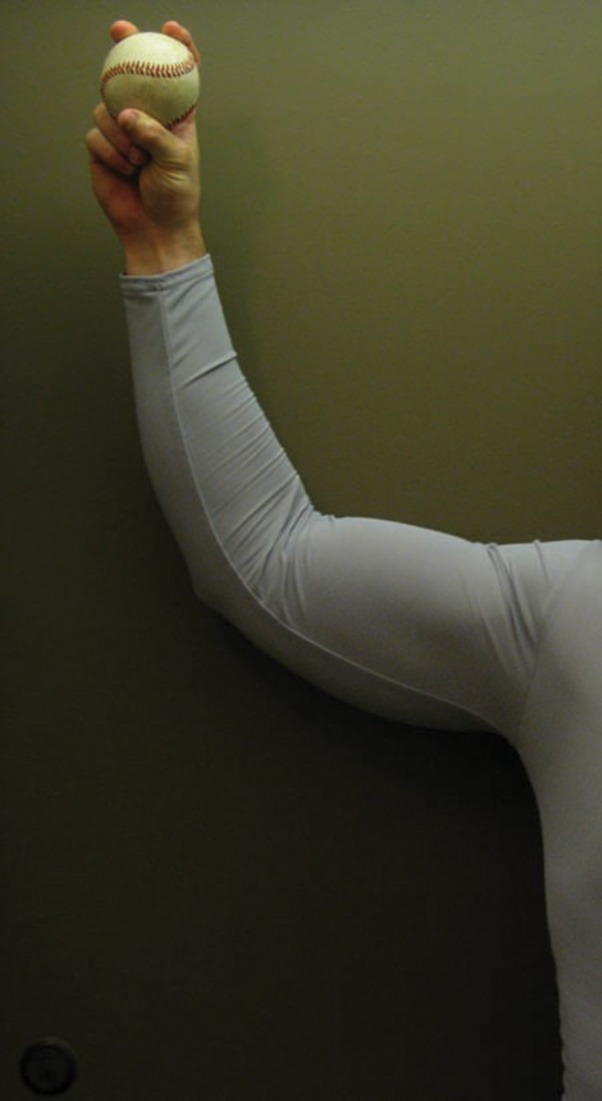
Finger position behind the baseball at ball release that generates backspin on the baseball.
Figure 11.
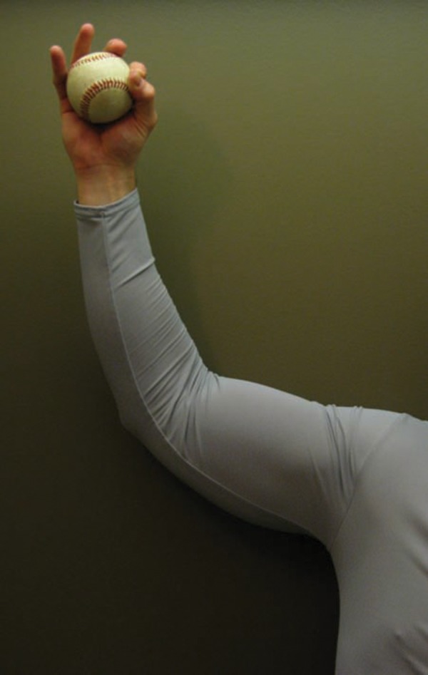
Hand position at ball release when throwing a changeup. The fingers remain behind the baseball, as in the fastball, with 3 fingers behind the ball.
Figure 12.
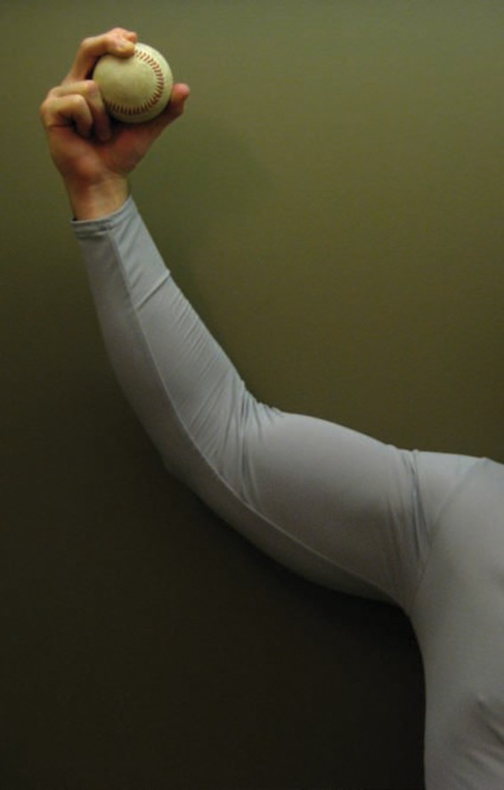
Hand position when throwing a curveball. The fingers at ball release are on top of the ball and generate forward spin on the baseball.
Escamilla et al7 studied the throwing mechanics in overhead throwers with respect to pitch type. Compared to the curveball and changeup, the fastball was thrown with more pelvis and upper torso angular velocities and stride length.7 The differences in peak shoulder angular velocities were significantly greater in the fastball and slider pitches than in the changeup pitches. Peak angular velocities of the pelvis and torso were greater when throwing the slider than the changeup. Compared to the other pitches, the curveball was thrown with more knee flexion, greater forward and lateral trunk tilt, and increased shoulder horizontal adduction at ball release.7 Shoulder abduction was greater during acceleration in the curveball versus fastball. Maximum elbow extension angular velocity and shoulder internal rotation angular velocity were greater with a curveball than a changeup.7 With the changeup, the lead knee continues to flex throughout much of acceleration and is greater at ball release.7 The researchers found subtle differences in 60% of the 26 kinematic parameters among the 4 pitches.
Conclusion
The baseball throwing motion is a complex and coordinated body event that culminates with a ballistic motion of the throwing extremity, exposing its muscular structures to supraphysiologic kinematic loads and motions. Kinematic, kinetic, and temporal variations in the throwing motion have been related to improved velocity and force generation. To generate high velocity, the overhand pitcher must optimize the coordinated use of muscle segments throughout the body to generate and sequentially transfer potential energy to the upper extremity for conversion to kinetic energy to propel the baseball toward home plate. Inefficiency or failure of the kinetic chain can increase the kinetic requirements of the shoulder to maintain top velocity and performance. Knowledge of the kinetic chain and key temporal parameters of the throwing motion can improve technique that can assist in performance enhancement, rehabilitation, and injury prevention.
Footnotes
No potential conflict of interest declared.
NATA Members: Receive 3 free CEUs each year when you subscribe to Sports Health and take and pass the related online quizzes! Not a subscriber? Not a member? The Sports Health–related CEU quizzes are also available for purchase. For more information and to take the quiz for this article, visit www.nata.org/sportshealthquizzes.
References
- 1. Burkhart SS, Morgan CD, Kibler WB. The disabled throwing shoulder: spectrum of pathology. Part I: pathoanatomy and biomechanics. Arthroscopy. 2003;19(4):404-420 [DOI] [PubMed] [Google Scholar]
- 2. Burkhart SS, Morgan CD, Kibler WB. The disabled throwing shoulder: spectrum of pathology. Part III: The SICK scapula, scapular dyskinesis, the kinetic chain, and rehabilitation. Arthroscopy. 2003;19(6):641-661 [DOI] [PubMed] [Google Scholar]
- 3. Decicco PV, Fisher MM. The effects of proprioceptive neuromuscular facilitation stretching on shoulder range of motion in overhand athletes. J Sports Med Phys Fitness. 2005;45(2):183-187 [PubMed] [Google Scholar]
- 4. DiGiovine NM, Jobe FW, Pink M, Perry J. An electromyographic analysis of the upper extremity in pitching. J Shoulder Elbow Surg. 1992;1(1):15-25 [DOI] [PubMed] [Google Scholar]
- 5. Dillman CJ, Fleisig GS, Andrews JR. Biomechanics of pitching with emphasis upon shoulder kinematics. J Orthop Sports Phys Ther. 1993;18(2):402-408 [DOI] [PubMed] [Google Scholar]
- 6. Downar JM, Sauers EL. Clinical measures of shoulder mobility in the professional baseball player. J Athl Train. 2005;40(1):23-29 [PMC free article] [PubMed] [Google Scholar]
- 7. Escamilla RF, Fleisig GS, Barrentine SW, Zheng N, Andrews JR. Kinematic comparisons of throwing different types of baseball pitches. J Appl Biomech. 1998;14:1-23 [Google Scholar]
- 8. Fleisig GS, Andrews JR, Dillman CJ, Escamilla RF. Kinetics of baseball pitching with implications about injury mechanisms. Am J Sports Med. 1995;23(2):233-239 [DOI] [PubMed] [Google Scholar]
- 9. Fleisig GS, Barrentine SW, Zheng N, Escamilla RF, Andrews JR. Kinematic and kinetic comparison of baseball pitching among various levels of development. J Biomech. 1999;32(12):1371-1375 [DOI] [PubMed] [Google Scholar]
- 10. Fleisig GS, Dillman CJ, Andrews JR. Biomechanics of the shoulder during throwing. In: Andrews JR, ed. The Athlete’s Shoulder. New York, NY: Churchill Livingstone; 1994:360-365 [Google Scholar]
- 11. Fleisig G, Escamilla RF, Andrews JR, Matsuo T, Satterwhite Y, Barrentine SW. Kinematic and kinetic comparison between baseball pitching and football passing. J Appl Biomech. 1996;12:207-224 [Google Scholar]
- 12. Fleisig GS, Escamilla RF, Barrentine SW. Biomechanics of pitching: mechanism and motion analysis. In: Andrews JR, Wilk KE, eds. Injuries in Baseball. Philadelphia, PA: Lippincott-Raven; 1998:3-22 [Google Scholar]
- 13. Glousman R, Jobe F, Tibone J, Moynes D, Antonelli D, Perry J. Dynamic electromyographic analysis of the throwing shoulder with glenohumeral instability. J Bone Joint Surg Am. 1988;70(2):220-226 [PubMed] [Google Scholar]
- 14. Gowan ID, Jobe FW, Tibone JE, Perry J, Moynes DR. A comparative electromyographic analysis of the shoulder during pitching: professional versus amateur pitchers. Am J Sports Med. 1987;15(6):586-590 [DOI] [PubMed] [Google Scholar]
- 15. Itoi E, Kuechle DK, Newman SR, Morrey BF, An KN. Stabilising function of the biceps in stable and unstable shoulders. J Bone Joint Surg Br. 1993;75(4):546-550 [DOI] [PubMed] [Google Scholar]
- 16. Jobe FW, Moynes DR, Tibone JE, Perry J. An EMG analysis of the shoulder in pitching: a second report. Am J Sports Med. 1984;12(3):218-220 [DOI] [PubMed] [Google Scholar]
- 17. Jobe FW, Tibone JE, Perry J, Moynes D. An EMG analysis of the shoulder in throwing and pitching: a preliminary report. Am J Sports Med. 1983;11(1):3-5 [DOI] [PubMed] [Google Scholar]
- 18. Kibler WB. The role of the scapula in athletic shoulder function. Am J Sports Med. 1998;26(2):325-337 [DOI] [PubMed] [Google Scholar]
- 19. Kibler WB, Chandler J. Baseball and tennis. In: Griffin LY, ed. Rehabilitation of the Injured Knee. St. Louis, MO: Mosby; 1995:219-226 [Google Scholar]
- 20. Matsen FA, Harryman DT, Sidles JA. Mechanics of glenohumeral instability. Clin Sports Med. 1991;10(4):783-788 [PubMed] [Google Scholar]
- 21. Matsuo T, Escamilla R, Fleisig GS, Barrentine SW, Andrews JR. Comparison of kinematic and temporal parameters between different pitch velocity groups. J Appl Biomech. 2001;17(1):1-13 [Google Scholar]
- 22. Meister K. Injuries to the shoulder in the throwing athlete. Part one: biomechanics/pathophysiology/classification of injury. Am J Sports Med. 2000;28(2):265-275 [DOI] [PubMed] [Google Scholar]
- 23. Mura N, O’Driscoll SW, Zobitz ME, et al. The effect of infraspinatus disruption on glenohumeral torque and superior migration of the humeral head: a biomechanical study. J Shoulder Elbow Surg. 2003;12(2):179-184 [DOI] [PubMed] [Google Scholar]
- 24. Myers JB, Laudner KG, Pasquale MR, Bradley JP, Lephart SM. Scapular position and orientation in throwing athletes. Am J Sports Med. 2005;33(2):263-271 [DOI] [PubMed] [Google Scholar]
- 25. Pappas AM, Zawacki RM, Sullivan TJ. Biomechanics of baseball pitching: a preliminary report. Am J Sports Med. 1985;13(4):216-222 [DOI] [PubMed] [Google Scholar]
- 26. Park SS, Loebenberg ML, Rokito AS, Zuckerman JD. The shoulder in baseball pitching: biomechanics and related injuries: part 1. Bull Hosp Jt Dis. 2002;61(1-2):68-79 [PubMed] [Google Scholar]
- 27. Putnam CA. Sequential motions of body segments in striking and throwing skill: descriptions and explanations. J Biomech. 1993;26(suppl 1):125-135 [DOI] [PubMed] [Google Scholar]
- 28. Sharkey NA, Marder RA. The rotator cuff opposes superior translation of the humeral head. Am J Sports Med. 1995;23(3):270-275 [DOI] [PubMed] [Google Scholar]
- 29. Sisto DJ, Jobe FW, Moynes DR, Antonelli DJ. An electromyographic analysis of the elbow in pitching. Am J Sports Med. 1987;15(3):260-263 [DOI] [PubMed] [Google Scholar]
- 30. Stodden DF, Campbell BM, Moyer TM. Comparison of trunk kinematics in trunk training exercises and throwing. J Strength Cond Res. 2008;22(1):112-118 [DOI] [PubMed] [Google Scholar]
- 31. Stodden DF, Fleisig GS, McLean SP, Andrews JR. Relationship of biomechanical factors to baseball pitching velocity: within pitcher variation. J Appl Biomech. 2005;21(1):44-56 [DOI] [PubMed] [Google Scholar]
- 32. Stodden DF, Fleisig GS, McLean SP, Lyman SL, Andrews JR. Relationship of pelvis and upper torso kinematics to pitched baseball velocity. J Appl Biomech. 2001;17(2):164-172 [Google Scholar]
- 33. Watkins RG, Dennis S, Dillin WH, et al. Dynamic EMG analysis of torque transfer in professional baseball pitchers. Spine. 1989;14(4):404-408 [DOI] [PubMed] [Google Scholar]
- 34. Werner SL, Guido JA, Stewart GW, McNeice RP, VanDyke T, Jones DG. Relationships between throwing mechanics and shoulder distraction in collegiate baseball pitchers. J Shoulder Elbow Surg. 2007;16(1):37-42 [DOI] [PubMed] [Google Scholar]
- 35. Werner SL, Suri M, Guido JA, Meister K, Jones DG. Relationships between ball velocity and throwing mechanics in collegiate baseball pitchers. J Shoulder Elbow Surg. 2008;17(6):905-908 [DOI] [PubMed] [Google Scholar]


